Search results (72 results)
-
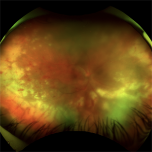 Acute Retinal Necrosis (ARN)
Jul 3 2025 by Heitor Nogueira
Fundus photograph of an 63-year-old woman who reported unilateral visual acuity loss for 10 days associated with ocular pain. He presented conjunctival hyperemia with temporal and nasal nodular scleritis, anterior chamber reaction 2+/4+, Koeppe nodules, granulomatous PKs, vitreitis 2+/4+, multiple areas of vasculitis in the arcades and periphery, associated with hemorrhages and necrotizing retinitis in the temporal, inferior and nasal periphery. Positive serology for Herpes Virus
Photographer: Heitor Nogueira, Penido Burnier Institute, Campinas, São Paulo, Brazil
Imaging device: Optos Daytona
Condition/keywords: ARN complications, Herpes, progressive outer retinal necrosis (PORN), Uveitis
-
 Acute Retinal Necrosis
Jul 3 2025 by Heitor Nogueira
Fundus photograph of an 53-year-old woman with patient who reported unilateral visual acuity loss for 10 days associated with ocular pain. She presented conjunctival hyperemia with temporal and nasal nodular scleritis, anterior chamber reaction 2+/4+, Koeppe nodules, granulomatous PKs, vitritis 2+/4+, multiple areas of vasculitis in arcades and periphery, associated with hemorrhages and necrotizing retinitis in temporal, inferior and nasal periphery. patient who reported unilateral visual acuity loss for 10 days associated with ocular pain. He presented conjunctival hyperemia with temporal and nasal nodular scleritis, anterior chamber reaction 2+/4+, Koeppe nodules, granulomatous PKs, vitreitis 2+/4+, multiple areas of vasculitis in the arcades and periphery, associated with hemorrhages and necrotizing retinitis in the temporal, inferior and nasal periphery. Positive serology for Herpes Virus.
Photographer: Heitor Nogueira, Penido Burnier Institute and CHOV, Campinas, São Paulo, Brazil
Imaging device: Optos Daytona
Condition/keywords: ARN complications, Herpes, progressive outer retinal necrosis (PORN)
-
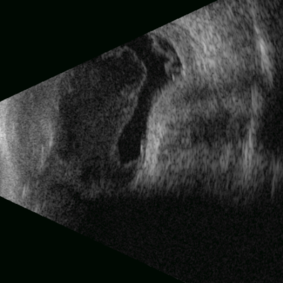 Posterior Nodular Scleritis
Apr 23 2025 by Gustavo Uriel Fonseca Aguirre
This B-mode ultrasound scan demonstrates a posterior scleral nodule accompanied by vitritis, serous retinal detachment, and annular choroidal detachment. The nodule appears as a localized hypoechoic scleral thickening, while the serous retinal detachment shows a smooth convex configuration. The choroidal detachment presents with the characteristic ring-shaped elevation, suggesting significant intraocular inflammation or hypotony.
Photographer: Gustavo U. Fonseca Aguirre, Hospital Conde de Valenciana, Ciudad de México
Condition/keywords: posterior nodular scleritis, posterior scleritis
-
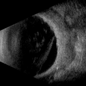 Necrotizing Scleritis USG
Apr 17 2025 by Gustavo Uriel Fonseca Aguirre
This B-mode transverse ultrasound scan reveals necrotizing scleritis with inferior perilimbal uveal tissue prolapse, demonstrating scleral thinning and irregular uveal protrusion. Vitreous cellularity and partial vitreous detachment are also observed, indicating associated intraocular inflammation. These findings collectively characterize this severe inflammatory condition.
Photographer: Gustavo U. Fonseca Aguirre, Hospital Conde de Valenciana, Ciudad de México
Condition/keywords: necrotizing scleritis
-
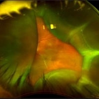
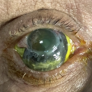 Necrotizing Scleritis
Apr 17 2025 by Gustavo Uriel Fonseca Aguirre
The clinical photograph shows necrotizing scleritis with perilimbal involvement, featuring marked scleral thinning and violaceous episcleral injection in the inferior quadrant. Focal uveal prolapse is visible at the area of maximal scleral necrosis, accompanied by peripheral ulcerative keratitis. Fluorescein staining residue is observed on the ocular surface. Associated findings include mild conjunctival chemosis and dilated episcleral vessels.
Photographer: Gustavo U. Fonseca Aguirre, Hospital Conde de Valenciana, Ciudad de México
Condition/keywords: necrotizing scleritis
-
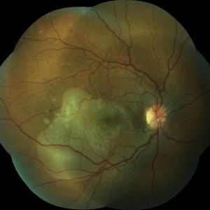
 Posterior Scleritis
Jan 4 2025 by Tejaswita Verma
Left eye fundus photo of a 49 year old female presenting with 5 days history of blurred vision, Vision was finger counting 2 mts. and nodular posterior scleritis with T sign on USG was present. OCT revealed altered foveal contour with septations and SRF pockets and bacillary layer detachment.
Photographer: DR. TEJASWITA VERMA
Imaging device: MIRANTE
Condition/keywords: posterior scleritis
-
 Posterior Scleritis
Aug 5 2024 by Aniruddh Soni, DO DNB FLVPEI
Fundus photo of a 35 year old female presenting with pain and defective vision.
Photographer: Dr Aniruddh Soni
Imaging device: Zeiss Visucam 500
Condition/keywords: posterior scleritis
-
 POSTERIOR SCLERITIS
Nov 1 2023 by ANKIT JAIN
USG B SACN image showing typical T-sign in axial horizontal view with increased thickening of the sclero-choroidal complex suggestive of posterior scleritis
Photographer: DR ANKIT JAIN
Condition/keywords: B scan ultrasound, posterior scleritis, ULTRASOUND
-
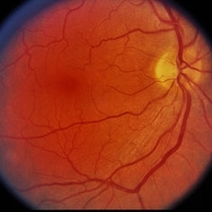
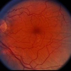
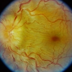 Posterior Scleritis
Sep 12 2023 by Ben Serar
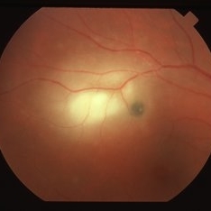
Fundus photograph of LE showing Disc edema with Choroidal folds in a case of Posterior Scleritis
Condition/keywords: chorioretinal folds, disc edema, posterior scleritis
-
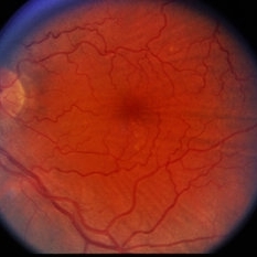
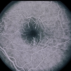
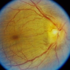 Posterior Scleritis
Sep 12 2023 by Ben Serar
Fundus photograph of RE showing Disc edema with Choroidal folds in a case of Posterior Scleritis.
Condition/keywords: chorioretinal folds, disc edema, Posterior scleritis
-
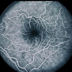
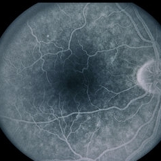
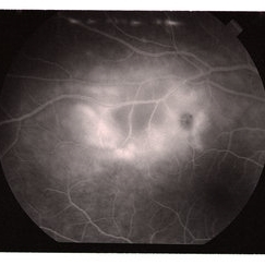 Posterior Scleritis
Jun 6 2019 by Gary R. Cook, MD, FACS
Mid-phase fluorescein angiogram image of acute posterior scleritis lesion beneath the superotemporal arcade OD; V.A. = 20/40-2
Imaging device: Topcon VT-50
Condition/keywords: FA mid phase, fluorescein angiogram (FA), posterior scleritis
-
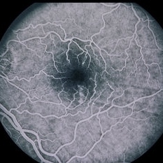
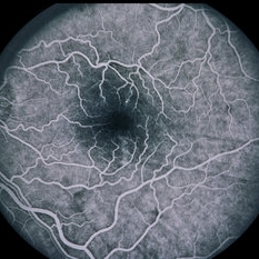
 Posterior Scleritis
Apr 2 2019 by Gary R. Cook, MD, FACS
White female with a focus of acute posterior scleritis beneath the superotemporal arcade OD; VA = 20/40-2
Imaging device: Topcon VT-50
Condition/keywords: posterior scleritis
-
 Slide 7-60
Feb 25 2019 by Lancaster Course in Ophthalmology
Marked thickening of the sclera in scleritis.
Condition/keywords: sclera, scleritis
-
 Slide 7-58
Feb 25 2019 by Lancaster Course in Ophthalmology
Scleritis showing marked granulomatous inflammation of the sclera.
Condition/keywords: sclera, scleritis
-
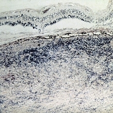
 Slide 1-29
Feb 19 2019 by Lancaster Course in Ophthalmology
Smudgy, "fibrinoid" necrosis of collagen in the sclera of a patient with rheumatoid scleritis. Some abscess formation is seen above, rimmed by a granulomatous reaction. (H&E stain)
Condition/keywords: fibrinoid, sclera, scleritis
-
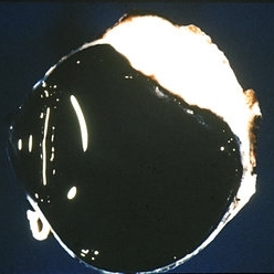
 Choroidal Detachment
Oct 4 2018 by Emily Cooper
Optos photograph of an 80-year-old man presenting with red, painful eye after heart surgery.
Photographer: Emily Cooper, Retina Specialists of Michigan, Grand Rapids MI
Imaging device: Optos
Condition/keywords: choroidal detachment, posterior scleritis
-
 Wegener's Disease
Feb 20 2015 by H. Michael Lambert, MD
Thinning and vascularization of the peripheral cornea, a sequelae of peripheral ulcerative keratitis and scleritis.
Condition/keywords: corneal furrow, Wegener's granulomatosis
-
 Scleritis with Chorioretinal Folds
Feb 13 2015 by David Callanan, MD
46-year-old white male, scleritis with chorio-retinal folds.
Condition/keywords: chorioretinal fold, scleritis
-
 Scleritis with Chorioretinal Folds
Feb 13 2015 by David Callanan, MD
46-year-old white male, scleritis with chorio-retinal folds.
Condition/keywords: chorioretinal fold, scleritis
-
 Scleritis with Chorioretinal Folds
Feb 13 2015 by David Callanan, MD
46-year-old white male, scleritis with chorio-retinal folds.
Condition/keywords: chorioretinal fold, scleritis
-
 Scleritis with Chorioretinal Folds
Feb 13 2015 by David Callanan, MD
46-year-old white male, scleritis with chorio-retinal folds.
Condition/keywords: chorioretinal fold, scleritis
-
 Scleritis with Chorioretinal Folds
Feb 13 2015 by David Callanan, MD
46-year-old white male, scleritis with chorio-retinal folds.
Condition/keywords: chorioretinal fold, scleritis
-
 Scleritis with Chorioretinal Folds
Feb 13 2015 by David Callanan, MD
46-year-old white male, scleritis with chorio-retinal folds.
Condition/keywords: chorioretinal fold, scleritis
-
 Scleritis with Chorioretinal Folds
Feb 13 2015 by David Callanan, MD
46-year-old white male, scleritis with chorio-retinal folds.
Condition/keywords: chorioretinal fold, scleritis
-
 Scleritis with Chorioretinal Folds
Feb 13 2015 by David Callanan, MD
46-year-old white male, scleritis with chorio-retinal folds.
Condition/keywords: chorioretinal fold, scleritis
 Loading…