Search results (453 results)
-
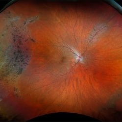
 Retinitis Pigmentosa
Dec 9 2025 by Kimberly Wakester
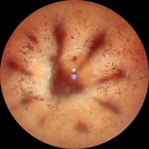
Optomap RGB and AF of an 78-year-old woman with Retinitis Pigmentosa. Patient is to continue follow up care every 6 months to monitor progression.
Photographer: Kimberly Wakester, COA, OCT-C
Imaging device: Optos California
Condition/keywords: retinitis pigmentosa
-
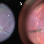
 Unilateral Pigmentary Retinopathy
Nov 9 2025 by Hrishikesh Naik, MS
Montage fundus photographs of a 47 year old female presenting with unilateral vision loss in the left eye. Fundoscopy revealed extensive intraretinal pigment clumps, waxy disc pallor, and marked vessel attenuation in the left eye with a normal fundus in the right. Electroretinography showed unilateral reduction in rod and cone function. Unilateral pigmentary retinopathy, an uncommon variant of retinitis pigmentosa (reported incidence ˜ 5%) presents with RP-like changes in one eye, the fellow eye being completely normal. Proposed causes include lyonization and somatic mosaicism. Conditions which mimic RP should be excluded, and any diagnoses should be supported with electrodiagnostic tests and autofluorescence imaging. Management parallels RP, focusing on cataract and macular complications and long-term follow-up to monitor possible bilateral progression.
Imaging device: Zeiss Visucam 224
Condition/keywords: montage, retinitis pigmentosa, unilateral
-
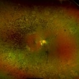
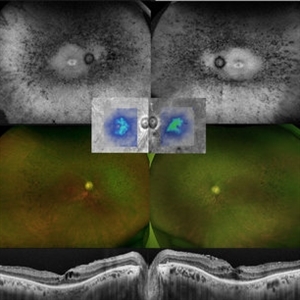
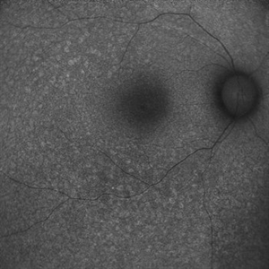
 Retinitis Pigmentosa: Now available in its Pericentral edition
Nov 7 2025 by SHRADDHA RAJ SHRIVASTAVA
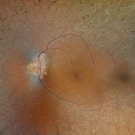
Left eye Green-FAF image, of a 50 year old patient, diagnosed with bilateral Pericentral variant of Retinitis Pigmentosa. The disease is characterized by pigmentary changes closer to the macula, and an earlier involvement of central visual acuity as compared to typical RP. We can see prominent, scalloped hypoautofluorescent lesions in the pericentral region, which corresponds to areas of severe RPE atrophy and photoreceptor cell loss. Macula shows preserved background autofluorescence, with darker areas corresponding to no detectable fluorescence due to macular atrophy (loss of melanin/lipofuscin).
Photographer: Dr. Shraddha Raj Shrivastava
Imaging device: Nidek Mirante SLO/OCT (Confocal scanning/Spectral domain OCT)
Condition/keywords: ATYPICAL RETINITIS PIGMENTOSA, fundus autofluorescence (FAF), pericentral retinitis pigmentosa, RP variant
-
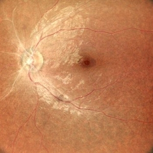
 Retinitis Pigmentosa: Now available in its Pericentral edition
Nov 7 2025 by SHRADDHA RAJ SHRIVASTAVA
Right eye fundus photo of a 50 year old patient, diagnosed with bilateral Pericentral variant of Retinitis Pigmentosa. True to the subtype, the pigmentation is closer to fixation. There are bony spicules like pigmentary changes and RPE atrophy seen around the macula and disc (posterior pole), just adjacent to the arcades, while the peripheral fundus appears unaffected. The macula shows severe macular atrophy and scarring. Similar changes were observed in the left eye.
Photographer: Dr. Shraddha Raj Shrivastava
Imaging device: Nidek Mirante SLO/OCT (Confocal scanning/Spectral domain OCT)
Condition/keywords: pericentral retinitis pigmentosa, retinitis pigmentosa (RP) dystrophy, Rod cone dystrophy, RP variant
-
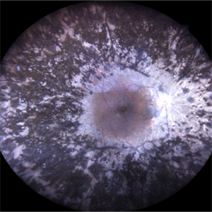
 Retinitis Pigmentosa
Oct 1 2025 by Shivankar Sen, MS, FVRS
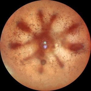
Typical Case of Retinitis Pigmentosa - Optos Green Auto Fluorescence highlighting Peripapillary Hypoautofluorescence; a classical ring hypoautofluorescence at the macula surrounding the fovea and speckled autofluorescence in the periphery corresponding to bony spicules
Photographer: Dr. Shivankar Sen
Imaging device: Optos Daytona
Condition/keywords: fundus autofluorescence (FAF), Optos, RETINITIS PIGMENTOSA
-
 Retinitis Pigmentosa with Macular Hole with Posterior Subcapsular Cataract
Apr 28 2025 by Malvika Singh
Fundus photograph of the left eye of a 31 year old with retinitis pigmentosa, showing the shadow of posterior subcapsular cataract over the fundus.
Photographer: Dr Malvika Singh, Retina Foundation, Ahmedabad, India
Imaging device: Mirante SLO/OCT
Condition/keywords: posterior subcapsular cataract, retinitis pigmentosa
-
 Retinitis Pigmentosa with Macular Hole with Posterior Subcapsular Cataract
Apr 28 2025 by Malvika Singh
Fundus photograph of the right eye of a 31 year old with retinitis pigmentosa with a macular hole, showing the shadow of posterior subcapsular cataract over the fundus.
Photographer: Dr Malvika Singh, Retina Foundation, Ahmedabad, India
Imaging device: Mirante SLO/OCT
Condition/keywords: macular hole, posterior subcapsular cataract, retinitis pigmentosa
-
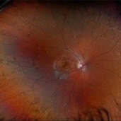
 Optic Disc Drusen, RP
Apr 21 2025 by Virginia Gebhart
28 year old male with stable retinitis pigmentosa and optic disc drusen OU. Bardet-Biedl variant identified in previous genetic testing. BCVA 20/50 OD, 20/30 OS
Photographer: Virginia Gebhart, Retina Consultants of Carolina
Imaging device: Optos California
Condition/keywords: Drusen, optic disc drusen, retinitis pigmentosa
-
 Retinitis Pigmentosa
Apr 17 2025 by Virginia Gebhart
Fundus autofluorescence of 75 year old female with Retinitis Pigmentosa. Pt diagnosed at age 53. Diffuse RPE atrophy with minimal central sparing present in both eyes. Stable and unchanged compared to previous exams. BCVA 20/200 OD, NLP OS
Photographer: Virginia Gebhart, Retina Consultants of Carolina
Imaging device: Optos California
Condition/keywords: autofluorescence imaging, bone spicule, retinitis pigmentosa, RP
-
 Retinitis Pigmentosa
Apr 9 2025 by Virginia Gebhart
35 year old female with stable sectoral RP and high myopia OU. RP has not progressed in either eye since initial visit in 2021. Will continue to observe. VA 20/20 OU
Photographer: Virginia Gebhart, Retina Consultants of Carolina
Imaging device: Optos California
Condition/keywords: high myopia, retinitis pigmentosa
-
 Retinitis Pigmentosa
Apr 1 2025 by Jordyn Beckman
63 year old woman with Retinitis Pigmentosa observed over time with peripheral loss. Over the span of 5 years BCVA changed from 20/25 to 20/50.
Photographer: Jordyn Beckman, Retina Consultants of Carolina, P.A.
Imaging device: Optos California
Condition/keywords: atrophy, bone spicules, retinitis pigmentosa
-
 Retinitis Pigmentosa
Mar 27 2025 by T. P . VIGNESH, MBBS,MS
Fundus photograph of a 52-year-old woman with retinitis pigmentosa with cystoid macular edema.
Photographer: Bharathi
Imaging device: EIDON
Condition/keywords: retinitis pigmentosa
-
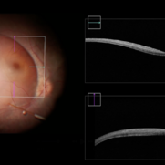 Completed Bleb with OCT Through Fovea
Mar 25 2025 by Robert Andrew Sisk, MD, FACS, FASRS
Color still from surgical video of subretinal delivery of laru-zova for X-linked retinitis pigmentosa. Live optical coherence tomography (OCT) with foveal tracking via the embedded software in the operating microscope allows monitoring foveal integrity for signs of stress. The contour of the fovea does not exceed the curvature of the bleb (e.g. no inversion). The tangential cannula angle facilitated steering of the bleb posteriorly. The bleb covers essentially the entire macula, which is the target area.
Imaging device: Zeiss Artevo 800
Condition/keywords: gene therapy, genetic disorder, optical coherence tomography (OCT), retinitis pigmentosa, subretinal injection
-
 Cannula Tip Pressure
Mar 25 2025 by Robert Andrew Sisk, MD, FACS, FASRS
Color stills from surgical videos of subretinal delivery of gene augmentation therapy with A) voretigene neparvovec-ryzl and B) laru-zova. In the left panel, the cannula is slightly bent, and the retina and RPE are blanched white around the cannula tip engagement. The bleb was challenging to form in this patient with advanced retinal degeneration, and the bleb is shallow and mostly clear. In the right panel, the cannula tip is gently engaged, the cannula is straight, and it follows the retinotomy as the retina is elevated by the injection fluid.
Imaging device: Leica Proveo 8
Condition/keywords: gene therapy, genetic disorder, Leber's congenital amaurosis, retinitis pigmentosa, subretinal injection
-
 Hereditary Retinal Dystrophy
Feb 27 2025 by Kimberly Wakester
Optomap RGB image of a 7-year-old girl with Hereditary retinal dystrophy. Biological mother is a CHM gene carrier and biological father is diagnosed with RP. Patient had genetic testing and was also confirmed to be a CHM gene carrier and also has the TTC21B gene. There is linear pigmentary changes on clinical exam and fundus photos. Atypical appearance of Retinitis Pigmentosa. Patient will continue follow up care with repeat imaging.
Photographer: Kimberly Wakester, COA
Imaging device: Optos California
Condition/keywords: CHM gene, hereditary retinal dystrophy, linear pigmentary changes
-
 Retinitis Pigmentosa
Feb 18 2025 by Drew Mitchell
FAF, Color, IR, OCT of Mild CME secondary to Retinitis Pigmentosa.
Photographer: Drew Mitchell OCT-C
Imaging device: Optos California
Condition/keywords: cystoid macular edema (CME), Optos, OPTOS CALIFORNIA, retinitis pigmentosa, RP
-
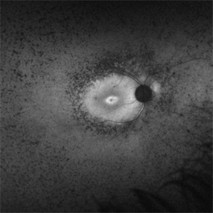 Retinitis Pigmentosa Bullseye Appearing Autofluorescence
Feb 4 2025 by Isaac Agranoff
Fundus Autofluorescence of a 14-year-old boy with suspected RP. ERG performed afterwards was almost flat. VA measured at 20/30 but with extensive constriction of confrontational visual fields. Currently awaiting genetic testing.
Photographer: Isaac Agranoff
Imaging device: Optos California
Condition/keywords: fundus autofluorescence (FAF), retinitis pigmentosa, RP
-
 Retinitis Pigmentosa with PPRPE - FAF-G
Jan 27 2025 by Vishal Agrawal, MD, FRCS,FACS,FASRS
16 year-old male patient presented with DOV, nyctalopia and nystagmus. Fundus revealed pigment clumping, pale disc and preserved para-arteriolar retinal pigment epithelium (PPRPE) in both eyes. Genetic testing revealed CRB1 gene mutation.
Photographer: Dr Ayushi Gupta
Imaging device: Clarus 700
Condition/keywords: retinitis pigmentosa
-
 Retinitis Pigmentosa with PPRPE
Jan 27 2025 by Vishal Agrawal, MD, FRCS,FACS,FASRS
16 year-old male patient presented with DOV, nyctalopia and nystagmus. Fundus revealed pigment clumping, pale disc and preserved para-arteriolar retinal pigment epithelium (PPRPE) in both eyes. Genetic testing revealed CRB1 gene mutation.
Photographer: Dr Ayushi
Imaging device: Clarus 700
Condition/keywords: retinitis pigmentosa
-
 Retinitis Pigmentosa
Jan 15 2025 by Virginia Gebhart
52 year old male with advanced RP OU. BCVA HM OD, LP OS. Referred to genetic specialist per pt request to discuss gene therapy.
Photographer: Virginia Gebhart, Retina Consultants of Carolina
Imaging device: Optos California
Condition/keywords: bone spicule, retinitis pigmentosa, retinitis pigmentosa (RP) dystrophy
-
 Retinitis Pigmentosa
Jan 11 2025 by rohan jain
A case of advance retinitis pigmentosa in a 56 year-old male with BCVA- hand movement.
Photographer: Dr. ROHAN JAIN
Condition/keywords: bone spicule, Night Blindness, retinitis pigmentosa, RP
-
 Asteroid Hyalosis in Retinitis Pigmentosa
Dec 9 2024 by Mauricio Bayram-Suverza, MD
A 54 year-old male patient presented with asteroid hyalosis. Retinal examination revealed the presence of bone spicules, primarily located in the mid-periphery. Genetic testing identified a pathogenic variant in the RHO gene.
Photographer: Mauricio Bayram-Suverza, Casey Eye Institute, OHSU.
Imaging device: Optos California
Condition/keywords: Asteroid hyalosis, retinal dystrophy, Retinitis Pigmentosa, vitreous
-
 Atypical RP with Typhoid Retinitis Sequelae with Old CRAO
Dec 5 2024 by Tejaswita Verma
FAF of a 20 year old female who presented with 2 months history of sudden painless vision loss, bilaterally light perception vision, s/o presumed atypical RP, bilateral old CRAO with typhoid retinitis sequelae.
Photographer: DR. TEJASWITA VERMA
Imaging device: MIRANTE
Condition/keywords: CRAO, retinitis pigmentosa, typhoid fever
-
 Atypical Retintis Pigmentosa With Old CRAO
Nov 15 2024 by Tejaswita Verma
Fundus image of a 20 year old female who presented with bilateral visual loss since 2 months following history of typhoid fever, UTI, showing pale disc, sclerosed vessels,altered foveal reflex and granular fundus. Vision was light perception in both eyes. CBC, MRI Brain +Orbit , carotid doppler tests were WNL. Brucella IgG,IgM negative. ERG and VEP were abnormal.
Photographer: DR. TEJASWITA VERMA
Imaging device: MIRANTE
Condition/keywords: ATYPICAL RETINITIS PIGMENTOSA, CRAO
-
 Advanced RP
Nov 5 2024 by rahul saradge
A man, 58, arrived complaining of BOV for both near and distance vision in both eyes, with a 6/9 BCVA in each eye. For a year, the patient had been taking medication for both diabetes and hypertension. In both eyes, the dilated ophthalmoscopic retina revealed waxy disc pallor paired with bony spicules in the mid-periphery. The patient was prescribed spectacles and given counseling regarding the nature of the illness.
Photographer: Lokesh Dukare ,Isha Netralaya Thane
Imaging device: optos
Condition/keywords: bone spicule, optic disc pallor, Optos, Retinitis Pigmentosa
 Loading…