Search results (162 results)
-
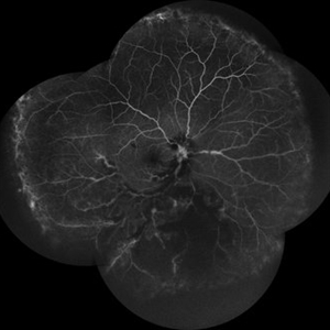
 Angiographic Storm: Fluorescein Leakage in Retinal Vasculitis
Nov 17 2025 by SHRADDHA RAJ SHRIVASTAVA
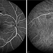
This left eye montage fundus fluorescein angiography (FFA) image of a 19 year old male with idiopathic retinal vasculitis, having skip vasculitic lesions predominantly involving retinal veins. There are areas of blocked fluorescence due to intraretinal hemorrhages, the involved veins have filling defects and occlusions, leading to formation of numerous collateral channels. The inflamed vessels also show perivascular fuzzy hyperfluorescent stain due to leakage of dye. We can also see multiple peripheral capillary non perfusion (CNP) areas, with a 'hot disc', suggestive of ongoing inflammation.
Photographer: Dr. Shraddha Raj Shrivastava
Imaging device: Nidek Mirante SLO/OCT (Confocal scanning/Spectral domain OCT)
Condition/keywords: FA late phase leakage, Fundus Fluorescein Angiography, idiopathic retinal vasculitis, optic disc leakage, VASCULITIS
-
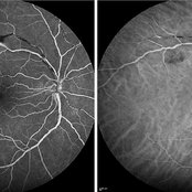
 Disc Edema with Vasculitis
Jul 15 2025 by rohan jain
Case of disc edema with vasculitis.
Photographer: Dr. ROHAN JAIN
Imaging device: mirante
Condition/keywords: disc edema, idiopathic retinal vasculitis, VASCULITIS
-
 Disc Edema with Vasculitis
Jul 15 2025 by rohan jain
Case of disc edema with vasculitis.
Photographer: Dr. ROHAN JAIN
Imaging device: mirante
Condition/keywords: disc edema, idiopathic retinal vasculitis, VASCULITIS
-
 Retinal Vasculitis
Mar 26 2025 by Korey Starkey
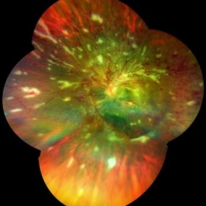
41 year-old patient presents with vascular FA findings of occlusive vasculitis with four quadrant Kyrieleis plaques OU showcases a possibly rare but reported atypical presentation of Behcet's Syndrome.
Photographer: Korey Starkey
Imaging device: Optos
Condition/keywords: FA early phase, Fundus Fluorescein Angiography, ischemia, Optos, retinal vasculitis, ultra-wide field imaging, venous beading
-
 Retinal Vasculitis
Mar 26 2025 by Korey Starkey
41 year-old patient presents with vascular FA findings of occlusive vasculitis with four quadrant Kyrieleis plaques OU showcases a possibly rare but reported atypical presentation of Behcet's Syndrome.
Photographer: Korey Starkey
Imaging device: Optos
Condition/keywords: Behcet's Disease, FA early phase, Fundus Fluorescein Angiography, Optos, retinal vasculitis, ultra-wide field imaging, venous beading
-
 Retinal Vasculitis/TAU
Jan 23 2025 by Virginia Gebhart
25 year-old female with vascular sheathing and traction, concern for possible tattoo-associated uveitis. Pt confirms ringing in ears and occasional rash on tattoos. Most recent lab workup revealed elevated ANA. Referred to rheumatologist, treatment pending. Recommended pt abstain from further tattoos.
Photographer: Virginia Gebhart, Retina Consultants of Carolina
Imaging device: Optos California
Condition/keywords: retinal vasculitis
-
 Early FA/ICG at 1 Minute of Atypical ANCA Associated Retinal Vasculitis
Nov 13 2024 by Deepak Sambhara, MD
Fluorescein and Indocyanine Green Angiography of a 49-year-old male with high ANA titer, atypical ANCA positivity, who presented to clinic with 1 month of vision loss. Exam revealed anterior chamber cell, mild vitreous cell, sclerotic vessels along arterioles. Early FA/ICG at 1 minute demonstrates absent arteriole fill.
Photographer: Killian Roberts, Micaela Hertz; Eye Clinic of Wisconsin
Imaging device: Heidelberg Spectralis
Condition/keywords: A-ANCA, autoimmune vasculitis, fluorescein angiogram (FA), indocyanine green (ICG) angiography, retinal vasculitis
-
 Late FA/ICG at 4 Minutes of Atypical ANCA Associated Retinal Vasculitis
Nov 13 2024 by Deepak Sambhara, MD
Fluorescein and Indocyanine Green Angiography of a 49-year-old male with high ANA titer, atypical ANCA positivity, who presented to clinic with 1 month of vision loss. Exam revealed anterior chamber cell, mild vitreous cell, sclerotic vessels along arterioles. Late FA/ICG at 4 minutes demonstrates absent arteriole fill with venular periphlebitis.
Photographer: Killian Roberts, Micaela Hertz; Eye Clinic of Wisconsin
Imaging device: Heidelberg Spectralis
Condition/keywords: A-ANCA, autoimmune vasculitis, fluorescein angiogram (FA), indocyanine green (ICG) angiography, retinal vasculitis
-
 Occlusive Retinal Vasculitis
Oct 3 2024 by Logan ryzenga
4 view ultra-widefield Optos fluorescein angiogram in the left eye of a 39 year old woman occlusive retinal vasculitis with four quadrant Kyrieleis plaques. This is a showcase of a suspected, rarely reported, and atypical presentation of Behcet's Syndrome.
Photographer: Logan Ryzenga
Imaging device: Optos California
Condition/keywords: Behcet's Disease, Behcet's uveitis, Fluorescein angiography, fluorescein leakage, kyrieleis plaques, non-perfusion, OPTOS, OPTOS CALIFORNIA, ultra-wide field imaging, Uveitis
-
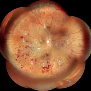
 Atypical Tubercular Occlusive Peripheral Retinal Vasculitis
Jun 21 2024 by Tejaswita Verma
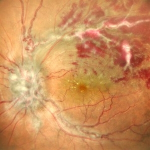
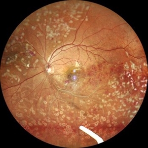
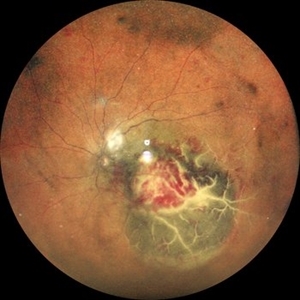
Follow up right eye fundus photograph of a 27 year old male with vision 6/12 , diagnosed with systemic tuberculosis(mediastinal lymphadenopathy on chest CT) on oral steroids, and started on ATT .We can see a parafoveal sub-ILM hemorrhage with vascular sheathing and hemorrhages in inferior and temporal quadrants . The patient was advised anti-VEGF intravitreal injection, later sectoral laser after resolution of inflammation
Photographer: DR. TEJASWITA VERMA
Imaging device: MIRANTE
Condition/keywords: obliterative peripheral vasculitis, ocular tuberculosis
-
 FFA in Atypical Tubercular Peripheral Occlusive Retinal Vasculitis
Jun 21 2024 by Tejaswita Verma
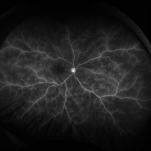
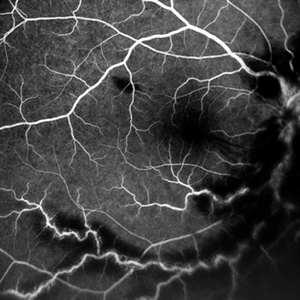
Right eye FFA montage of a 27 year male with peripheral occlusive tubercular vasculitis, showing CNP areas inferiorly and temporally, leakages and blocked fluorescence due to hemorrhages. The patient was advised intravitreal anti-VEGF injection and later sectoral laser once inflammation subsides.
Photographer: DR. TEJASWITA VERMA
Imaging device: MIRANTE
Condition/keywords: obliterative peripheral vasculitis, ocular tuberculosis
-
 Atypical Tubercular Peripheral Occlusive Retinal Vasculitis
Jun 21 2024 by Tejaswita Verma
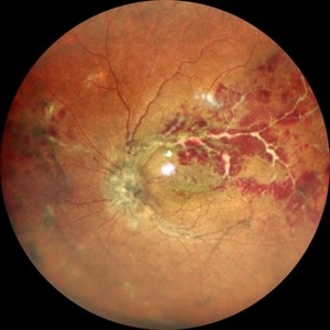
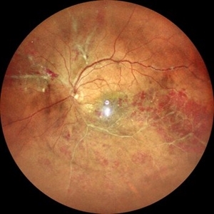
Fundus montage of the right eye of a 27 year old male with macula threatening occlusive vasculitis showing hemorrhages in inferior, temporal quadrant with vascular sheathing. The patient was Mantoux positive (20 mm induration) and IGRA (TB-GOLD)positive and started on oral steroids. The case was atypical due to no vitritis at presentation which is unusual of tuberculosis. Behcet's disease was ruled out as there was no panuveitis like picture.
Photographer: DR. TEJASWITA VERMA
Imaging device: MIRANTE
Condition/keywords: occlusive vasculitis, ocular tuberculosis
-
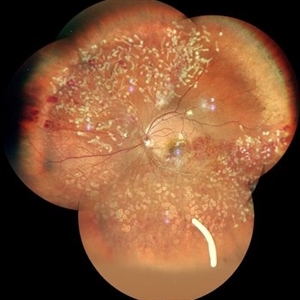 Idiopathic Retinal Vasculitis
Jun 9 2024 by Anjana Mirajkar, MS Ophthalmology
A color photo montage of an 32 year old male of LE showing laser marks in inferior and superior half with an floating ozurdex implant (inferiorly) in a case of idiopathic retinal vasculitis.
Photographer: Dr. Anjana Mirajkar -Retina Foundation, Ahmedabad
Imaging device: Mirante-Nidek
Condition/keywords: idiopathic retinal vasculitis, laser photocoagulation, Ozurdex implant
-
 Idiopathic Retinal Vasculitis
Jun 9 2024 by Anjana Mirajkar, MS Ophthalmology
A widefield image of a 32 year old male of LE showing laser marks in inferior and superior half with an floating ozurdex implant (inferiorly) in a case of idiopathic retinal vasculitis.
Photographer: Dr. Anjana Mirajkar -Retina Foundation, Ahmedabad
Imaging device: Mirante-Nidek
Condition/keywords: idiopathic retinal vasculitis, laser photocoagulation, Ozurdex implant, pan-retinal photocoagulation (PRP)
-
 Idiopathic Retinal Vasculitis
Jun 9 2024 by Anjana Mirajkar, MS Ophthalmology
A widefield image of a 32 year old male of LE showing sclerosed vessels more prominent inferiorly with superficial hemorrhages noted in all quadrants along with sheathing of vessels noted in superiorly.
Photographer: Dr. Anjana Mirajkar -Retina Foundation, Ahmedabad
Imaging device: Mirante-Nidek
Condition/keywords: Eales disease
-
Tractional RD-Making the Decision When and Where to Stop
May 23 2024 by ARVIND JAIN M
This is a young gentlemen with defective vision for 3 months in his right eye. He gave the history of recurrent redness of the right past few months. he was diagnosed to have right eye vasculitis with tractional detachment. He underwent uveitic workup and under steroid cover right eye paraplana vitrectomy with membrane peeling with endolaser with c3f8 gas was planned. patient improved significantly. this surgical video demonstrates when and where to stop during membrane peeling and get good results.
Condition/keywords: Eales disease, retinal vasculitis, tractional retinal detachment
-
 Retinal Vasculitis
Aug 22 2023 by Karen Santamaría
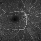
Fluorescein angiography of patient of a 23 year-old man diagnosed with retinal vasculitis and juvenile glaucoma.
Photographer: Karen Santamaría, Hospital Militar de Especialidades Oftalmológicas - Servicio de Glaucoma, Ciudad de México
Imaging device: Optos California
Condition/keywords: juvenile glaucoma, retina, vasculitis
-
 Idiophatic retinal vasculitis
Jul 9 2023 by Luiz A Zago, PhD
Idiophatic retinal vasculitis in a 45 year old woman
Photographer: Luiz Zago, PhD.
Imaging device: Topcon 50IX
Condition/keywords: diffuse vasculitis, intraretinal hemorrhage, optic disc leakage
-
 Idiophatic retinal vasculitis
Jul 9 2023 by Luiz A Zago, PhD
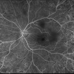
Mid phase angiography of a 45 woman with a idiophatic vasculitis. She is been followed in the last 10 years. No sistemic association was found beside face rosacea, This case was first described in Diagnostic and Therapeutic Challenges - Retina Journal
Photographer: Luiz Zago, PhD.
Imaging device: Topcon 50IX
Condition/keywords: anomalous foveal avascular zone, Choroidal Folds, intraretinal microvascular abnormalities, microaneurysms, optic neuritis, Vaculitis
-
 Idiopathic Vasculitis
Feb 4 2023 by Aditya S Kelkar, MS, FRCS, FASRS,FRCOphth
Color fundus photograph of the left eye showing idiopathic retinal vasculitis.
Photographer: Dr. Pranali Surawase. National Institute of Ophthalmology, Pune, India.
Imaging device: Zeiss Clarus 500
Condition/keywords: retinal vasculitis, vasculitis
-
 Choroidal Abscess with occlusive retinal vasculitis
Jan 28 2023 by Anjana Mirajkar, MS Ophthalmology
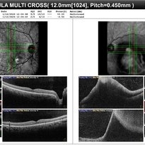
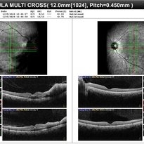
OCT BE of a 55 year old male a case of Choroidal abscess with occlusive retinal vasculitis.
Photographer: Dr. Anjana Mirajkar -Retina Foundation, Ahmedabad
Condition/keywords: Choroidal abscess
-
 Choroidal Abscess with occlusive retinal vasculitis
Jan 28 2023 by Anjana Mirajkar, MS Ophthalmology
Widefield color image of LE of a 55 year old male a case of Choroidal abscess with occlusive retinal vasculitis.
Photographer: Dr. Anjana Mirajkar -Retina Foundation, Ahmedabad
Condition/keywords: choroidal abscess
-
 Occlusive Vasculitis
Jan 28 2023 by Anjana Mirajkar, MS Ophthalmology
Central FA picture of a 40 year old female a case of occlusive retinal vasculitis.
Photographer: Dr. Anjana Mirajkar -Retina Foundation, Ahmedabad.
Condition/keywords: occlusive retinal vasculitis
-
 Occlusive Vasculitis
Jan 28 2023 by Anjana Mirajkar, MS Ophthalmology
OCT image of BE of a 40 year old female a case of occlusive retinal vasculitis.
Photographer: Dr. Anjana Mirajkar -Retina Foundation, Ahmedabad
Condition/keywords: occlusive retinal vasculitis
-
 Occlusive Vasculitis
Jan 28 2023 by Anjana Mirajkar, MS Ophthalmology
Wide field FA image of RE of a 40 year old female a case of occlusive retinal vasculitis.
Photographer: Dr. Anjana Mirajkar -Retina Foundation, Ahmedabad
Condition/keywords: occlusive retinal vasculitis
 Loading…