Search results (130 results)
-
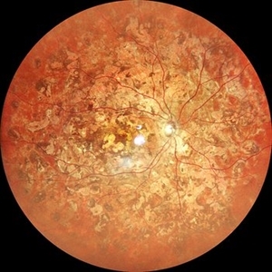
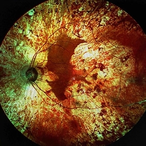
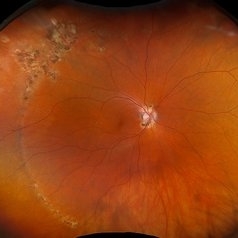
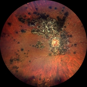
 Healed AZOOR- Multiple White Dot Syndrome
Aug 29 2025 by Aditya S Kelkar, MS, FRCS, FASRS,FRCOphth
Fundus photograph of a 58 year old woman with multiple, well-defined, punched-out chorioretinal scars scattered throughout the posterior pole and mid-periphery. The macular area shows a large, confluent, yellowish-white scar involving the fovea.
Photographer: Dr. Muskan Mangal
Imaging device: Optos Daytona
Condition/keywords: multifocal choroiditis, multiple evanescent white dot syndrome (MEWDS), punctate inner choroidopathy (PIC)
-
 Retinal Aneurysms
Aug 6 2025 by Korey Starkey
54 year-old patient presents with scattered peripheral aneurysms with exudates. FA was performed showing peripheral nonperfusion and aneurysms. Treated patient with PRP and focal laser to aneurysms and continued observation.
Photographer: Kore Starkey
Imaging device: Optos
Condition/keywords: aneurysm, branch retinal vein occlusion (BRVO), chorioretinal scar, circinate ring, exudates, fundus photography, lesion, Optos, retinal aneurysms
-
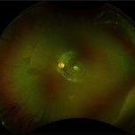
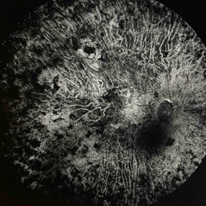
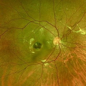
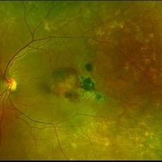
 Ghost Map Retina
Aug 4 2025 by Malvika Singh
Fundus photograph of a 50 year old male showing extensive chorioretinal scarring.
Photographer: Dr Malvika Singh, Retina Foundation, Ahmedabad, India
Imaging device: Mirante SLO/OCT
Condition/keywords: healed choroiditis
-
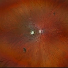
 Chorioretinal Macula Scar (Macula View)
May 12 2025 by Briana Hernandez
Zoomed in Macular View of Chorioretinal Macular Scar in 9-year-old female patient.
Photographer: Briana Hernandez, Hilton Head Retina Insitute
Imaging device: Optos
Condition/keywords: chorioretinal scar
-
 Chorioretinal Macula Scar (Ultrawide View)
May 12 2025 by Briana Hernandez
Ultra wide Optos image of Chorioretinal Macular Scar in 9-year-old female patient.
Photographer: Briana Hernandez, Hilton Head Retina Institute
Imaging device: Optos
Condition/keywords: chorioretinal scar, macular scar, ultra-wide field imaging
-
 Retinocoroiditis Inactiva Por Toxoplasmosis
Apr 28 2025 by Paulina Araujo
Fundus photography demonstrates a 2-disc-diameter chorioretinal scar in the superior temporal arcade, consistent with inactive toxoplasmic retinochoroiditis. The lesion exhibits pigmented borders and central atrophy, with adjacent splinter hemorrhages and vascular sheathing. No vitreous inflammation or active satellite lesions are present.
Photographer: Paulina D.Araujo Martínez, Asociación para Evitar la Ceguera en México I.A.P., Hospital Dr Luis Sánchez Bulnes.
Condition/keywords: toxoplasmosis chorioretinitis
-
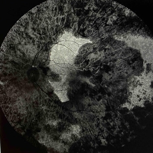 Extensive Chorioretinal Scarring With Partial Macular Sparing
Apr 22 2025 by Maxwell J Wingelaar, MD
Fundus autofluorescence of extensive chorioretinal scarring in the left eye.
Photographer: Killian Roberts
Imaging device: Heidelberg Spectralis AF
Condition/keywords: chorioretinal atrophy, chorioretinal inflammations
-
 Extensive Chorioretinal Scarring with Partial Macular Sparring
Apr 22 2025 by Maxwell J Wingelaar, MD
A multicolor photo showing chorioretinal scarring with partial macular sparing in the left eye.
Photographer: Killian Roberts
Imaging device: Heidelberg Spectralis Multicolor Photo
Condition/keywords: chorioretinal atrophy, chorioretinal inflammations
-
 Extensive Chorioretinal Scarring in the Right Eye
Apr 22 2025 by Maxwell J Wingelaar, MD
Fundus autofluorescence of Extensive chorioretinal scarring in the right eye.
Photographer: Killian Roberts
Imaging device: Heidelberg Spectralis AF
Condition/keywords: chorioretinal atrophy, chorioretinal inflammations
-
 Extensive Chorioretinal Scarring in the Right Eye
Apr 22 2025 by Maxwell J Wingelaar, MD
A multicolor photo showing chorioretinal scarring with macular involvement in the right eye
Photographer: Killian Roberts
Imaging device: Heidelberg Spectralis Multicolor Photo
Condition/keywords: chorioretinal atrophy, chorioretinal inflammations
-
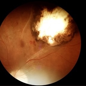
 Inadvert Globe Perfuration After Peribulbar Block
Mar 13 2025 by Bruno B Ribeiro
Fundus photograph of a 74-year-old woman who underwent pars plana vitrectomy OS due to rhegmatogenous retinal detachment. A horseshoe retinal tear can be seen at 5h. Intraoperative evaluation revealed a chorioretinal scar with the shape of the needle track at the same location. Despite rare, globe perfuration after peri or retrobulbar block may happen, even by the most experienced anesthesiologist.
Photographer: Bruno Barbosa Ribeiro, Angelina Meireles
Imaging device: Optos California
Condition/keywords: retinal detachment
-
 Inadvert Globe Perfuration After Peribulbar Block
Mar 13 2025 by Bruno B Ribeiro
Fundus photograph of a 74-year-old woman who underwent pars plana vitrectomy OS due to rhegmatogenous retinal detachment. A horseshoe retinal tear can be seen at 5h. Intraoperative evaluation revealed a chorioretinal scar with the shape of the needle track at the same location. Despite rare, globe perfuration after peri or retrobulbar block may happen, even by the most experienced anesthesiologist.
Photographer: Bruno Barbosa Ribeiro, Angelina Meireles
Imaging device: Optos California
Condition/keywords: hemmorhage
-
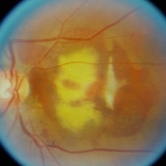
 Inactive Chorioretinal Scars
Dec 11 2024 by Virginia Gebhart
30 year old female with chorioretinal and macula scars. Appears post-infectious, most likely toxoplasmic. No active inflammatory changes or choroidal neovascularization. Will continue to monitor. Central vision limited by macula scar, BCVA 20/100
Photographer: Virginia Gebhart, Retina Consultants of Carolina
Imaging device: Optos California
Condition/keywords: chorioretinal scar, inactive toxoplasmosis
-
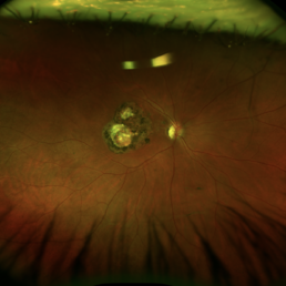
 POHS/Schlaegel Lines
Sep 19 2024 by Virginia Gebhart
46 year old female with h/o Histoplasmosis. Multiple punched out chorioretinal scars with Schlaegel lines. No evidence of CNV or active inflammation. VA 20/20
Photographer: Virginia Gebhart, Retina Consultants of Carolina
Imaging device: Optos California
Condition/keywords: chorioretinal scar, histoplasmosis, presumed ocular histoplasmosis syndrome (POHS)
-
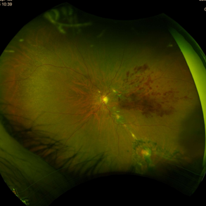
 Intriguing Web
Aug 28 2024 by Hemanth Murthy, MBBS, MD, FASRS
Right eye of a 43 year female patient came with blurring of vision of right eye since 2 years. There was loose redundant skin in the neck and axilla. Angiod streaks were in a spider web appearance .Vision was 1/60 in right eye and 6/9 in left eye. Right macula showed a sub retinal scar with pigmentation.
Photographer: Mr Veda Vyas
Imaging device: Optos Daytona
Condition/keywords: Angiod streaks in Pseudoxanthoma elasticum
-
 Large PEHCR causing an exudative inferior detachment in a patient with AMD
Apr 15 2024 by David A Reichstein, MD
(A) Ultra-widefield color fundus photograph demonstrates a temporal PEHCR causing minimal intra- and subretinal hemorrhage along with lipid exudation. There is an associated inferior detachment due to the dependent nature of the exudation. Note the lipid exudation at the posterior edge of the detachment indicating chronicity of the lesion. Drusen in the macula are also appreciated. (B) Ultra-widefield FA in early stage demonstrates hypofluorescence temporally and inferiorly. (C) Ultra-widefield color fundus photograph taken after 1 year of monthly anti-VEGF therapy demonstrates resolution of the exudative detachment and resultant chorioretinal scarring.
Condition/keywords: peripheral exudative hemorrhagic chorioretinopathy (PEHCR)
-
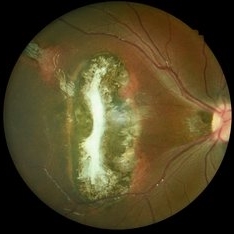
 Toxoplasmosis Macular Scar
Mar 8 2024 by Andre Beckenkamp
Optos image of a patient with extensive choriorretinal scar due to toxoplasmosis infection.
Photographer: Andre Beckenkamp,MD , Prevent Senior
Condition/keywords: toxoplasmosis
-
 Chorio Retinal Scar
Feb 19 2024 by Sanauddin Samejo , Diploma (Ophthalmic Technician Training Course)
A patient came in to Clinic of Dr Madhav Rao (VR Surgeon)
Photographer: Sanauddin Samejo, Burjeel Hospital, Abu Dhabi, UAE.
Imaging device: Optos Silverstone
Condition/keywords: chorioretinal scar
-
 Chorioretinal Scar
Feb 19 2024 by Sanauddin Samejo , Diploma (Ophthalmic Technician Training Course)
A patient came in to the clinic of Dr Madhav Rao (VR Surgeon).
Photographer: Sanauddin Samejo, Burjeel Hospital, Abu Dhabi, UAE.
Imaging device: Optos Silverstone
Condition/keywords: retinal scar
-
 Subretinal haemorrhage
Sep 26 2023 by Ben Serar
Fundus photograph showing subretinal bleed with subretinal scarring and fibrosis as sequelae.
Condition/keywords: Subretinal haemorrhage
-
 Multifocal choroiditis secondary to sarcoidosis- Quiescent
May 6 2023 by Niloofar Piri, MD
Montage fundus photograph of the left eye in a patient with sarcoidosis demonstrating peripheral inactive multifocal chorioretinal scars after systemic immunomodulatory therapy.
Photographer: Sean Kelso, Saint Lousi University
Condition/keywords: multifocal chorioretinitis (MCP), multifocal choroiditis, sarcoid uveitis, sarcoidosis
-
 Chorioretinitis with Overlying Vitreous Stranding/Vitritis
Mar 23 2023 by Isaac Agranoff
Fundus photograph of a 37-year-old woman presenting with chorioretinitis with overlying vitreous stranding/vitritis that has remained unchanged for multiple years. Patient presented with irritation and blurred vision and her vision was 20/40 OD. The OCT revealed evidence of low-grade inflammation and the recommend treatment was anti-inflammatory eye drops at this time and to obtain second opinion with another physician in the office.
Photographer: Isaac Agranoff, Technician
Imaging device: Optos California
Condition/keywords: chorioretinal scar, chorioretinitis, inflammation, Optos, ultra-wide field imaging, vitritis
-
 Tuberculosis-related serpiginous-like choroiditis
Nov 22 2022 by Ricardo Leitão Guerra
True color BLFI of a 60-year-old male presenting chorioretinal scars from a tuberculosis-related serpiginous-like choroiditis.
Photographer: Ricardo Leitão Guerra
Imaging device: Zeiss Clarus 700
Condition/keywords: serpiginous choroiditis, tuberculosis
-
 Subretinal Hemorrhage with Chorioretinal Macular Scars
Sep 28 2022 by Denica Rodriguez
Ultra-widefield pseudocolor fundus photograph of a 59 year old female with Subretinal Hemorrhage with Chorioretinal Macular Scars affecting her left eye. The physician presumes the etiology is CNV from adjacent scarring/choroidal rupture. Patient has history of ocular trauma with cricket ball at age 10-12 years old. She suspects that she might have suffered choroidal rupture, which has resulted in secondary CNV and hemorrhage that we are seeing today. She recommends treatment with Eylea sample injection in a series of 4 at a 4-5 week interval. The patient's vision at the time of her appointment was Dcc20/40-2 PHNI.
Photographer: Denica Rodriguez, COA
Imaging device: Optos California
Condition/keywords: antiVEGF therapy, chorioretinal scar, choroidal neovascular membrane (CNVM), fundus photography, left eye, macular scar, Optos, peripheral drusen, pseudocolor, secondary CNV, subretinal hemorrhage, ULTRA WIDE FIELD, ultra-wide field imaging
-
 Toxoplasmosis
Nov 24 2021 by Catarina Monteiro
Fundus photograph of a 7-year-old child with a chorioretinal scar likely due to congenital toxoplasmosis.
Photographer: Catarina Monteiro
Condition/keywords: chorioretinal scar, congenital toxoplasmosis, ocular toxoplasmosis
 Loading…