Search results (77 results)
-
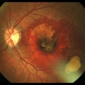 RPE Tear After Anti-VEGF Injection
Mar 17 2021 by RAFAEL REIS PEREIRA, MD
Retinal pigment epithelium (RPE) tear is a rare devastating complication of age-related macular degeneration (AMD). The believed mechanism lies in an adherence of the neovascularization to the undersurface of the RPE creating a contractile force that increases after VEGF therapy and causes the tear.
Photographer: Rafael Reis, Retina Clinic, São Paulo
Condition/keywords: retinal pigment epithelium (RPE) contracture
-
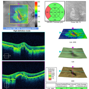
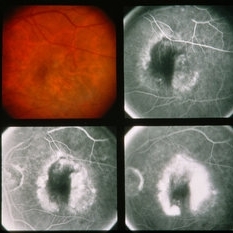
 RPE Micro Rip in Central Serous Chorioretinopathy
Jun 26 2016 by Rameez N Hussain, MD
SD OCT image of a case of central serous retinopathy showing RPE micro rip (RPE leak).
Photographer: DR RAMEEZ N HUSSAIN
Imaging device: Dense scan mode - Heidelberg Spectralis
Condition/keywords: central serous chorioretinopathy (CSCR), focal laser, leakage, retinal pigment epithelium (RPE) tear, Spectralis
-
 RPE tear in an 82-Year-Old Woman
Dec 7 2015 by Roy Schwartz, MD
An RPE tear in an 82-year-old woman, found incidentally after a year of visual deterioration. She hasn't visited an ophthalmologist since then. She has never underwent an intravitreal injection.
Photographer: Galit Yair Pur
Condition/keywords: retinal pigment epithelium (RPE) tear
-
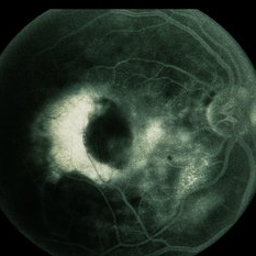
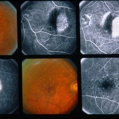
 RIP 2 FAF
Oct 7 2015 by Roberto Gallego-Pinazo, MD, PhD, DiSSO
Multicolor and autofluorescence sequence of a retinal pigment epithelium tear following intravitreal anti-VEGF injection.
Photographer: Rosa Dolz-Marco, University and Polytechnic Hospital La Fe, Valencia, Spain
Condition/keywords: age-related macular degeneration (AMD), autofluorescence imaging, choroidal neovascularization (CNV), multicolor, retinal pigment epithelium (RPE) tear
-
 RIP2
RIP2
Oct 7 2015 by Roberto Gallego-Pinazo, MD, PhD, DiSSO
Multicolor and autofluorescence sequence of a retinal pigment epithelium tear following intravitreal anti-VEGF injection.
Photographer: Rosa Dolz-Marco, University and Polytechnic Hospital La Fe, Valencia, Spain
Condition/keywords: age-related macular degeneration (AMD), autofluorescence imaging, choroidal neovascularization (CNV), multicolor, retinal pigment epithelium (RPE) tear
-
 RIP 1 FAF
Oct 7 2015 by Roberto Gallego-Pinazo, MD, PhD, DiSSO
Multicolor and autofluorescence sequence of a retinal pigment epithelium tear following intravitreal anti-VEGF injection.
Photographer: Rosa Dolz-Marco, University and Polytechnic Hospital La Fe, Valencia, Spain
Condition/keywords: age-related macular degeneration (AMD), autofluorescence imaging, choroidal neovascularization (CNV), multicolor, retinal pigment epithelium (RPE) tear
-
 RIP1
RIP1
Oct 7 2015 by Roberto Gallego-Pinazo, MD, PhD, DiSSO
Multicolor and autofluorescence sequence of a retinal pigment epithelium tear following intravitreal anti-VEGF injection.
Photographer: Rosa Dolz-Marco, University and Polytechnic Hospital La Fe, Valencia, Spain
Condition/keywords: age-related macular degeneration (AMD), autofluorescence imaging, choroidal neovascularization (CNV), multicolor, retinal pigment epithelium (RPE) tear
-
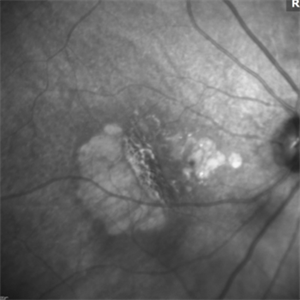
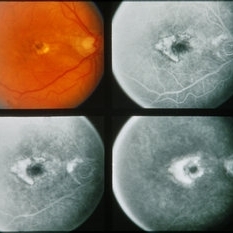
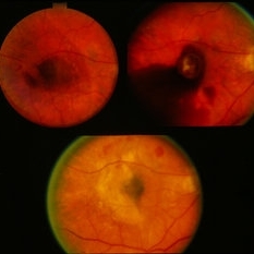
 RPE Tear: OCT
May 2 2015 by Thomas A. Ciulla, MD, MBA, FASRS
OCT revealed the RPE tear with some scolled redundant RPE more centrally.
Condition/keywords: choroidal neovascular membrane (CNVM), retinal pigment epithelium (RPE) tear, wet age-related macular degeneration (wet AMD)
-
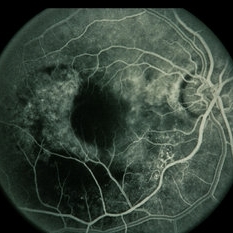
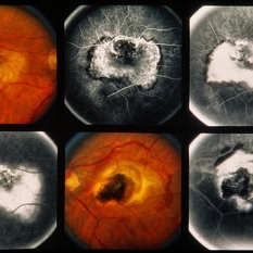
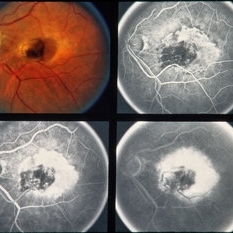
 RPE Tear: Fluorescein Angiography
May 2 2015 by Thomas A. Ciulla, MD, MBA, FASRS
Mid Phase Fluorescein Angiogram: The scrolled and redundant RPE just temporal to the fovea blocks underlying choroidal fluorescence. The absent RPE, more temporally, results in a window defect with intense hyperfluorescence.
Photographer: Stuart Alfred
Condition/keywords: choroidal neovascular membrane (CNVM), retinal pigment epithelium (RPE) tear, wet age-related macular degeneration (wet AMD)
-
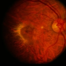
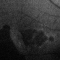 RPE Tear: Autofluorescence
May 2 2015 by Thomas A. Ciulla, MD, MBA, FASRS
Autofluorescence image: Note the discrete hypo-autofluorescence due to the absent RPE temporally. Note also the hypo-autofluorescence due to geographic atrophy centrally.
Photographer: Stuart Alfred
Condition/keywords: choroidal neovascular membrane (CNVM), retinal pigment epithelium (RPE) tear, wet age-related macular degeneration (wet AMD)
-
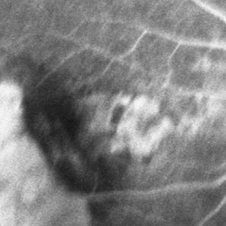
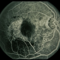
 RPE Tear: Infrared Photo
May 2 2015 by Thomas A. Ciulla, MD, MBA, FASRS
Infrared image: Note the scrolled and redundant RPE just temporal to the fovea and the absent RPE more temporally where there are visible larger choroidal vessels.
Photographer: Stuart Alfred
Condition/keywords: choroidal neovascular membrane (CNVM), retinal pigment epithelium (RPE) tear, wet age-related macular degeneration (wet AMD)
-
 ARMD / CNVM / PED With RPE Tear
Nov 7 2014 by David Callanan, MD
59-year-old male, ARMD / CNVM / PED with RPE Tear.
Condition/keywords: choroidal neovascular membrane (CNVM), pigment epithelial detachment (PED), retinal pigment epithelium (RPE) tear
-
 ARMD / CNVM / PED With RPE Tear
Nov 7 2014 by David Callanan, MD
59-year-old male, ARMD / CNVM / PED with RPE Tear.
Condition/keywords: choroidal neovascular membrane (CNVM), pigment epithelial detachment (PED), retinal pigment epithelium (RPE) tear
-
 ARMD / CNVM / PED With RPE Tear
Nov 7 2014 by David Callanan, MD
59-year-old male, ARMD / CNVM / PED with RPE Tear.
Condition/keywords: choroidal neovascular membrane (CNVM), pigment epithelial detachment (PED), retinal pigment epithelium (RPE) tear
-
 ARMD / CNVM / PED With RPE Tear
Nov 7 2014 by David Callanan, MD
59-year-old male, ARMD / CNVM / PED with RPE Tear.
Condition/keywords: choroidal neovascular membrane (CNVM), pigment epithelial detachment (PED), retinal pigment epithelium (RPE) tear
-
 ARMD / CNVM / PED With RPE Tear
Nov 7 2014 by David Callanan, MD
59-year-old male, ARMD / CNVM / PED with RPE Tear.
Condition/keywords: choroidal neovascular membrane (CNVM), pigment epithelial detachment (PED), retinal pigment epithelium (RPE) tear
-
 RPE Tear
-
 RPE Tear
Oct 6 2014 by Howard Schatz, MD
80-year-old white female. RPE Tear. RE 20/100 LE 4/200.
Condition/keywords: retinal pigment epithelium (RPE) tear
-
 RPE Tear
Oct 6 2014 by Howard Schatz, MD
80-year-old white female. RPE Tear. Re 4/200 LE 20/25.
Condition/keywords: retinal pigment epithelium (RPE) tear
-
 RPE Tear
Oct 6 2014 by Howard Schatz, MD
80-year-old white female. RPE Tear. Re 10/200 LE 20/20.
Condition/keywords: retinal pigment epithelium (RPE) tear
-
 RPE Tear
Oct 6 2014 by Howard Schatz, MD
74-year-old white male. RPE Tear. 20/200 and 1/200.
Condition/keywords: retinal pigment epithelium (RPE) tear
-
 RPE Tear
Oct 6 2014 by Howard Schatz, MD
73-year-old white male. RPE Tear. 20/20 and 1/200.
Condition/keywords: retinal pigment epithelium (RPE) tear
-
 RPE Tear
Oct 6 2014 by Howard Schatz, MD
77-year-old white female. RPE Tear. 20/30 and 20/200.
Condition/keywords: retinal pigment epithelium (RPE) tear
-
 RPE Tear
Oct 6 2014 by Howard Schatz, MD
76-year-old white female. RPE Tear. RE 20/30 LE 20/200.
Condition/keywords: retinal pigment epithelium (RPE) tear
-
 RPE Tear
Oct 6 2014 by Howard Schatz, MD
74-year-old white female. 2/200 and 20/100-. RPE Tear.
Condition/keywords: retinal pigment epithelium (RPE) tear
 Loading…