Search results (127 results)
-
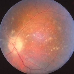
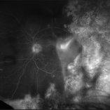
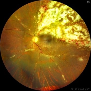 Cheese Pizza Pie Appearance in CMV Retinitis
Mar 30 2024 by KANWALJEET HARJOT MADAN, M.S. (Ophthalmology), FAICO (Vitreous - Retina)
This is Fundus Photograph of left eye of 53 year male depicting an area of Retinal Necrosis with few Retinal Haemorrhages suggestive of CMV Retinitis. Areas of Perivascular Exudation also seen. On investigations, the patient was found to be HIV positive. He was started on Anti Retro Viral treatment after physician opinion.
Photographer: Dr. Kanwaljeet Harjot Madan, Thind Eye Hospital, Jalandhar City (Punjab) INDIA.
Imaging device: Zeiss Fundus Camera
Condition/keywords: AIDS, cytomegalovirus (CMV), retinitis
-
 Pigmented KPs
Dec 8 2023 by Nassim Alejandro Abreu Arbaje, MD
Anterior segment photograph of a 27 year old female diagnosed with an Acute Retinal Necrosis. In the picture we can see mutton fat keratic precipitates already pigmented.
Photographer: Nassim Abreu
Imaging device: Alcon NGenuity Systems
Condition/keywords: Acute Retinal Necrosis, mutton-fat keratic precipitates (KP), Uveitis
-
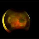
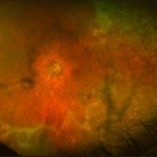
 CMV retintis
Sep 14 2023 by Ben Serar
Fundus photograph of LE showing superficial flame-shaped haemorrhages with surrounding retinal necrosis at the posterior pole along the superotemporal arcade in a case of fulminant type of CMV retinitis.
Condition/keywords: CMV retinitis, fulminant retinitis, pizza-pie appearance, viral retinitis
-
 Acute Retinal Necrosis with Retinal Detachment
Jan 13 2022 by Tuan Tran, MBBS, MMed (OphthSc), FRANZCO, DRCPSC
Acute Retinal Necrosis with Retinal Detachment.
Photographer: Tuan Tran
Condition/keywords: Acute Retinal Necrosis with Retinal Detachment, retinal necrosis
-
 Left Acute Retinal Necrosis
Jan 11 2022 by Tuan Tran, MBBS, MMed (OphthSc), FRANZCO, DRCPSC
84 year-old gentleman presenting with left acute retinal necrosis.
Photographer: Tuan Tran
Imaging device: Optos widefield
Condition/keywords: ARN complications
-
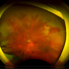
 Acute Retinal Necrosis
May 31 2021 by Aditya S Kelkar, MS, FRCS, FASRS,FRCOphth
Fundus photograph of 43-year-old female with left eye acute retinal necrosis.
Imaging device: Clarus 500
Condition/keywords: acute retinal necrosis
-
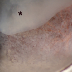 Toxoplasmic Acute Retinal Necrosis
May 18 2020 by McGill University Health Centre
Toxoplasmosis can have several manifestations in the eye, of which toxoplasmic acute retinal necrosis has the worst prognosis. This enucleation specimen shows extensive retinal necrosis with multiple coalescent foci. The vitreous is hazy (*).
Condition/keywords: acute retinal necrosis, toxoplasmosis
-
 CMV Retinitis in AIDS Patient
Dec 12 2019 by McGill University Health Centre
Fundus photograph of a 32-year-old man with HIV infection and 100 CD4+ cells count. Several areas of retinal necrosis interspersed with areas of hemorrhage around blood vessels can be observed.
Photographer: Miguel N. Burnier, McGill University Health Center-McGill University Ocular Pathology & Translational Research Laboratory
Imaging device: Fundoscopy
Condition/keywords: AIDS, cytomegalovirus (CMV), HIV, retinitis
-
 Primary Intraocular Lymphoma
Nov 20 2019 by McGill University Health Centre
73-year-old man with retinal vasculitis and acute retinal lesions of the left eye. Optic nerve and retinal infiltrates consistent with acute retinal necrosis.
Condition/keywords: acute retinal necrosis, primary intraocular lymphoma
-
 Progressive Outer Retinal Necrosis
Nov 5 2019 by Nichole Lewis
86-year-old male with progressive outer retinal necrosis, significant retinitis, retinal whitening, intraretinal hemorrhages and peripheral rpe changes. FA showed occlusive vasculitis with non-perfusion. Patient is immuno-suppressed with a history of renal transplant. VA 20/60.
Photographer: Nichole Lewis
Imaging device: Optos
Condition/keywords: intraretinal hemorrhage, occlusive vasculitis, progressive outer retinal necrosis (PORN), retinal pigment epithelium (RPE) changes, retinal whitening, retinitis
-
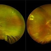 ARN (#3) This is comparison between the latest visit (left) and one week prior (which is the right photo, and same one as photo #2)
May 27 2019 by John S. King, MD
60-year-old African American female who had been treated for iridocyclitis for at least a week sent in for vitritis and a nasal fundus lesion. Complaints included redness, floaters, photophobia, and decreased vision. Husband had recent shingles. Acuity was 20/60-2 with IOP of 12, and small KP in Art's triangel, 1-2+ a/c cell, 2-3+ ant vit cell, diffuse arteriolar sheathing, multiple areas of retinal whitening in periphery and mid-periphery (see Photo #1). PCR of a/c was performed, and intravitreal GCV administered, and VACV 2g qid and ASA started.... PCR positive for HZV, pred taper was started two days after presentation as the infection had begun to stablize..... Five days from presentation the vision was 20/60, inflammation and areas of retinal whitening had improved (see Photo #2).... One week later acuity was 20/30, the a/c was quiet and KP resolved; ant vitreous cell decreased; and there was further improvement in retinal appearance without any signs of retinal holes or detachment; she is now on low dose maint VACV (see photo#3)
Photographer: Maysee Yang
Imaging device: Optos CA
Condition/keywords: acute retinal necrosis, Herpes zoster
-
 ARN (#2) Five Days Since Initial Visit
May 27 2019 by John S. King, MD
60-year-old African American female who had been treated for iridocyclitis for at least a week sent in for vitritis and a nasal fundus lesion. Complaints included redness, floaters, photophobia, and decreased vision. Husband had recent shingles. Acuity was 20/60-2 with IOP of 12, and small KP in Art's triangel, 1-2+ a/c cell, 2-3+ ant vit cell, diffuse arteriolar sheathing, multiple areas of retinal whitening in periphery and mid-periphery (see Photo #1). PCR of a/c was performed, and intravitreal GCV administered, and VACV 2g qid and ASA started.... PCR positive for HZV, pred taper was started two days after presentation as the infection had begun to stablize..... Five days from presentation the vision was 20/60, inflammation and areas of retinal whitening had improved (see Photo #2).... One week later acuity was 20/30, the a/c was quiet and KP resolved; ant vitreous cell decreased; and there was further improvement in retinal appearance without any signs of retinal holes or detachment; she is now on low dose maint VACV (see photo#3)
Photographer: Maysee Yang
Imaging device: Optos CA
Condition/keywords: acute retinal necrosis, Herpes zoster
-
 ARN (#1) Initial Photo
May 27 2019 by John S. King, MD
60-year-old African American female who had been treated for iridocyclitis for at least a week sent in for vitritis and a nasal fundus lesion. Complaints included redness, floaters, photophobia, and decreased vision. Husband had recent shingles. Acuity was 20/60-2 with IOP of 12, and small KP in Art's triangel, 1-2+ a/c cell, 2-3+ ant vit cell, diffuse arteriolar sheathing, multiple areas of retinal whitening in periphery and mid-periphery (see Photo #1). PCR of a/c was performed, and intravitreal GCV administered, and VACV 2g qid and ASA started.... PCR positive for HZV, pred taper was started two days after presentation as the infection had begun to stablize..... Five days from presentation the vision was 20/60, inflammation and areas of retinal whitening had improved (see Photo #2).... One week later acuity was 20/30, the a/c was quiet and KP resolved; ant vitreous cell decreased; and there was further improvement in retinal appearance without any signs of retinal holes or detachment; she is now on low dose maint VACV (see photo#3)
Photographer: Maysee Yang
Imaging device: Optos CA
Condition/keywords: acute retinal necrosis, Herpes zoster
-
 Acute Retinal Necrosis with Proliferative Vitreoretinopathy and Total Retinal Detachment
Mar 26 2019 by Gary R. Cook, MD, FACS
Same WF patient 9 weeks after initial presentation with Acute Retinal Necrosis now with proliferative vitreoretinopathy and a total combined traction & rhegmatogenous retinal detachment
Imaging device: Topcon VT-50
Condition/keywords: acute retinal necrosis, proliferative vitreoretinopathy (PVR), tractional retinal detachment
-
 Acute Retinal Necrosis
Mar 26 2019 by Gary R. Cook, MD, FACS
Left eye of same patient with acute retinal necrosis who developed rhegmatogenous RD seven weeks after presentation.
Imaging device: Topcon VT-50
Condition/keywords: acute retinal necrosis
-
 Acute Retinal Necrosis
Mar 26 2019 by Gary R. Cook, MD, FACS
Middle-aged white female with peripheral retinal lesions of acute retinal necrosis OS at presentation.
Imaging device: Topcon VT-50
Condition/keywords: acute retinal necrosis
-
 Acute Retinal Necrosis
Mar 26 2019 by Gary R. Cook, MD, FACS
Middle-aged white female with ARN OS showing additional involvement within 2 weeks. Patient was seen prior to the availability of anti-viral therapies.
Imaging device: Topcon VT-50
Condition/keywords: acute retinal necrosis
-
 Slide 13-10
Mar 4 2019 by Lancaster Course in Ophthalmology
High-power view of a retinoblastoma undergoing necrosis.
Condition/keywords: retinal necrosis, retinoblastoma
-
 Slide 12-23
Feb 27 2019 by Lancaster Course in Ophthalmology
Pigmentary glaucoma. Necrosis of the iris pigment epithelium, macrophages filled with pigment in the iris stroma, and atrophy and hyperplasia of the iris dilator muscle are present (H&E x101).
Condition/keywords: glaucoma, hyperplasia, retinal necrosis
-
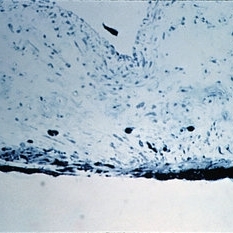
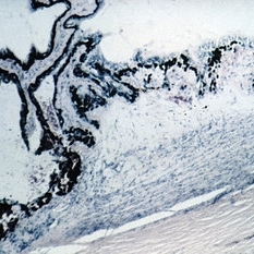
 Slide 12-8
Feb 27 2019 by Lancaster Course in Ophthalmology
A necrotic iris is shown, with loss of stroma as well as dilator and sphincter muscles (H&E x21).
Condition/keywords: retinal necrosis
-
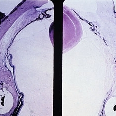 Slide 9-75
Feb 26 2019 by Lancaster Course in Ophthalmology
Ischemic necrosis of iris and ciliary body (right) in eye following scleral buckling procedure with the use of a polyethylene tube.
Condition/keywords: ciliary, retinal necrosis
-
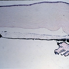
 Slide 7-118
Feb 25 2019 by Lancaster Course in Ophthalmology
Hemorrhage and necrosis anterior to a scleral buckle.
Condition/keywords: hemorrhage, retinal necrosis, scleral buckle
-
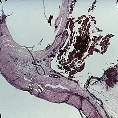
 Slide 7-115
Feb 25 2019 by Lancaster Course in Ophthalmology
Cyclocryotherapy produces necrosis of the ciliary body.
Condition/keywords: ciliary, cyclocryotherapy, retinal necrosis
-
 Progressive Outer Retinal Necrosis
Nov 30 2018 by Nichole Lewis
Fluorescein angiogram of an 86-year-old male with progressive outer retinal necrosis and chronic cystoid macular edema. This patient has occlusive vasculitis with non-perfusion, significant retinitis, retinal whitening and intra-retinal hemorrhages. Patient is immunosupressed with a history of kidney transplantation. VA 20/60. Patient was treated with intravitreal foscarnet and admitted to the hospital for an infectious disease and transplant team consultation.
Photographer: Nichole Lewis
Condition/keywords: cystoid macular edema (CME), intragel hemorrhage, non-perfusion, occlusive vasculitis, progressive outer retinal necrosis (PORN), retinal whitening, retinitis
 Loading…