Search results (35 results)
-
 Retinal Detachment and Lattice Degeneration
Mar 25 2025 by Korey Starkey
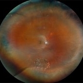
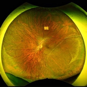
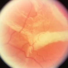
26 year-old patient presented at first visit with rhegmatogenous macula involving retinal detachment of the left eye. Underwent prompt surgical repair. Both eyes also present with lattice degeneration with atrophic holes.
Photographer: Korey Starkey
Condition/keywords: atrophic retinal hole, fundus photography, lattice degeneration, montage photo, Optos, OPTOS CALIFORNIA RGB, retinal detachment, retinal holes, rhegmatogenous retinal detachment, ultra-wide field imaging
-
 Lattice Degeneration With Atrophic Retinal Holes
Jan 30 2025 by Kimberly Wakester
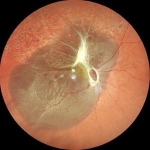
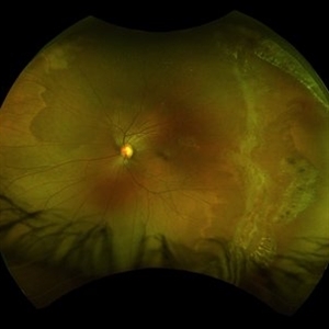
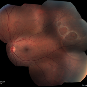
Ultra-wide field montage fundus photograph of a 56-year-old woman with lattice degeneration with atrophic holes statues post laser. Patient also has a small CHRPE temporal to macula and trace ERM that is not visually significant. Will continue follow up care to monitor and treat as needed.
Photographer: Kimberly Wakester, COA
Imaging device: Optos California
Condition/keywords: atrophic retinal hole, CHRPE, epiretinal membrane (ERM), lattice degeneration, montage photo
-
 The Bullet Ridden Retina
Feb 17 2024 by SHISHIR VERGHESE, MS, FVRS, FAICO (Retina)
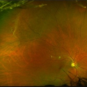
Fundus image obtained of a case of lasered branch retinal vein occlusion (BRVO) with fibrovascular proliferation (FVP) where the laser marks have given way to multiple small retinal holes due to traction from the same.
Photographer: DIVYA SHAJI
Imaging device: NIDEK MIRANTE
Condition/keywords: BRVO, chronic retinal detachment
-
 Peripheral retinal degenerations
Jan 29 2024 by Anupama Kiran Kumar
Fundus photo of a young man who underwent barrage laser after he presented to the clinic with floaters and was diagnosed to have lattices with horse shoe tears and retinal holes.
Photographer: Dr Anupama Kiran Kumar DNB FVR , Narayana Nethralaya Bangalore
Imaging device: Mirante SLO/OCT (Nidek Co., Gamagori, Japan)
Condition/keywords: lattice degeneration, peripheral retinal degeneration
-
 macular hole
Jan 28 2024 by Anjana Mirajkar, MS Ophthalmology
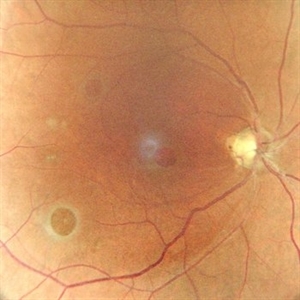
Fundus photograph of a 40 year old female showing macular hole with 2 retinal holes surrounding it
Photographer: Dr. Anjana Mirajkar -Retina Foundation, Ahmedabad
Imaging device: Mirante-Nidek
Condition/keywords: macular hole, multiple retinal holes
-
 Rhegmatogenous retinal detachment in a Young Patient
May 18 2023 by Jesus Lozano, MD
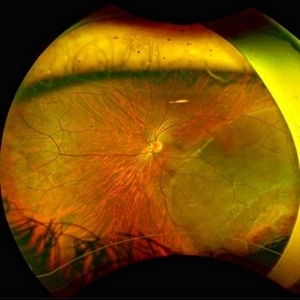
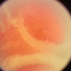
25 year old male patient. High Myope. Arrived to my clinic with a VA Fc 2mts. - Optos Image : Rhegmatogenous Retinal Detachment Mácula Off with ínfero temporal retinal holes in an area of lattice degeneration. Visual Acuity FC 2mts.
Imaging device: Optos
Condition/keywords: retinal detachment with retinal defect
-
 1 year Follow Up after Scleral Buckle Surgery in a Young Patient
May 18 2023 by Jesus Lozano, MD
25 year old man after Scleral Buckle Surgery + laser Retinopexy do to RRD macula off with ínfero temporal mid peripheral retinal holes in an area of lattice degeneration. Final VA 6/9.
Imaging device: Optos
Condition/keywords: scleral buckle
-
 Total retinal Detachment multiple holes
Sep 26 2022 by Denica Rodriguez
60 year old Male presented with two week old Macula off Retinal detachment with multiple tears.
Photographer: Denica Rodriguez
Imaging device: Optos California
Condition/keywords: color fundus photograph, color photo, macula-off, optos, pseudocolor, Retinal detachment, retinal holes, retinal tear, Retinal tear with detachment, superior arcade, superior field, superior retina, total retinal detachment
-
 Retinal Holes, Demarcation Line
Jul 19 2021 by RUSHIK PATEL
Utlrawide pseudo-color fundus photograph of 28-year-old boy with 2 retinal hole surrounded by subretinal fluid less than 1 disc diameter and demarcation line.
Photographer: Rushik Patel, Netralaya Super Speciality Eye Hospital
Condition/keywords: retinal hole
-
 Myopia with Lattice Degeneration and White Without Pressure in the Setting of Marfan's Syndrome
Aug 31 2020 by Sophia El Hamichi, MD
A 1-year-old female with Marfan's syndrome, myopia OU, congenital nystagmus and exotopia OD. Ultra-wide field imaging of her left eye showed lattice degeneration with atrophic retinal holes temporally, in addition to multiple sections of white without pressure.
Imaging device: Optos
Condition/keywords: atrophic retinal hole, lattice degeneration, Marfan's syndrome, myopia, Optos, ultra-wide field imaging
-
 Bullous Retinoschisis with Outer Retinal Holes
Jun 15 2020 by Olivia Rainey
Ultra-widefield pseudocolor fundus photograph of a 56-year-old female with bullous retinoschisis with outer retinal holes affecting her right eye. The physician noted superotemporal retinoschisis in her monoculcar functioning eye. There was no demarcation line and no inner or outer layer breaks at her first appointment in February of 2020. On 6/15/20 she had a new onset outer holes and SRF tracking inferiorly. The physician recommended observation, however if this continues to progress we have discussed indications for barrier laser.
Photographer: Olivia Rainey, OCT-C, COA
Imaging device: Optos California
Condition/keywords: bullous retinoschisis, Optos, outer layer breaks, outer layer hole, pseudocolor, subretinal fluid, superior retina, ultra-wide field imaging
-
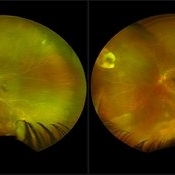 ARN (#3) This is comparison between the latest visit (left) and one week prior (which is the right photo, and same one as photo #2)
May 27 2019 by John S. King, MD
60-year-old African American female who had been treated for iridocyclitis for at least a week sent in for vitritis and a nasal fundus lesion. Complaints included redness, floaters, photophobia, and decreased vision. Husband had recent shingles. Acuity was 20/60-2 with IOP of 12, and small KP in Art's triangel, 1-2+ a/c cell, 2-3+ ant vit cell, diffuse arteriolar sheathing, multiple areas of retinal whitening in periphery and mid-periphery (see Photo #1). PCR of a/c was performed, and intravitreal GCV administered, and VACV 2g qid and ASA started.... PCR positive for HZV, pred taper was started two days after presentation as the infection had begun to stablize..... Five days from presentation the vision was 20/60, inflammation and areas of retinal whitening had improved (see Photo #2).... One week later acuity was 20/30, the a/c was quiet and KP resolved; ant vitreous cell decreased; and there was further improvement in retinal appearance without any signs of retinal holes or detachment; she is now on low dose maint VACV (see photo#3)
Photographer: Maysee Yang
Imaging device: Optos CA
Condition/keywords: acute retinal necrosis, Herpes zoster
-
 ARN (#2) Five Days Since Initial Visit
May 27 2019 by John S. King, MD
60-year-old African American female who had been treated for iridocyclitis for at least a week sent in for vitritis and a nasal fundus lesion. Complaints included redness, floaters, photophobia, and decreased vision. Husband had recent shingles. Acuity was 20/60-2 with IOP of 12, and small KP in Art's triangel, 1-2+ a/c cell, 2-3+ ant vit cell, diffuse arteriolar sheathing, multiple areas of retinal whitening in periphery and mid-periphery (see Photo #1). PCR of a/c was performed, and intravitreal GCV administered, and VACV 2g qid and ASA started.... PCR positive for HZV, pred taper was started two days after presentation as the infection had begun to stablize..... Five days from presentation the vision was 20/60, inflammation and areas of retinal whitening had improved (see Photo #2).... One week later acuity was 20/30, the a/c was quiet and KP resolved; ant vitreous cell decreased; and there was further improvement in retinal appearance without any signs of retinal holes or detachment; she is now on low dose maint VACV (see photo#3)
Photographer: Maysee Yang
Imaging device: Optos CA
Condition/keywords: acute retinal necrosis, Herpes zoster
-
 ARN (#1) Initial Photo
May 27 2019 by John S. King, MD
60-year-old African American female who had been treated for iridocyclitis for at least a week sent in for vitritis and a nasal fundus lesion. Complaints included redness, floaters, photophobia, and decreased vision. Husband had recent shingles. Acuity was 20/60-2 with IOP of 12, and small KP in Art's triangel, 1-2+ a/c cell, 2-3+ ant vit cell, diffuse arteriolar sheathing, multiple areas of retinal whitening in periphery and mid-periphery (see Photo #1). PCR of a/c was performed, and intravitreal GCV administered, and VACV 2g qid and ASA started.... PCR positive for HZV, pred taper was started two days after presentation as the infection had begun to stablize..... Five days from presentation the vision was 20/60, inflammation and areas of retinal whitening had improved (see Photo #2).... One week later acuity was 20/30, the a/c was quiet and KP resolved; ant vitreous cell decreased; and there was further improvement in retinal appearance without any signs of retinal holes or detachment; she is now on low dose maint VACV (see photo#3)
Photographer: Maysee Yang
Imaging device: Optos CA
Condition/keywords: acute retinal necrosis, Herpes zoster
-
 Retinal Detachment with Retinal Holes
Apr 8 2019 by Gary R. Cook, MD, FACS
Retinal detachment with retinal holes OS
Imaging device: Topcon VT-50
-
 Outer Layer Holes with Rolled Edges in Retinoschisis
Apr 2 2019 by Gary R. Cook, MD, FACS
Another view of a 71-year-old white female with rolled edges of outer layer retinal holes in bullous, adult-type retinoschisis OS.
Condition/keywords: bullous retinoschisis, retinoschisis
-
 Outer Layer Holes with Rolled Edges in Retinoschisis
Apr 2 2019 by Gary R. Cook, MD, FACS
71-year-old white female with outer layer retinal holes with rolled edges in an area of bullous, adult-type retinoschisis OS
Condition/keywords: bullous retinoschisis, retinoschisis
-
 Slide 8-9
Mar 4 2019 by Lancaster Course in Ophthalmology
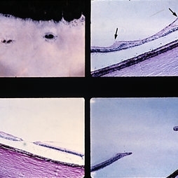
Lattice degeneration of retina with retinal holes. Microscopic appearance at three levels (upper, right, and lower) shows discontinuity of the internal limiting membrane, an overlying pocket of fluid vitreous, atrophy of inner retinal layers, hole formation, and vitreoretinal adhesion (arrows) at margin of lesion. (E.P. No. 30544)
Condition/keywords: atrophy, lesion, retinal degeneration, retinal hole, vitreoretinal adhesion
-
 Retinal Holes
Mar 27 2018 by Nichole Lewis
Retinal holes.
Photographer: Nichole Lewis
Condition/keywords: retinal hole
-
 Multiple Retinal Holes
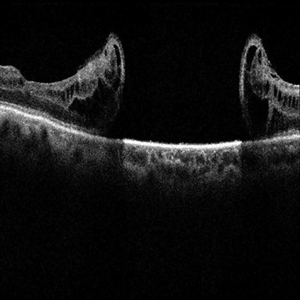
Sep 10 2017 by JEFFERSON R SOUSA, Tecg.º (Biomedical Systems Technology)
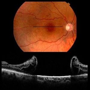
Patient 57-years-old, male, attended the clinic with complaint of low visual acuity and history of already having undergone a surgical procedure in another service. In previous evaluation of retinal mapping and retinography, being confirmed in optical coherence tomography, several retinal holes were observed in the posterior pole.
Photographer: JEFFERSON R SOUSA - Study Center and Ophthalmological Research Dr. Andre M V Gomes, Dr. Suel Abujamra Institute São Paulo-Brazil
Imaging device: OCT CIRRUS 4000, Protocol Horizontal line.
Condition/keywords: retinal hole
-
 Multiple Retinal Holes
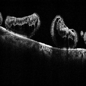
Sep 10 2017 by JEFFERSON R SOUSA, Tecg.º (Biomedical Systems Technology)
Patient 57-years-old, male, attended the clinic with complaint of low visual acuity and history of already having undergone a surgical procedure in another service. In previous evaluation of retinal mapping and retinography, being confirmed in optical coherence tomography, several retinal holes were observed in the posterior pole.
Photographer: JEFFERSON R SOUSA - Study Center and Ophthalmological Research Dr. Andre M V Gomes, Dr. Suel Abujamra Institute São Paulo-Brazil
Imaging device: OCT CIRRUS 4000, Protocol Line, transverse 125 degrees.
Condition/keywords: retinal hole
-
 Multiple Retinal Holes
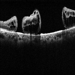
Sep 10 2017 by JEFFERSON R SOUSA, Tecg.º (Biomedical Systems Technology)
Patient 57-years-old, male, attended the clinic with complaint of low visual acuity and history of already having undergone a surgical procedure in another service. In previous evaluation of retinal mapping and retinography, being confirmed in optical coherence tomography, several retinal holes were observed in the posterior pole.
Photographer: JEFFERSON R SOUSA - Study Center and Ophthalmological Research Dr. Andre M V Gomes, Dr. Suel Abujamra Institute São Paulo-Brazil
Imaging device: OCT CIRRUS 4000, Protocol Horizontal line.
Condition/keywords: retinal hole
-
 Multiple Retinal Holes
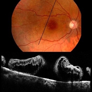
Sep 10 2017 by JEFFERSON R SOUSA, Tecg.º (Biomedical Systems Technology)
Patient 57-years-old, male, attended the clinic with complaint of low visual acuity and history of already having undergone a surgical procedure in another service. In previous evaluation of retinal mapping and retinography, being confirmed in optical coherence tomography, several retinal holes were observed in the posterior pole.
Photographer: JEFFERSON R SOUSA - Study Center and Ophthalmological Research Dr. Andre M V Gomes, Dr. Suel Abujamra Institute São Paulo-Brazil
Imaging device: Topcon TRC-50 DX, Imaginet, campo de 50 graus. Flash 75 / Optical coherence tomography system OCT CIRRUS 4000, Protocol Line 125 degrees.
Condition/keywords: retinal hole
-
 Multiple Retinal Holes
Sep 10 2017 by JEFFERSON R SOUSA, Tecg.º (Biomedical Systems Technology)
Patient 57-years-old, male, attended the clinic with complaint of low visual acuity and history of already having undergone a surgical procedure in another service. In previous evaluation of retinal mapping and retinography, being confirmed in optical coherence tomography, several retinal holes were observed in the posterior pole.
Photographer: JEFFERSON R SOUSA - Study Center and Ophthalmological Research Dr. Andre M V Gomes, Dr. Suel Abujamra Institute São Paulo-Brazil
Imaging device: OCT CIRRUS 4000, Protocol Horizontal line.
Condition/keywords: retinal hole
-
 Multiple Retinal Holes
Sep 10 2017 by JEFFERSON R SOUSA, Tecg.º (Biomedical Systems Technology)
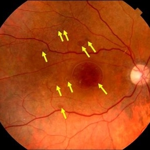
Patient 57-years-old, male, attended the clinic with complaint of low visual acuity and history of already having undergone a surgical procedure in another service. In previous evaluation of retinal mapping and retinography, being confirmed in optical coherence tomography, several retinal holes were observed in the posterior pole. Each arrow represents a hole. Nine retinal holes in the posterior pole
Photographer: JEFFERSON R SOUSA - Study Center and Ophthalmological Research Dr. Andre M V Gomes, Dr. Suel Abujamra Institute São Paulo-Brazil
Imaging device: Retinografo Topcon TRC-50 DX, Imaginet, campo de 50 graus. Flash 75
Condition/keywords: retinal hole
 Loading…