Search results (100 results)
-
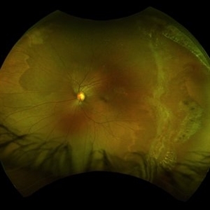
 Retinal Detachment and Lattice Degeneration
Mar 25 2025 by Korey Starkey
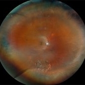
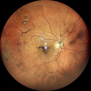
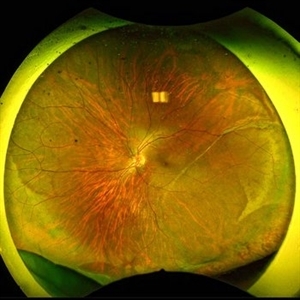
26 year-old patient presented at first visit with rhegmatogenous macula involving retinal detachment of the left eye. Underwent prompt surgical repair. Both eyes also present with lattice degeneration with atrophic holes.
Photographer: Korey Starkey
Condition/keywords: atrophic retinal hole, fundus photography, lattice degeneration, montage photo, Optos, OPTOS CALIFORNIA RGB, retinal detachment, retinal holes, rhegmatogenous retinal detachment, ultra-wide field imaging
-
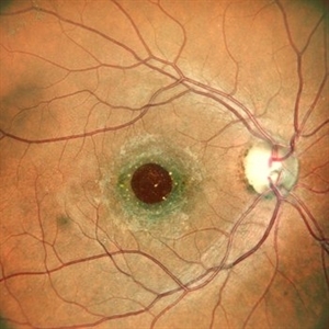
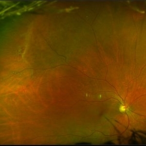
 Lattice Degeneration With Atrophic Retinal Holes
Jan 30 2025 by Kimberly Wakester
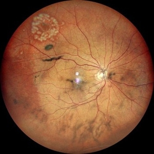
Ultra-wide field montage fundus photograph of a 56-year-old woman with lattice degeneration with atrophic holes statues post laser. Patient also has a small CHRPE temporal to macula and trace ERM that is not visually significant. Will continue follow up care to monitor and treat as needed.
Photographer: Kimberly Wakester, COA
Imaging device: Optos California
Condition/keywords: atrophic retinal hole, CHRPE, epiretinal membrane (ERM), lattice degeneration, montage photo
-
 Barrage Laser
Nov 5 2024 by Dr Bilal Mir
Freshly barrage lasered fundus picture of a young myope.
Photographer: Dr BILAL AHMED MIR, Mbbs Ms ophthalmology
Condition/keywords: retinal hole
-
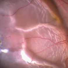 Retinal Detachment Secondary to Retinal Hole
Sep 28 2024 by Anjana Mirajkar, MS Ophthalmology
An intra operative image showing retinal detachment involving the macula secondary to retinal hole.
Photographer: Dr. Anjana Mirajkar -Retina Foundation, Ahmedabad
Condition/keywords: retinal hole, rhegmatogenous retinal detachment
-
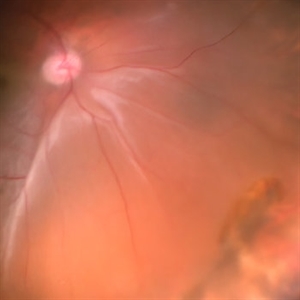 Retinal Detachment With Retinal Break
Jul 4 2024 by Anjana Mirajkar, MS Ophthalmology
Intra operative image showing us a bullous retinal detachment with retinal break noted inferiorly.
Photographer: Dr. Anjana Mirajkar -Retina Foundation, Ahmedabad
Condition/keywords: retinal hole, rhegmatogenous retinal detachment
-
 The Bullet Ridden Retina
Feb 17 2024 by SHISHIR VERGHESE, MS, FVRS, FAICO (Retina)
Fundus image obtained of a case of lasered branch retinal vein occlusion (BRVO) with fibrovascular proliferation (FVP) where the laser marks have given way to multiple small retinal holes due to traction from the same.
Photographer: DIVYA SHAJI
Imaging device: NIDEK MIRANTE
Condition/keywords: BRVO, chronic retinal detachment
-
 Retinal Hole
Feb 11 2024 by Anjana Mirajkar, MS Ophthalmology
A color photo of RE of a 50 year old male showing retinal hole superiorly with vitreous degeneration.
Photographer: Dr. Anjana Mirajkar -Retina Foundation, Ahmedabad
Imaging device: Mirante-Nidek
Condition/keywords: retinal hole
-
 Retinal Hole
Feb 11 2024 by Anjana Mirajkar, MS Ophthalmology
A color photo of RE of a 50 year old male showing lasered retinal hole superiorly with vitreous degeneration.
Photographer: Dr. Anjana Mirajkar -Retina Foundation, Ahmedabad
Imaging device: Mirante-Nidek
Condition/keywords: full thickness retinal hole
-
 Lattice With Holes
Feb 6 2024 by Thirumalesh Mochi Basavaraj, MD
25 year old myopic patient with extensive lattice degeneration with multiple atrophic holes.
Photographer: Puttaswamy
Condition/keywords: atrophic retinal hole, High Myopia, peripheral lattice degeneration
-
 Peripheral retinal degenerations
Jan 29 2024 by Anupama Kiran Kumar
Fundus photo of a young man who underwent barrage laser after he presented to the clinic with floaters and was diagnosed to have lattices with horse shoe tears and retinal holes.
Photographer: Dr Anupama Kiran Kumar DNB FVR , Narayana Nethralaya Bangalore
Imaging device: Mirante SLO/OCT (Nidek Co., Gamagori, Japan)
Condition/keywords: lattice degeneration, peripheral retinal degeneration
-
 macular hole
Jan 28 2024 by Anjana Mirajkar, MS Ophthalmology
Fundus photograph of a 40 year old female showing macular hole with 2 retinal holes surrounding it
Photographer: Dr. Anjana Mirajkar -Retina Foundation, Ahmedabad
Imaging device: Mirante-Nidek
Condition/keywords: macular hole, multiple retinal holes
-
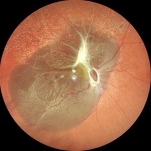
 LARGE OPERCULATED HOLE
Oct 16 2023 by ANKIT JAIN
Left Eye Montage of 50 years old male with high myopia with large operculated hole
Photographer: DR ANKIT JAIN
Imaging device: MIRANTE
Condition/keywords: High Myopia, myopia, operculated retinal hole
-
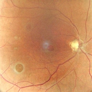
Peripheral Retinal Hole with OCT Co-localization
Sep 26 2023 by Bradley T. Smith, MD, FASRS
Peripheral asymptomatic atrophic retinal hole with OCT co localization demonstrating small cuff of sub retinal fluid. Near infrared imaging shows hyper reflectivity through hole.
Condition/keywords: atrophic hole, lattice degeneration, OCT
-
 Rhegmatogenous retinal detachment in a Young Patient
May 18 2023 by Jesus Lozano, MD
25 year old male patient. High Myope. Arrived to my clinic with a VA Fc 2mts. - Optos Image : Rhegmatogenous Retinal Detachment Mácula Off with ínfero temporal retinal holes in an area of lattice degeneration. Visual Acuity FC 2mts.
Imaging device: Optos
Condition/keywords: retinal detachment with retinal defect
-
 1 year Follow Up after Scleral Buckle Surgery in a Young Patient
May 18 2023 by Jesus Lozano, MD
25 year old man after Scleral Buckle Surgery + laser Retinopexy do to RRD macula off with ínfero temporal mid peripheral retinal holes in an area of lattice degeneration. Final VA 6/9.
Imaging device: Optos
Condition/keywords: scleral buckle
-
 Full Thickness Macular Hole
Oct 16 2022 by Pramod Kumar Suman, MBBS, MD
A 32 years old female presents with complains of diminution of vision in right eye with Full thickness retinal hole involving the fovea.
Photographer: Dr Pramod Kumar Suman
Imaging device: Mirante
Condition/keywords: full thickness macular hole
-
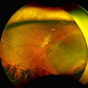
 Total retinal Detachment multiple holes
Sep 26 2022 by Denica Rodriguez
60 year old Male presented with two week old Macula off Retinal detachment with multiple tears.
Photographer: Denica Rodriguez
Imaging device: Optos California
Condition/keywords: color fundus photograph, color photo, macula-off, optos, pseudocolor, Retinal detachment, retinal holes, retinal tear, Retinal tear with detachment, superior arcade, superior field, superior retina, total retinal detachment
-
 Macular Hole
Dec 19 2021 by Eduardo Javier Pinuer Alvarado
Fundus photograph of a 62-year-old woman with macular hole grade 3-4.
Photographer: Eduardo Pinuer A, Universidad Austral de Chile.
Imaging device: CR-2 AF Digital Non-Mydriatic Retinal Camera, Canon.
Condition/keywords: atrophic retinal hole, macular, retina
-
 Retinal Holes, Demarcation Line
Jul 19 2021 by RUSHIK PATEL
Utlrawide pseudo-color fundus photograph of 28-year-old boy with 2 retinal hole surrounded by subretinal fluid less than 1 disc diameter and demarcation line.
Photographer: Rushik Patel, Netralaya Super Speciality Eye Hospital
Condition/keywords: retinal hole
-
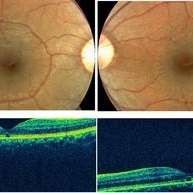 Welders Maculopathy
Apr 27 2021 by Priya Rasipuram Chandrasekaran, MBBS, DO, DNB, FRCS
Fundus photo of both eyes showing absent foveal reflex and orange red discolouration surrounded by a pigmentary halo. The corresponding OCT image of the macula shows outer retinal hole extending from the inner layer of retinal pigment epithelium to the external limiting membrane which inturn corresponds to IS/OS junction.
Condition/keywords: Welder's maculopathy
-
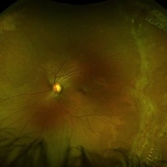 Ultra-Wide Field Fundus Photography Showing Lattice Degeneration
Mar 22 2021 by Sophia El Hamichi, MD
Lattice degeneration, atrophic holes, white without pressure OS in a 19-year-old female.
Condition/keywords: atrophic retinal hole, Optos, peripheral lattice degeneration, ultra-wide field imaging, white without pressure
-
 Myopia with Lattice Degeneration and White Without Pressure in the Setting of Marfan's Syndrome
Aug 31 2020 by Sophia El Hamichi, MD
A 1-year-old female with Marfan's syndrome, myopia OU, congenital nystagmus and exotopia OD. Ultra-wide field imaging of her left eye showed lattice degeneration with atrophic retinal holes temporally, in addition to multiple sections of white without pressure.
Imaging device: Optos
Condition/keywords: atrophic retinal hole, lattice degeneration, Marfan's syndrome, myopia, Optos, ultra-wide field imaging
-
 Bullous Retinoschisis with Outer Retinal Holes
Jun 15 2020 by Olivia Rainey
Ultra-widefield pseudocolor fundus photograph of a 56-year-old female with bullous retinoschisis with outer retinal holes affecting her right eye. The physician noted superotemporal retinoschisis in her monoculcar functioning eye. There was no demarcation line and no inner or outer layer breaks at her first appointment in February of 2020. On 6/15/20 she had a new onset outer holes and SRF tracking inferiorly. The physician recommended observation, however if this continues to progress we have discussed indications for barrier laser.
Photographer: Olivia Rainey, OCT-C, COA
Imaging device: Optos California
Condition/keywords: bullous retinoschisis, Optos, outer layer breaks, outer layer hole, pseudocolor, subretinal fluid, superior retina, ultra-wide field imaging
-
 Operculated Hole with Barrier Laser
Nov 5 2019 by Nichole Lewis
66-year-old female with an operculated retinal hole s/p barrier laser treatment. Choroidal Nevus Inferior.
Photographer: Nichole Lewis
Imaging device: Optos
Condition/keywords: barrier laser, choroidal nevus, operculated retinal hole, operculated tear
-
 ARN (#3) This is comparison between the latest visit (left) and one week prior (which is the right photo, and same one as photo #2)
May 27 2019 by John S. King, MD
60-year-old African American female who had been treated for iridocyclitis for at least a week sent in for vitritis and a nasal fundus lesion. Complaints included redness, floaters, photophobia, and decreased vision. Husband had recent shingles. Acuity was 20/60-2 with IOP of 12, and small KP in Art's triangel, 1-2+ a/c cell, 2-3+ ant vit cell, diffuse arteriolar sheathing, multiple areas of retinal whitening in periphery and mid-periphery (see Photo #1). PCR of a/c was performed, and intravitreal GCV administered, and VACV 2g qid and ASA started.... PCR positive for HZV, pred taper was started two days after presentation as the infection had begun to stablize..... Five days from presentation the vision was 20/60, inflammation and areas of retinal whitening had improved (see Photo #2).... One week later acuity was 20/30, the a/c was quiet and KP resolved; ant vitreous cell decreased; and there was further improvement in retinal appearance without any signs of retinal holes or detachment; she is now on low dose maint VACV (see photo#3)
Photographer: Maysee Yang
Imaging device: Optos CA
Condition/keywords: acute retinal necrosis, Herpes zoster
 Loading…