Search results (660 results)
-
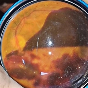
 Angiographic Storm: Fluorescein Leakage in Retinal Vasculitis
Nov 17 2025 by SHRADDHA RAJ SHRIVASTAVA
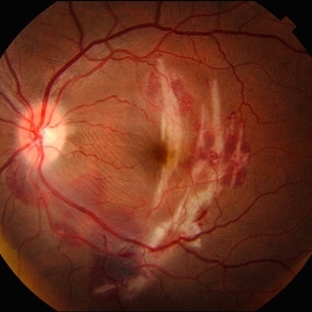
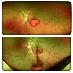
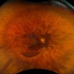
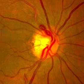
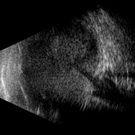
This left eye montage fundus fluorescein angiography (FFA) image of a 19 year old male with idiopathic retinal vasculitis, having skip vasculitic lesions predominantly involving retinal veins. There are areas of blocked fluorescence due to intraretinal hemorrhages, the involved veins have filling defects and occlusions, leading to formation of numerous collateral channels. The inflamed vessels also show perivascular fuzzy hyperfluorescent stain due to leakage of dye. We can also see multiple peripheral capillary non perfusion (CNP) areas, with a 'hot disc', suggestive of ongoing inflammation.
Photographer: Dr. Shraddha Raj Shrivastava
Imaging device: Nidek Mirante SLO/OCT (Confocal scanning/Spectral domain OCT)
Condition/keywords: FA late phase leakage, Fundus Fluorescein Angiography, idiopathic retinal vasculitis, optic disc leakage, VASCULITIS
-
 Choroidal Rupture
Sep 30 2025 by César Adrián Gomez Valdivia, MD
This fundus photograph shows curvilinear streaks of choroidal rupture radiating from the fovea, associated with subretinal hemorrhage. The rupture lines appear as crescent-shaped, whitish streaks representing a break in Bruch’s membrane, choriocapillaris, and retinal pigment epithelium.
Photographer: @eyemissu2
Imaging device: TOPCON TRX
Condition/keywords: Choroidal, Rupture
-
 Type 1 Aneurysmal Neovascularization
Sep 22 2025 by Gustavo Uriel Fonseca Aguirre
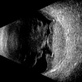
This transverse B-scan demonstrates vitreous hemorrhage, a bullous retinal detachment involving the macula, and dense subretinal hemorrhage, consistent with type 1 aneurysmal neovascularization. The scan reveals significant exudative activity with multi-level bleeding.
Photographer: Gustavo U. Fonseca Aguirre, Hospital Conde de Valenciana, Ciudad de México
Condition/keywords: polypoidal choroidal vasculopathy (PCV), Type 1 Aneurysmal Neovascularization
-
 Lower Temporal Branch Retinal Vein Occlusion
Sep 16 2025 by Seif Allah Anwar
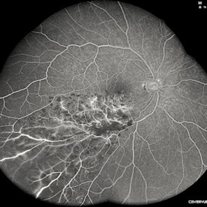
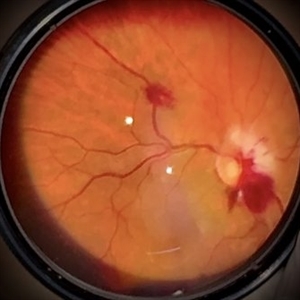
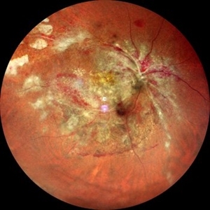
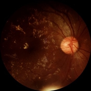
FFA of A 46-year old hypertensive male patient showing blocked fluorescence by retinal hemorrhages along the area drained by the obstructed vein with areas of retinal capillary drop-out.
Photographer: Dr Seif Anwar , FRCSEd
Imaging device: CENTERVUE EIDON
Condition/keywords: Lower temporal branch retinal vein occlusion
-
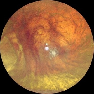
 Papillophlebitis Salauno
Sep 3 2025 by Pablo Angel Garcia Uribe
Fundus photograph of a 24-year-old woman, previously healthy, with a history of recreational inhaled cannabis use, presented with a 24-hour history of photopsias and mild decrease in visual acuity, associated with a subtle relative central scotoma in the right eye. On ophthalmic examination, the anterior segment of both eyes was unremarkable. Best-corrected visual acuity was slightly reduced in the right eye and normal in the left. Fundus biomicroscopy of the right eye revealed moderate disc edema with hyperemia and well-defined margins, accompanied by venous engorgement and tortuosity, predominantly affecting the venules. No retinal hemorrhages were observed. Additionally, retinal thickening was noted along the temporal arcades, with apparent foveal sparing. The left eye showed no pathological findings. Based on the patient’s age, the acute onset of symptoms, the fundoscopic features, and the absence of systemic risk factors, the clinical presentation was consistent with papillophlebitis.
Photographer: Clínica Oftalmológica Salauno
Imaging device: Visucam 524, Carl Zeiss Meditec AG, Jena, Germany
Condition/keywords: papillophlebitis
-
 YAG Laser Hyaloidotomy
Aug 31 2025 by Giriraj Vibhute
A 24-year-old young man presented with sudden loss of vision in left eye following history of rigorous coughing. Visual acuity in RE was 6/6, LE was 6/60p. Fundoscopy showed bilateral multiple small intraretinal hemorrhages with LE large premacular subhyaloid hemorrhage just covering the fovea suggestive of bilateral valsalva retinopathy changes. Nd:YAG laser hyaloidotomy was performed to left eye the same day (A250; 2mJ;6 SHOTS). Visual acuity improved to 6/9 immediately following the procedure. After 1 week, the subhyaloid hemorrhage had completely cleared with dispersed intragel hemorrhage in the inferior vitreous cavity with visual acuity of 6/6 in left eye
Photographer: Dr Vani S. MM Joshi eye institute, Hubli
Condition/keywords: valsalva retinopathy, YAG HYALOIDOTOMY
-
 Bilateral Roth Spot in the Setting of Mitral Valve Endocarditis
Aug 18 2025 by Helder Vasconcelos
A 55-year-old man with chronic alcoholism presented with wasting and fever. The symptoms were preceded by a recent tooth extraction and gingivitis. Fundus examination in the ICU showed a retinal hemorrhage with a white spot (Roth spot) associated with peripapillary hemorrhage and cotton wool exudate. A similar Roth spot was observed in the contralateral eye.
Photographer: Helder Vasconcelos
Imaging device: Smartphone Fundoscopy
Condition/keywords: Infectious endocarditis, Roth Spots
-
 Commotio Retinae
Aug 7 2025 by Gabriel Costa Andrade, PhD
Color fundus photograph of a 13-year-old girl who was hit by accidental discharge of gel bullet in her right eye. She presented with retinal whitening with intraretinal hemorrhages in temporal inferior area of the peripheral retina.
Photographer: Gabriel Andrade
Condition/keywords: macula, Retina, Trauma
-
 Sub-ILM Hemorrhage
Aug 6 2025 by Virginia Gebhart
28 year old female with sub-ILM hemorrhage and questionable cotton wool spot. Pt states sudden vision loss after being hit in the eye with laser lights at a nightclub. Laser lights also damaged the camera in her iPhone. Will give hemorrhage more time to clear on its own, will discuss treatment options if no improvement at next visit.
Photographer: Virginia Gebhart, Retina Consultants of Carolina
Imaging device: Topcon TRC 50DX
Condition/keywords: retinal hemorrhage, sub ILM hemorrhage
-
 Central Retinal Vein Occlusion with Macular Edema
Aug 4 2025 by Virginia Gebhart
58 year old female with Central Retinal Vein Occlusion with Macular Edema. Diffuse retinal hemorrhages, central SRF and sub-hyaloid/vitreous hemorrhage, no obvious active NV. Will start patient on series of 4 monthly intravitreal anti-VEGF injections, followed by T&E protocol once stable.
Photographer: Virginia Gebhart, Retina Consultants of Carolina
Imaging device: Optos California
Condition/keywords: central retinal vein occlusion, CRVO, CRVO with macular edema, retinal hemorrhage, vitreous hemorrhage
-
 CMV Retinitis: Turning Retina into Abstract Art Since Immunosuppression
Aug 4 2025 by rohan jain
We report a case of 34 years old HIV positive male who presented with Diminution of vision in OD since 1 month .Examination of OD showed hazy media due to vitritis, diffuse yellowish-whitish retinal necrosis and retinal hemorrhages around the disc and attenuated retinal vessels.
Photographer: Dr. ROHAN JAIN
Imaging device: mirante
Condition/keywords: CMV chorioretinitis, CMV retinitis, cytomegalovirus (CMV), Cytomegalovirus Retinitis
-
 CMV Retinitis: Turning Retina into Abstract Art Since Immunosuppression
Aug 4 2025 by rohan jain
We report a case of 34 years old HIV positive male who presented with Diminution of vision in OD since 1 month. Examination of OD showed hazy media due to vitritis, diffuse yellowish-whitish retinal necrosis and retinal hemorrhages around the disc and attenuated retinal vessels.
Photographer: Dr. ROHAN JAIN
Imaging device: mirante
Condition/keywords: CMV chorioretinitis, CMV retinitis, cytomegalovirus (CMV), Cytomegalovirus Retinitis
-
 Large Subhyaloid Hemorrhage
Jul 11 2025 by Jessilla Phou
This is a fundus photograph depicting a large subhyaloid hemorrhage in the mid periphery of the left eye. The patient, a 53-year-old female, presented with a sudden onset of floaters, headache, and blurred vision. The image also demonstrates associated optic disc hemorrhage, vitreous hemorrhage, retinal hemorrhage, and venous tortuosity. Despite the extensive workup performed and the severity of the hemorrhage, no underlying cause was determined.
Photographer: Jessilla Phou
Imaging device: Optos California
Condition/keywords: fundus photograph, optic disc hemorrhage, retinal hemorrhage, venous tortuosity, vitreous hemorrhage
-
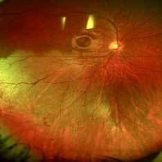
 Prepapillary Vascular Loop
Jul 4 2025 by KANWALJEET HARJOT MADAN, M.S. (Ophthalmology); FAICO (Vitreous - Retina)
This is the fundus picture of right eye of a young 32 years female depicting pre papillary vascular loop. A prepapillary vascular loop is a congenital anomaly of the optic disc that presents as an elevated and twisted bundle of vessels projecting into the vitreous cavity. It is a benign condition, usually unilateral but can be bilateral. It is asymptomatic and discovered during routine eye examination. This anomaly can sometimes cause complications like branch retinal artery occlusion, vitreous hemorrhage, or sub retinal hemorrhage.
Photographer: Dr. Kanwaljeet Harjot Madan, Thind Eye Hospital, Jalandhar City (Punjab) INDIA.
Imaging device: Zeiss Fundus Camera
Condition/keywords: branch retinal artery occlusion (BRAO), optic disc, Prepapillary Vascular Loop, SUB RETINAL HEMORRHAGE, Vitreous hemorrhage
-
 Diabetic Retinopathy
Jun 4 2025 by Paulina Araujo
The 55-degree central fundus photograph of the right eye demonstrates numerous hard exudates, dot intraretinal hemorrhages, and microaneurysms.
Photographer: Paulina D.Araujo Martínez, Asociación para Evitar la Ceguera en México I.A.P., Hospital Dr Luis Sánchez Bulnes.
Condition/keywords: diabetic retinopathy
-
 Macular Edema
Jun 4 2025 by Paulina Araujo
The composite fundus photograph of the right eye demonstrates circinate hard exudates in the thickened macular area, along with flame-shaped intraretinal hemorrhages along the inferior temporal arcade.
Photographer: Paulina D.Araujo Martínez, Asociación para Evitar la Ceguera en México I.A.P., Hospital Dr Luis Sánchez Bulnes.
Condition/keywords: macular edema
-
 Retinal Macroaneurysm (Left Eye)
Apr 29 2025 by Daniela Bogenschutz
72 year-old female has visual complaints of central vision changes ongoing for 4 days. Patient was acutely symptomatic with an intraretinal hemorrhage due to the retinal macroaneurysm. We had a fun little laugh as this retinal macroaneurysm form a shape of a tick in her left eye. This photo is a side-by-side of the color photos and the autofluorescence done. She is being treated by her general doctor for elevated blood pressure.
Photographer: Daniela Bogenschutz, OSC; Retina Consultants of Carolina, P.A.
Imaging device: Optos
Condition/keywords: retinal macroaneurysm
-
 Advanced Proliferative Diabetic Retinopathy
Apr 9 2025 by Gustavo Uriel Fonseca Aguirre
B-mode ultrasound of a patient with long-standing poorly controlled diabetes demonstrates characteristic findings of advanced proliferative diabetic retinopathy. The examination reveals moderate vitreous hemorrhage appearing as diffuse hyperechoic opacities throughout the vitreous cavity, along with a posterior hyaloid membrane densely infiltrated by hemorrhagic material, showing irregular thickening and increased reflectivity. A mild subhyaloid hemorrhage is visible as a subtle hyphema-like space anterior to the retinal surface. The study documents a total tractional retinal detachment, evidenced by rigid retinal folds with clear insertion points of vitreous strands, accompanied by a significant subretinal hemorrhage seen as a prominent hyperechoic collection beneath the elevated retina. These findings collectively illustrate the severe vitreoretinal interface pathology characteristic of end-stage diabetic eye disease, with predominant tractional components and distinct echographic stratification of hemorrhagic layers - from anterior vitreous involvement to deeper subretinal blood accumulation.
Photographer: Gustavo U. Fonseca Aguirre, Hospital Conde de Valenciana, Ciudad de México
Condition/keywords: diabetic retinopathy, tractional retinal detachment, Vitreous hemorrhage
-
 Negative Retinal Detachment
Apr 7 2025 by Gustavo Uriel Fonseca Aguirre
B-mode ultrasound of a 7-month-old premature infant with a history of perinatal supplemental oxygen therapy reveals a total funnel-shaped retinal detachment appearing as a hypoechoic structure, accompanied by significant hyperechoic subretinal hemorrhage. This distinctive echographic pattern creates the characteristic appearance of a "negative retinal detachment."
Photographer: Gustavo U. Fonseca Aguirre, Hospital Conde de Valenciana, Ciudad de México
Condition/keywords: retinopathy of prematurity
-
 Papillary Hemorrhage and Associated Vitreous Hemorrhage
Mar 3 2025 by Juan J. Prados-Carmona
Juxtapapillary thrombosis with retinal hemorrhage and associated vitreous hemorrhage localized in the posterior pole.
Photographer: Juan J. Prados-Carmona; Hospital Universitario Reina Sofía (Córdoba), España.
Condition/keywords: hemorrhage
-
 Choroidal Melanoma Masquerading as PEHCR
Mar 3 2025 by Tejaswita Verma
A 65 year old diabetic male presented with large nasal retinal mass giving the appearance of organized dehaemoglobinized subretinal hemorrhage with breakthrough vitreous hemorrhage , with 6/6P vision. Enucleation specimen showed histopathology confirmed choroidal melanoma.
Photographer: DR. TEJASWITA VERMA
Imaging device: MIRANTE
Condition/keywords: vitreous hemorrhage
-
 Central Retinal Vein Occlusion
Feb 25 2025 by Prithvi Chandrakanth
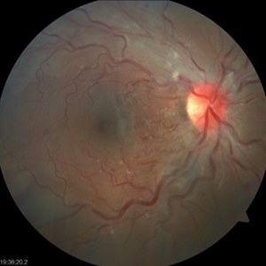
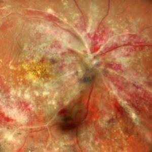
A 61-year-old woman with a history of hypertension noticed a sudden painless blurring of vision in her left eye. Over the next few days, the blurriness persisted, and she experienced a mild central scotoma. On examination, Fundoscopic evaluation revealed dilated, tortuous retinal veins, retinal hemorrhages, and macular oedema.
Photographer: DR.PRITHVI CHANDRAKANTH, DR.CHANDRAKANTH NETHRALAYA, KOZHIKODE
Imaging device: EIDON
Condition/keywords: CRVO, CRVO WITH MACULA EDEMA, flame shaped retinal hemorrhage
-
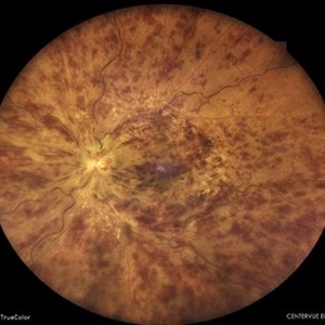
 Coat's Disease with Exudative RD
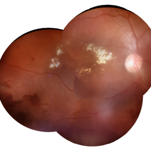
Feb 12 2025 by Tejaswita Verma
Fundus photo of a 7 year old boy with vision Counting fingers close to face in the right eye and intermittent outward deviation of the right eye observed by parents. Fundus examination shows subretinal exudates, telengiectatic vessels in superotemporal quadrant, intraretinal hemorrhages, macular scar, NVD.
Photographer: DR. TEJASWITA VERMA
Imaging device: MIRANTE
Condition/keywords: Coats' disease, exudative retinal detachment
-
 Choroidal Melanoma Masquerading as Subretinal Hemorrhage With Breakthrough VH
Jan 23 2025 by Tejaswita Verma
A 65 year old diabetic male presented with large nasal retinal mass giving the appearance of organized dehaemoglobinized subretinal hemorrhage with breakthrough vitreous hemorrhage , with 6/6P vision. Enucleation specimen showed histopathology confirmed choroidal melanoma.
Photographer: DR. TEJASWITA VERMA
Imaging device: MIRANTE
-
 Subhyaloid Hemorrhage
Jan 22 2025 by DR Rohit Gupta
48 year old female presented with right eye diminution of vision, on fundus examination a large hemorrhage was seen in subhyaloid space with multiple retinal hemorrhages. Patients was known case of diabetes with uncontrolled blood sugar level.
Photographer: Dr Rohit gupta
Imaging device: Samsung S21
Condition/keywords: SUB ILM hemorrhage, subhyaloid blood, subhyaloid hemorrhage
 Loading…