Search results (73 results)
-
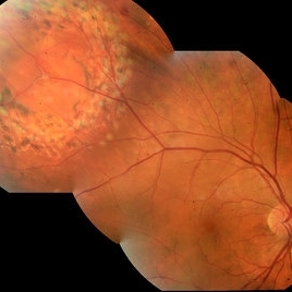
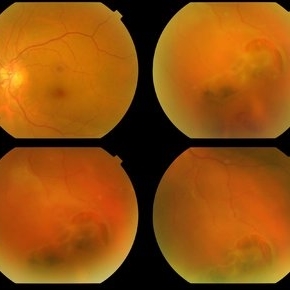
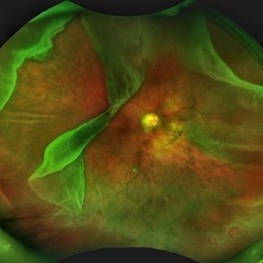 Giant Retinal Tear with Multiple Retinal Breaks
Apr 21 2025 by Hrishikesh Naik, MS
A 28 year old high myope with retinal detachment associated with a supero-temporal giant retinal tear in addition to multiple peripheral horseshoe tears and an additional supero-nasal retinal tear.
Photographer: Hrishikesh Naik
Imaging device: Optos Daytona
Condition/keywords: giant retinal tear, High Myopia, horseshoe tear, retinal break, retinal detachment
-
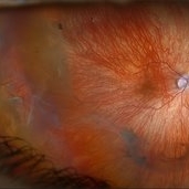
 Hosreshoe Tears on Posterior Pole
Mar 22 2025 by Deepak Bhojwani, MS
A fundus image of an asymptomatic 64 year old male with large horseshoe shaped breaks in inferonasal quadrant on posterior pole, an unusual location for retinal breaks.
Photographer: DR DEEPAK BHOJWANI
Condition/keywords: horseshoe tear, posterior pole break, retinal break
-
 Retinal Detachment
Mar 5 2025 by Kimberly Wakester
Optomap RGB image of an 9-year-old boy with a retinal detachment with retinal break at 9:00 in the right eye. Surgery was recommended. Patient is to continue follow up care post operatively.
Photographer: Kimberly Wakester, COA
Imaging device: Optos California
Condition/keywords: myopic eye, Retinal Detachment, retinal tear
-
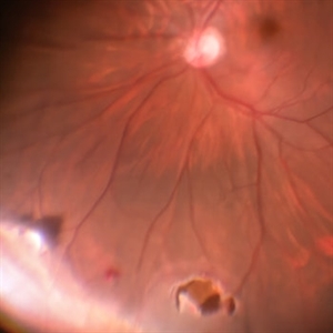 Retinal Break Inferiorly
Jul 5 2024 by Anjana Mirajkar, MS Ophthalmology
An Intra operative still showing a break inferiorly.
Photographer: Dr. Anjana Mirajkar -Retina Foundation, Ahmedabad
Condition/keywords: retinal break
-
 Retinal Detachment With Retinal Break
Jul 4 2024 by Anjana Mirajkar, MS Ophthalmology
Intra operative image showing us a bullous retinal detachment with retinal break noted inferiorly.
Photographer: Dr. Anjana Mirajkar -Retina Foundation, Ahmedabad
Condition/keywords: retinal hole, rhegmatogenous retinal detachment
-
 Subretinal Gas After Pneumatic Retinopexy
Mar 6 2024 by James P Dossett, MD
Pseudocolor fundus photograph of a 68-year-old man who presented with a macula-on rhegmatogenous retinal detachment with a single horseshoe tear at 12 o'clock. Pneumatic retinopexy was performed with cryopexy to the retinal break. He returned to clinic three days later and the entire SF6 gas bubble was noted to have migrated to the subretinal space through the retinal break. Pars plana vitrectomy was performed that day with retinal reattachment and improvement in vision to 20/40 now 6 months postoperatively.
Imaging device: Optos
Condition/keywords: pneumatic retinopexy, subretinal gas bubble
-
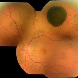
 Retinal detachment
Nov 23 2023 by Anand Temkar
LE color photo montage of a 50 years old male with supero-nasal retinal detachment (with break) and we can see horseshoe tear temporally with sub-retinal fluid.
Photographer: Dr.Anand Temkar- Retina Foundation, Ahmedabad
Imaging device: Mirante
Condition/keywords: RD, retinal break
-
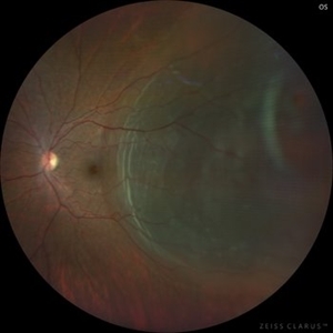
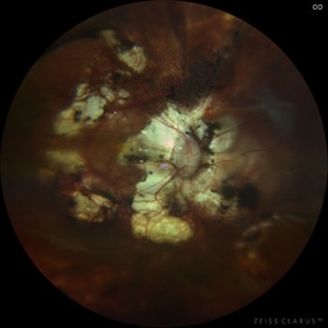 Pathological Myopia with posterior pole retinal detachment & new open break
Jul 31 2023 by Harsh Vardhan Singh, MS
45-year female with redetachment & new break
Photographer: Dr Harsh Vardhan Singh, AIIMS, Guwahati
Imaging device: Zeiss Clarus 700
Condition/keywords: pathologic myopia, posterior staphyloma, retinal break, rrd
-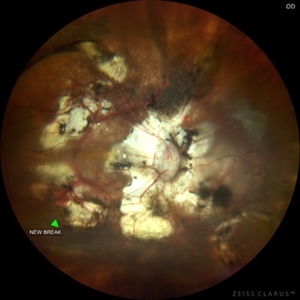
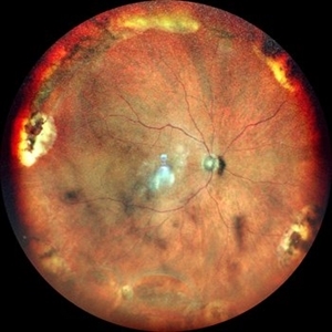 RETINAL BREAKS
Nov 21 2022 by Akansha Sharma
COLOUR FUNDUS PHOTOGRAPH OF A 60 YEAR OLD MALE PATIENT WITH RETINAL BREAK STATUS POST SCLERAL BUCKLING 25 YEARS AGO
Photographer: Dr. Akansha Sharma-Retina Foundation, Ahmedabad
Condition/keywords: retinal break, scleral buckle
-
 Retinal Detachment
Sep 9 2022 by Vishal Agrawal, MD, FRCS,FACS,FASRS
24-year-old female patient presented with sudden decrease in vision. On examination there was a left eye subtotal retinal detachment involving macula.
Photographer: Vishal Agrawal MD
Imaging device: Clarus 700
Condition/keywords: bullous retinal detachment, retinal break
-
 Retinal Detachment with Proliferative Vitreoretinopathy
Jan 31 2022 by Ahmad B. Tarabishy, MD
Ultra wide-field fundus photograph of a 55-year-old gentleman who had previously underwent laser retinopexy for multiple inferior retinal breaks. He presented with a macula-off retinal detachment from a new temporal break with proliferative vitreoretinopathy with fixed folds noted temporally and superonasally.
Photographer: Megan McLandsborough, Lakeland Eye Clinic
Imaging device: Optos California UWF Camera
Condition/keywords: laser retinopexy, macula off Retinal Detachment, proliferative retinopathy, proliferative vitreoretinopathy (PVR), Retinal Detachment, retinal detachment with retinal defect
-
 Ultra-Widefield Image of Tractional-Rhegmatogenous Retinal Detachment Sparing Fovea
Jul 16 2021 by Kushal S Delhiwala, MBBS, MS, FMRF,FICO, FAICO
Ultra-widefield fundus photograph of an 45-year-old phakic male with superior tractional-rhegmatogenous retinal detachment sparing fovea. Retinal break was observed at the base of fibrous proliferation. Scattered whitish outer retinal spots were noted in area of retinal detachment.
Photographer: Kushal Delhiwala, Netralaya superspeciality eye hospital, Ahmedabad, Gujarat,India
Imaging device: Optos Daytona
Condition/keywords: fibrovascular proliferation, fibrovascular tissue, outer retinal white spots, tractional retinal detachment, ultra-wide field imaging
-
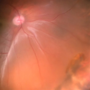
 Rhegmatogenous Retinal Detachment in Left Eye
Apr 4 2021 by MOHIT GUPTA
Fundus picture of left eye of 32-year-old female with rhegmatogemous retinal detachment with small break between 2 -3 'o' clock and subretinal fluid just encroaching the macula .
Photographer: Dr Mohit Gupta
Imaging device: Zeiss Clarus
Condition/keywords: retinal break
-
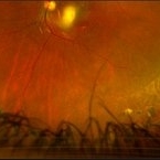
 Proliferative Sickle Cell Retinopathy
Apr 30 2020 by Jordan M Burnham, MD
This ultra-widefield fundus photo of the right eye demonstrates proliferative sickle cell retinopathy resulting in severe visual loss for a young man eventually requiring vitrectomy. Central vitreous hemorrhage and subhyaloid hemorrhage covers the macula (white arrow), causing profound vision loss. A fibrotic, regressed, sea fan neovascularization complex is present in the temporal periphery (green arrow). Subretinal fluid is present in the temporal retinal periphery within the area between the fibrosed sea fan lesion and the posterior preretinal hemorrhage (yellow arrow), likely due to traction or a retinal break obscured by the heme.
Condition/keywords: sickle cell retinopathy
-
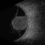 Large Choroidal Melanoma With Retinal Breakthrough - B Scan
Feb 13 2020 by Michael Seider, MD
Atypical presentation of a large pigmented choroidal melanoma in an individual of Caucasian descent. The melanoma has an irregular shape and has broken through the retina superior to the macula of the right eye. The lesion partially overlies the optic nerve. The tumor is not associated with the subretinal fluid/exudative retinal detachment often seen with choroidal melanoma because of the primary location within the vitreous chamber. The tumor is very hypo-autofluorescent as no retinal pigment epithelial tissue is overlying it. Some hyper-autofluorescent signal is seen in the macula from previous subretinal fluid. Optical coherence tomography confirms no subretinal fluid in the macula, mild epiretinal membrane, and outer retinal loss nasally from previous subretinal fluid. B-Scan ultrasonography shows moderate internal reflectivity (more common when considering deeply pigmented melanomas when compared to “classic” low internal reflectivity) and tenting of the hyaloid from the tumor (not commonly seen in uveal melanoma as it usually does not project into the vitreous chamber). No retinal detachment is present.
-
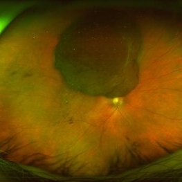
 Large Choroidal Melanoma With Retinal Breakthrough - Widefield FAF
Feb 13 2020 by Michael Seider, MD
Atypical presentation of a large pigmented choroidal melanoma in an individual of Caucasian descent. The melanoma has an irregular shape and has broken through the retina superior to the macula of the right eye. The lesion partially overlies the optic nerve. The tumor is not associated with the subretinal fluid/exudative retinal detachment often seen with choroidal melanoma because of the primary location within the vitreous chamber. The tumor is very hypo-autofluorescent as no retinal pigment epithelial tissue is overlying it. Some hyper-autofluorescent signal is seen in the macula from previous subretinal fluid. Optical coherence tomography confirms no subretinal fluid in the macula, mild epiretinal membrane, and outer retinal loss nasally from previous subretinal fluid. B-Scan ultrasonography shows moderate internal reflectivity (more common when considering deeply pigmented melanomas when compared to “classic” low internal reflectivity) and tenting of the hyaloid from the tumor (not commonly seen in uveal melanoma as it usually does not project into the vitreous chamber). No retinal detachment is present.
-
 Large Choroidal Melanoma With Retinal Breakthrough - Widefield Color
Feb 13 2020 by Michael Seider, MD
Atypical presentation of a large pigmented choroidal melanoma in an individual of Caucasian descent. The melanoma has an irregular shape and has broken through the retina superior to the macula of the right eye. The lesion partially overlies the optic nerve. The tumor is not associated with the subretinal fluid/exudative retinal detachment often seen with choroidal melanoma because of the primary location within the vitreous chamber. The tumor is very hypo-autofluorescent as no retinal pigment epithelial tissue is overlying it. Some hyper-autofluorescent signal is seen in the macula from previous subretinal fluid. Optical coherence tomography confirms no subretinal fluid in the macula, mild epiretinal membrane, and outer retinal loss nasally from previous subretinal fluid. B-Scan ultrasonography shows moderate internal reflectivity (more common when considering deeply pigmented melanomas when compared to “classic” low internal reflectivity) and tenting of the hyaloid from the tumor (not commonly seen in uveal melanoma as it usually does not project into the vitreous chamber). No retinal detachment is present.
-
 Large Choroidal Melanoma With Retinal Breakthrough - OCT
Feb 13 2020 by Michael Seider, MD
Atypical presentation of a large pigmented choroidal melanoma in an individual of Caucasian descent. The melanoma has an irregular shape and has broken through the retina superior to the macula of the right eye. The lesion partially overlies the optic nerve. The tumor is not associated with the subretinal fluid/exudative retinal detachment often seen with choroidal melanoma because of the primary location within the vitreous chamber. The tumor is very hypo-autofluorescent as no retinal pigment epithelial tissue is overlying it. Some hyper-autofluorescent signal is seen in the macula from previous subretinal fluid. Optical coherence tomography confirms no subretinal fluid in the macula, mild epiretinal membrane, and outer retinal loss nasally from previous subretinal fluid. B-Scan ultrasonography shows moderate internal reflectivity (more common when considering deeply pigmented melanomas when compared to “classic” low internal reflectivity) and tenting of the hyaloid from the tumor (not commonly seen in uveal melanoma as it usually does not project into the vitreous chamber). No retinal detachment is present.
-
 Retinal Break
Feb 12 2020 by DIEGO TOLENTINO
Retinal break at vascular junction and laser barricade.
Photographer: Diego Tolentino, CEOP
Condition/keywords: barrier laser, retinal break
-
 CHRPE
CHRPE
Oct 8 2019 by DIEGO TOLENTINO
CHRPE plus laser barricade around retinal break
Photographer: Diego Tolentino
Condition/keywords: congenital hypertrophy of the retinal pigment epithelium (CHRPE)
-
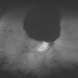
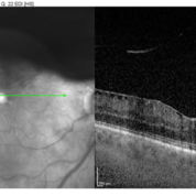
 Giant Retinal Tear (GRT)
Mar 8 2019 by Abdulaziz A. Alshamrani, MD
A 52-year-old male with a moderate myopia (-3.50 sphere) OU complaining of floaters OS since 1 month. BCVA is 20/20 OU. No macular subretinal fluid by OCT.
Condition/keywords: full thickness retinal tear, giant retinal tear, retinal break
-
 Multiple Retinal Horse Shoe Tears
Jun 27 2018 by Hosam Attia, MD
58-year-old African American, monocular, S/P Prophylactic Laser Retinopexy for multiple horse shoe tears OS
Imaging device: Optos - California
Condition/keywords: full thickness retinal tear, laser retinopexy, retinal break, retinal tear
-
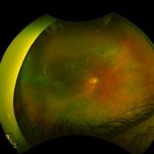
 Horseshoe Retinal Break
Apr 3 2018 by Wesam Safwat
Fundus photograph of an 40-year-old woman with lower temporal horseshoe retinal tear associated with lower sub total retinal detachment not involving macula.
Photographer: Wesam Safwat, Elferdaws eye hospital , Zagazig, Egypt.
Imaging device: Topcon
-
 Buckle intrusion with Retinal detachment
Feb 8 2018 by Manish Nagpal, MD, FRCS (UK), FASRS
Patient operated on 10 years back for a scleral buckling surgery presented with decreased vision and had a superonasal retinal detachment along with intrusion of the scleral buckle.
Photographer: Mehul Prajapati
Condition/keywords: acute retinal detachment, retinal break, scleral buckle
 Loading…