Search results (33 results)
-
 Coats Disease
Sep 29 2024 by Tejaswita Verma
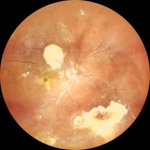
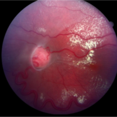
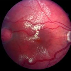
Fundus photo of the RE of a 14 y/o female ,nil premorbid presented with reduced vision in the RE ,diagnosed incidentally on ophthalmological examination elsewhere .Vision was finger counting 3 meters in the RE . Fundus picture reveals macular scar , subretinal and intraretinal exudation ,with scattered hemorrhages esp. in STQ, sclerosed vessels in superior, superonasal quadrant ,nasal, inferonasal quadrant, CR scars inferiorly, Telengiectatic vessels S/O Coat's disease. She was advised RE anti VEGF x1 + laser PRP + PST kenacort under GA with guarded prognosis.
Photographer: DR. TEJASWITA VERMA
Imaging device: MIRANTE
Condition/keywords: Coats' disease
-
 Macula-Involving Rhegmatogenous Retinal Detachment
Feb 17 2024 by Nikhil K Bommakanti, MD
A middle-aged man with a history of rhegmatogenous retinal detachment repair in the fellow eye several years prior presented with reduced vision, which he had noticed two days before.
Condition/keywords: retinal detachment of the macula, rhegmatogenous retinal detachment
-
 Bietti's Crystalline Dystrophy
Oct 3 2020 by SHISHIR VERGHESE, MS, FVRS, FAICO (Retina)
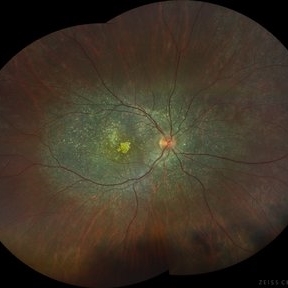
Fundus photo of the right eye of a 21-year-old gentleman with complaints of reduced vision, nyctalopia and visual field reduction. Fundus photograph showing numerous small glistening yellow-white retinal crystalline deposits in the retina.
Photographer: Shishir Verghese, Aravind Eye Hospital, Coimbatore
Condition/keywords: Bietti's crystalline dystrophy, hereditary retinal degeneration, heredomacular degeneration
-
 Bietti's Crystalline Dystrophy
Oct 3 2020 by SHISHIR VERGHESE, MS, FVRS, FAICO (Retina)
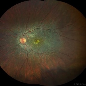
Fundus photo of the left eye of a 21-year-old gentleman with complaints of reduced vision, nyctalopia and visual field reduction. Fundus photograph showing numerous small glistening yellow-white retinal crystalline deposits in the retina.
Photographer: Shishir Verghese, Aravind Eye Hospital, Coimbatore
Condition/keywords: Bietti's crystalline dystrophy, heredomacular degeneration, retinal degeneration
-
 Solitary Retinal Capillary Hemangioblastoma
Apr 29 2019 by Michael A. Novak, MD
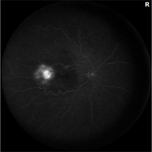
17-year-old young lady presented with reduced vision OD for several months. Her vision was 20/50.
Condition/keywords: retinal capillary hemangioblastoma, Von Hippel-Lindau
-
 Solitary Capillary Retinal Hemangioblastoma
Apr 29 2019 by Michael A. Novak, MD
17-year-old young lady presented with reduced vision OD for several months. Her vision was 20/50.
Condition/keywords: retinal capillary hemangioblastoma, Von Hippel-Lindau
-
 Solitary Retinal Capillary Hemangioblastoma
Apr 29 2019 by Michael A. Novak, MD
17-year-old young lady presented with reduced vision OD for several months. Her vision was 20/50.
Condition/keywords: retinal capillary hemangioblastoma, Von Hippel-Lindau
-
 Progressive Outer Retinal Necrosis
Nov 19 2017 by Navneet Mehrotra, DNB
42-year-old HIV positive male with reduced vision in right eye for two weeks.
Photographer: Mehul Prajapati
Imaging device: Topcon
Condition/keywords: HIV, progressive outer retinal necrosis (PORN)
-
 Endophthalmitis
Jul 12 2014 by Philip J. Polkinghorne, MD
A 75-year-old man who presented with reduced vision and pain 6 years following mitomycin assisted trabeculectomy.
Photographer: Philip Polkinghorne
Condition/keywords: endophthalmitis, post-trabeculectomy
-
 Leptospirosis Neuroretinitis
May 30 2014 by Mitzy E Torres Soriano, MD
50-year-old man, presented with sudden onset of reduced vision in the left eye. Visual acuity (VA) was count fingers. Fundoscopic examination revealed soft exudation adjacent the optic nerve, macular edema with hard exudates in star shape arrangement and retinal vasculitis. OCT confirmed macular edema. There were no systemic symptoms. History of alcoholism and crack cocaine addiction. Systemic work up revealed a positive leptospira. He was treated with oral doxycicline (100mg twice daily) and prednisona (1mg/kg with gradual taper) for two weeks. Follow up at six months showed an improvement of VA to 20/60 with partial resolution of clinical findings at fundoscopic exam. Leptospirosis should be ruled out in every case of neuroretinitis.
Photographer: Mitzy E. Torres Soriano, MD; Centro medico Cagua-Estado Aragua. Venezuela
Imaging device: Retinal Camera TRC-NW8, TOPCON
Condition/keywords: leptospirosis, macular star, neuroretinitis, retinal vasculitis
-
---thumb.jpg/image-square;max$300,300.ImageHandler) Reduced Vision
Reduced Vision
Feb 4 2014 by Maurice F. Rabb
69-year-old female with a detachment of the pigment epithelium in the right eye with hemorrhagic and early fibrous proliferative changes. The left eye contained a large, turbid detachment of the pigment epithelium with patchy atrophy, a very shallow evident overlying sensory retinal detachment, but no subretinal hemorrhage. The visual acuity in each eye was 20/400.
Condition/keywords: fibrous proliferation, patchy atrophy, pigment epithelial detachment (PED), reduced vision
 Loading…