Search results (109 results)
-
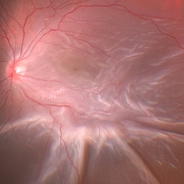
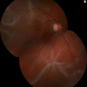
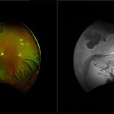 Radiation Retinopathy with Rhegmatogenous Retinal Detachment
Oct 20 2025 by Meng-Hsin Chen
Fundus photo of a 54-year-old woman showing chronic radiation retinopathy from in-utero exposure with rhegmatogenous retinal detachment at the 3:00 -9:00 region, atrophic retina, and PVR. Lattice degeneration is present superiorly and inferiorly with a neovascular frond.
Photographer: Meng-Hsin Chen
Condition/keywords: atrophic retina, lattice degeneration, neovascular frond, proliferative vitreoretinopathy (PVR), radiation retinopathy, retinal detachment
-
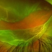
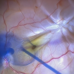
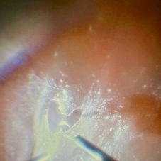 Love Through the Lens of Retinal Detachment
Jun 27 2025 by Claudio Brancato, MD
The image depicts a case of rhegmatogenous retinal detachment where the vitreous was extremely adherent to the retina. The primary surgeon was performing membrane peeling using a surgical loop, while the assisting surgeon was captivated by the intricate procedure. In a moment of affectionate dedication, the primary surgeon carefully peeled the membrane to form a heart shape, symbolizing both his passion for surgery and perhaps a personal gesture towards the assisting surgeon. This delicate and precise maneuver highlights the complexity and artistry involved in vitreoretinal surgery, showcasing the blend of technical skill and emotional expression within the operating room.
Photographer: Claudio Brancato, ARNAS CIVICO Hospital, Palermo, Italy
Imaging device: Zeiss Artevo 800
Condition/keywords: finesse, peeling, proliferative vitreoretinopathy (PVR), Retina detachment
-
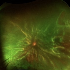
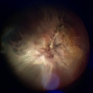
 Complex Retinal Detachment with PVR and Starfold
Jun 6 2025 by Jenn Geelan
57 year old male with a Complex Retinoschisis related retinal detachment with PVR and a Posterior Star Fold
Photographer: Jenn Geelan, Retina-Vitreous Surgeons of CNY
Imaging device: Optos California
Condition/keywords: proliferative vitreoretinopathy (PVR), rare, Retinal Detachment, retinoschisis, Starfolds, subretinal fluid
-
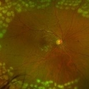
 Rhegmatogenous Retinal Detachment
May 19 2025 by Saarang Hansraj
Rhegmatogenous retinal detachment with grade C subretinal PVR
Condition/keywords: proliferative vitreoretinopathy (PVR), Rhegmatogenous retinal detachment
-
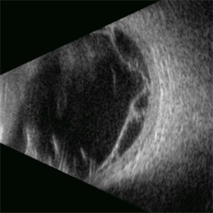 Proliferative Vitreoretinopathy
Apr 17 2025 by Gustavo Uriel Fonseca Aguirre
This B-mode transverse ultrasound scan depicts a post-vitrectomy eye with recurrent retinal detachment in a patient with diabetic retinopathy history. The image reveals fresh vitreous cavity hemorrhage and subretinal bleeding, along with subretinal proliferative bands (PVR strands). These findings indicate complicated tractional re-detachment with active hemorrhagic components.
Photographer: Gustavo U. Fonseca Aguirre, Hospital Conde de Valenciana, Ciudad de México
Condition/keywords: proliferative vitreoretinopathy (PVR)
-
 Rhegmatogenous Retinal Detachment with Gd C PVR Changes
Mar 28 2025 by Shrishti mishra
Fundus photograph of a 58 year old male who had undergone a pneumatic retinopexy elsewhere presented to us with a total retinal detachment with a retinal tear in the superotemporal quadrant and grade c pvr changes.
Photographer: Mrs Vinutha
Imaging device: Optos nikon
Condition/keywords: proliferative vitreoretinopathy (PVR), retinal tear with detachment, rhegmatogenous retinal detachment
-
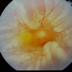
 A Large Break at the Posterior Pole With RD With PVR (S/p Old Blunt Trauma)
Jan 16 2025 by Anand Temkar
Right eye widefield fundus color photo of a 10 year old kid who noticed diminution of vision in right eye since a month. We can see the large break at the posterior pole with rolled up margins associated with retinal detachment and PVR changes.
Photographer: Dr.Anand Temkar- Retina Foundation, Ahmedabad
Imaging device: Mirante
Condition/keywords: posterior pole break, proliferative vitreoretinopathy (PVR), Retinal Detachment
-
 A Large Break at the Posterior Pole With RD With PVR (S/p Old Blunt Trauma)
Jan 16 2025 by Anand Temkar
Right eye central fundus color photo of a 10 year old kid who noticed diminution of vision in right eye since a month. We can see the large break at the posterior pole with rolled up margins associated with retinal detachment and PVR changes.
Photographer: Dr.Anand Temkar- Retina Foundation, Ahmedabad
Imaging device: Mirante
Condition/keywords: Posterior pole break, proliferative vitreoretinopathy (PVR), Retinal Detachment
-
 Repaired Retinal Detachment with Grade C PVR
Dec 23 2024 by Virginia Gebhart
61 year old male 1 day s/p retinectomy/SO exchange. Retina is attached under SO with good laser to retinectomy edge.
Photographer: Virginia Gebhart, Retina Consultants of Carolina
Imaging device: Optos California
Condition/keywords: gas bubble, proliferative vitreoretinopathy (PVR), retinectomy, silicone oil, total retinal detachment
-
 Retinectomy in Complex Retinal Detachment
Oct 28 2024 by Andreas Paulo Di Luciano Rojas, MD
Postop patient with giant tear and anterior PVR. Retinectomy was done.
Photographer: Andreas Di- Luciano, MD
Imaging device: Optos
Condition/keywords: proliferative vitreoretinopathy (PVR), retinectomy
-
 Giant Tear
Oct 28 2024 by Andreas Paulo Di Luciano Rojas, MD
Giant retinal tear secondary to trauma.
Photographer: Andreas Di-Luciano, MD
Imaging device: Optos
Condition/keywords: giant retinal tear, ocular trauma, proliferative vitreoretinopathy (PVR), retinectomy, Trauma
-
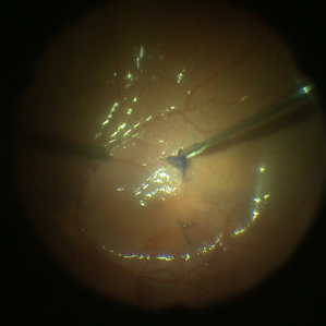
 Star Folds in a Chronic Retinal Detachment
Jul 3 2024 by Anjana Mirajkar, MS Ophthalmology
Intra-operative still RE showing a star fold at the parafoveal area causing traction at the macula. Brilliant blue dye being injected to the stain the ILM.
Photographer: Dr. Anjana Mirajkar -Retina Foundation, Ahmedabad
Condition/keywords: brilliant blue staining, proliferative vitreoretinopathy (PVR), star folds
-
 Rhegmatogenous Retinal Detachment + Proliferative Vitreoretinopathy Type C A12
Jun 28 2024 by Hector Gabriel Moreno Solano, MD, MHA
A 47-year-old female patient with a diagnosis of depression and poor support from the family network who suffered rhegmatogenous retinal detachment 2 years ago, refusing surgical treatment, came again requesting surgery and found grade C A12 proliferative vitreretinopathy.
Photographer: Héctor Gabriel Moreno-Solano, MD, MHA
Condition/keywords: proliferative vitreoretinopathy (PVR), Retina detachment, rhegmatogenous retinal detachment
-
 Retinal Detachment With PVR Changes
Jun 16 2024 by Anjana Mirajkar, MS Ophthalmology
An intra operative still of RE of a 32 year old female case of retinal detachment with Star fold along the supero temporal quadrant.
Photographer: Dr. Anjana Mirajkar -Retina Foundation, Ahmedabad
Condition/keywords: proliferative vitreoretinopathy (PVR)
-
 Proliferative Vitreoretinopathy
Jun 9 2024 by Marcelo Zas, MD PhD
We present a case of a 20-year-old patient who underwent surgery for congenital cataract when he was born and 20 years after he developed a retinal detachment with proliferative vitreoretinopathy. Proliferative vitreoretinopathy (PVR), a major complication of rhegmatogenous retinal detachment (RRD), is an abnormal process whereby proliferative, contractile cellular membranes form in the vitreous and on both sides of the retina, resulting in tractional retinal detachment with fixed retinal folds. PVR arises in an estimated 5-10% of RRD cases, and therefore represents a major complication of retinal detachment. The best treatment of PVR is its prevention. Clinical factors associated with increased risk of PVR include: • Chronic RRD • 2 o more horseshoe retinal tears and RRD exposing three-disc diameters or more of RPE • RD associated with giant retinal • RD associated with choroidal detachment • Ocular Trauma • RRD associated with vitreous hemorrhage • Aphakia and RRD • Failure of previous surgery or multiple retinal surgeries • Aggressive retinitis, etc.
Photographer: Luciano Scorsetti MD
Condition/keywords: proliferative vitreoretinopathy (PVR)
-
 Chronic Regmatogenous Retinal Detachment
Mar 21 2024 by Mauricio Bayram-Suverza, MD
A 65-year-old man came to our department with a complaint of chronic visual loss in his right eye. He had undergone cataract surgery in the same eye at another facility 8 years ago. During the examination, it was observed that the patient had no light perception in the affected eye. Further, slit lamp examination revealed a chronic anteriorized retinal detachment.
Photographer: Mauricio Bayram-Suverza, Fundación Hospital Nuestra Señora de la Luz
Imaging device: iphone
Condition/keywords: proliferative vitreoretinopathy (PVR), vitreoretinal surgery
-
 Chronic Retinal Detachment with Proliferative Vitreoretinopathy
Jan 25 2024 by Isaac Agranoff
Widefield fundus photography of a 24 year old male presenting with subtotal retinal detachment with circumferential anterior proliferative vitreoretinopathy. The detachment is bullous inferiorly with atrophic retina and subretinal bands. There are also scattered patches of lattice with atrophic holes and associated detachment in the periphery. Patient presented with flashes for 2 years with worsening vision over the past 6-8 months, measured at 20/100 ph 20/60 OS.
Photographer: Isaac Agranoff, Ashley Rigdon
Imaging device: Optos California
Condition/keywords: atrophic hole, chronic retinal detachment, lattice degeneration, proliferative vitreoretinopathy (PVR), subretinal bands
-
 Retinal Detachment with PVR
Jan 25 2024 by Virginia Gebhart
62 year old female with history of RD repair with scleral buckle 30 years ago. Low scleral buckle with cryo scar, attached anteriorly. New RD repaired with gas bubble
Photographer: Virginia Gebhart
Imaging device: Topcon
Condition/keywords: proliferative vitreoretinopathy (PVR), Retinal Detachment
-
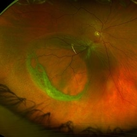
 Napkin ring membrane
Sep 14 2023 by Ben Serar
Fundus photograph showing circumferential contraction of an annular napkin-ring subretinal band, in a case retinal detachment with proliferative vitreoretinopathy.
Condition/keywords: Napkin ring membrane, proliferative vitreoretinopathy (PVR)
-
 Proliferative Vitreoretinopathy
Jun 11 2023 by Ethan K Sobol, MD
Intraoperative view of a retinal detachment with extensive inferior proliferative vitreoretinopathy, prior to successful retinal re-attachment.
Condition/keywords: pars plana vitrectomy (PPV), proliferative vitreoretinopathy (PVR)
-
 Proliferative Vitreoretinopathy
Jun 11 2023 by Ethan K Sobol, MD
Intraoperative view of inferior PVR prior to successful retinal re-attachment
Condition/keywords: pars plana vitrectomy (PPV), proliferative vitreoretinopathy (PVR)
-
 Funnel Retinal Detachment
Jun 11 2023 by Ethan K Sobol, MD
Intraoperative view of a funnel retinal detachment with proliferative vitreoretinoapthy in an eye with previous open globe injury. PVR membranes were peeled, and the retina was flattened and re-attached with an inferior relaxing retinotomy and silicone oil tamponade
Condition/keywords: intraoperative, open funnel RD, open globe injury, proliferative vitreoretinopathy (PVR)
-
 Inferior retinectomy
Jun 11 2023 by Ethan K Sobol, MD
Intraoperative view under air showing attached retina after a scleral buckle, lensectomy, and vitrectomy with an inferior 180 degree retinectomy
Condition/keywords: proliferative vitreoretinopathy (PVR), retinectomy
-
 Proliferative Vitreoretinopathy
Jun 11 2023 by Ethan K Sobol, MD
Intraoperative view of inferior PVR prior to successful retinal re-attachment
Condition/keywords: proliferative vitreoretinopathy (PVR)
-
 Peeling Under PFO
May 7 2023 by Maxwell J Wingelaar, MD
Peeling under PFO
Condition/keywords: ILM peeling, ILM staining, proliferative vitreoretinopathy (PVR)
 Loading…