Search results (79 results)
-
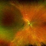
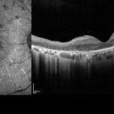
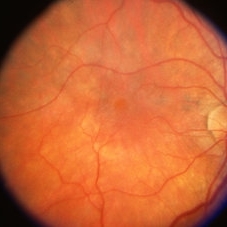
 Autosomal Recessive Bestrophinopathy
Apr 7 2022 by Nassim Alejandro Abreu Arbaje, MD
Color fundus photo of a 22 year old boy with foveal pigment changes and some creamy white lesions associated with localized serous detachment.
Photographer: Nassim Abreu
Imaging device: Topcon Triton Plus
Condition/keywords: Autosomal recessive bestrophinopathy
-
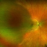
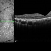
 Autosomal Recessive Bestrophinopathy
Apr 7 2022 by Nassim Alejandro Abreu Arbaje, MD
Color fundus photo collage of a 22 year old boy with foveal pigment changes and some creamy white lesions associated with localized serous detachment
Photographer: Nassim Abreu
Imaging device: Topcon Triton Plus
Condition/keywords: Autosomal recessive bestrophinopathy
-
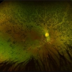
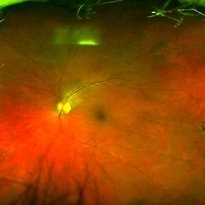
 Autosomal Recessive Bestrophinopathy
Apr 7 2022 by Nassim Alejandro Abreu Arbaje, MD
Color fundus photo collage of a 22 year old boy with foveal pigment changes and some creamy white lesions associated with localized serous detachment
Photographer: Nassim Abreu
Imaging device: Topcon Triton Plus
Condition/keywords: Autosoma recessive bestrophinopathy
-
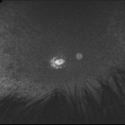
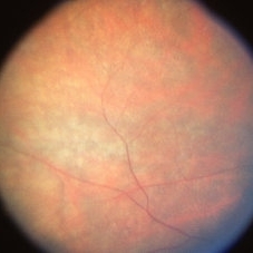
 Autosomal Recessive Bestrophinopathy
Apr 7 2022 by Nassim Alejandro Abreu Arbaje, MD
Color fundus photo of a 22 year old boy with foveal pigment changes and some creamy white lesions associated with localized serous detachment
Photographer: Nassim Abreu
Imaging device: Topcon Triton Plus
Condition/keywords: Autosomal recessive bestrophinopathy
-
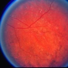
 Retinoschisis with Outer Layer Break
Nov 4 2020 by Thomas A. Ciulla, MD, MBA, FASRS
Inferior temporal retinoschisis extending posteriorly to arcade, associated with outer layer break and related pigment changes from chronicity.
Condition/keywords: bullous retinoschisis, inferotemporal retinoschisis, outer layer breaks
-
 Isolated Choroidal Melanocytosis - Montage
Oct 27 2019 by John S. King, MD
23-year-old white female consulted for a large pigmented choroidal lesion in the right eye. Healthy, no history of glaucoma, 20/20 OU without scleral pigment changes; large, flat pigmented choroidal lesion that is sectoral (temporally) and pigment appears to spare the major choroidal vessels. On OCT (not shown) there is mildly increased choroidal thickening in the area of the lesion.
Photographer: Shelly Blair
Imaging device: Optos CA
Condition/keywords: choroidal melanocytosis, ocular melanocytosis
-
 Isolated Choroidal Melanocytosis
Oct 27 2019 by John S. King, MD
23-year-old white female consulted for a large pigmented choroidal lesion in the right eye. Healthy, no history of glaucoma, 20/20 OU without scleral pigment changes; large, flat pigmented choroidal lesion that is sectoral (temporally) and pigment appears to spare the major choroidal vessels. On OCT (not shown) there is mildly increased choroidal thickening in the area of the lesion.
Photographer: Shelly Blair
Imaging device: Optos CA
Condition/keywords: choroidal melanocytosis, ocular melanocytosis
-
 Siderosis
Apr 2 2019 by Gary R. Cook, MD, FACS
27-year-old white male demonstrating pigment changes OS secondary to siderosis; V.A. = 20/30
Condition/keywords: secondary pigmentary degeneration, siderosis
-
 Pigmentary Retinal Dystrophy
Mar 29 2019 by Jessica Norkus
Optos ultra wide field image of 41-year-old male patient with pigmentary retinal dystrophy. Atypical findings due to unilateral presentation. Patient has been experiencing symptoms for 15 years, notes significant nyctalopia.
Photographer: Jessica Norkus
Imaging device: Optos Ultra Wide Field Camera
Condition/keywords: abnormal fundus, bone spicule, color fundus photograph, color photo, fundus photograph, Optos, peripheral bone spicules, pigment changes, ultra-wide field imaging, unilateral blindness
-
 Pigmentary Retinal Dystrophy
Mar 29 2019 by Jessica Norkus
Optos ultra wide field image of 41-year-old male patient with pigmentary retinal dystrophy. Atypical findings due to unilateral presentation. Patient has been experiencing symptoms for 15 years, notes significant nyctalopia.
Photographer: Jessica Norkus
Imaging device: Optos Ultra Wide Field Camera
Condition/keywords: abnormal fundus, bone spicule, color fundus photograph, color photo, fundus autofluorescence (FAF), fundus photograph, Optos, peripheral bone spicules, pigment changes, ultra-wide field imaging, unilateral blindness
-
 Pigmentary Retinal Dystrophy
Mar 29 2019 by Jessica Norkus
Heidelberg Spectralis image of 41-year-old male patient with pigmentary retinal dystrophy. Atypical findings due to unilateral presentation. Patient has been experiencing symptoms for 15 years, notes significant nyctalopia.
Photographer: Jessica Norkus
Imaging device: Heidelberg Spectralis
Condition/keywords: bone spicule, Heidelburg Spectralis, optical coherence tomography (OCT), pigment changes, unilateral blindness
-
 Post Traumatic ERM With Large Retinal Tear
Apr 9 2018 by Navneet Mehrotra, DNB
A 22-year-old male presented with epiretinal membrane with large retinal tear and pigmentary changes, two months following blunt trauma
Photographer: Mehul Choudhary
Condition/keywords: epiretinal membrane (ERM), pigment changes, retinal tear, trauma
-
 Retinal Pigment Changes After Blunt Ocular Trauma
Jun 27 2016 by Rita Couceiro, MD, MS
A 19-year-old man suffered blunt trauma of the left eye with a ball during soccer practice. At day 3 after trauma (upper pictures) the retinal area superior to the fovea looked pale and visual acuity was reduced to 20/32. This area revealed hypersignaling of retinal layers on OCT and the foveal area showed a localized disruption of retinal layers above the RPE. At day 30 (lower pictures) the retinal area of pallor showed pigmentary changes and OCT revealed atrophy of the external retinal layers. However the localized subfoveal retinal disruption was improved and only a slight disruption was seen on OCT at the ellipsoid level. Visual acuity of the left eye was restored to 20/20 although a scotoma remained.
Photographer: Rita Couceiro, Serviço de Oftalmologia do Hospital de Santa Maria, Lisboa, Portugal
Condition/keywords: blunt trauma, commotio retinae, pigment changes
-
 Pigment Dispersion Syndrome
May 25 2016 by M. Reza Razeghinejad
Fundus photograph of a 57-year-old woman with pigment dispersion syndrome and retinal perivascular pigmentation
Condition/keywords: pigment changes
-
 Pigmentary Change
Apr 14 2014 by Dipankar Barua, M.Sc
Male patient, 56-years-old with vision of boths eye is normal with a complaint of watering. It seems to be a case of pigmentary change.
Photographer: Dipankar Barua
Imaging device: TRC 50 DX (IA)
Condition/keywords: macula, pigment changes
-
 Pigmentary Change
Apr 14 2014 by Dipankar Barua, M.Sc
Male patient, 56-years-old with vision of the both eyes is normal with a complaint of watering. It seems to be a case of pigmentary change
Photographer: Dipankar Barua
Imaging device: TRC 50 DX (IA)
Condition/keywords: pigment changes
-
---thumb.jpg/image-square;max$300,300.ImageHandler) Pattern Dystrophy
Pattern Dystrophy
Aug 9 2013 by From the Collections of Thomas M. Aaberg, MD and Thomas M. Aaberg Jr., MD
Central pigment changes.
Condition/keywords: pattern macular dystrophy, pigment changes
-
 Choroideremia Carrier
Aug 1 2013 by From the Collections of Thomas M. Aaberg, MD and Thomas M. Aaberg Jr., MD
FA of choroideremia carrier with diffuse mottled fluorescence from pigment changes
Condition/keywords: choroideremia, pigment changes
-
 Choroideremia Carrier
Aug 1 2013 by From the Collections of Thomas M. Aaberg, MD and Thomas M. Aaberg Jr., MD
FA of choroideremia carrier with diffuse mottled fluorescence from pigment changes.
Condition/keywords: choroideremia
-
 Choroideremia Carrier
Aug 1 2013 by From the Collections of Thomas M. Aaberg, MD and Thomas M. Aaberg Jr., MD
Choroideremia carrier with diffuse mottled fluorescence from pigment changes.
Condition/keywords: choroideremia carrier
-
 Choroideremia Carrier
Aug 1 2013 by From the Collections of Thomas M. Aaberg, MD and Thomas M. Aaberg Jr., MD
FA of choroideremia carrier with diffuse mottled fluorescein from pigment changes.
Condition/keywords: choroideremia carrier
-
 Choroideremia With Periperal Pigment Changes, Drusenoid Flecks, Patchy Atrophy
Aug 1 2013 by From the Collections of Thomas M. Aaberg, MD and Thomas M. Aaberg Jr., MD
Choroideremia with periperal pigment changes, drusenoid flecks, patchy atrophy.
Condition/keywords: choroideremia, drusenoid flecks, patchy atrophy
-
 Choroideremia With Periperal Pigment Changes, Drusenoid Flecks, Patchy Atrophy
Aug 1 2013 by From the Collections of Thomas M. Aaberg, MD and Thomas M. Aaberg Jr., MD
Choroideremia with periperal pigment changes, drusenoid flecks, patchy atrophy.
Condition/keywords: atrophy, choroideremia, drusenoid flecks
 Loading…