Search results (45 results)
-
 The Great Disc-guise
Nov 12 2025 by SHRADDHA RAJ SHRIVASTAVA
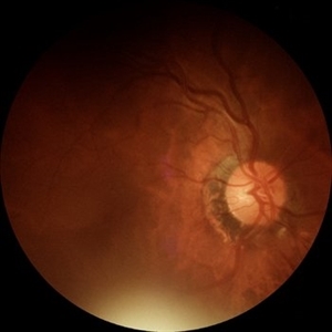
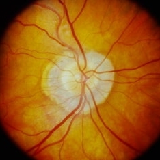
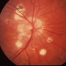
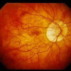
Right eye pseudocolor fundus photo of a 20 year old with Both eyes Pathological Myopia (spherical refractive error of - 18.00 DS in BE), showing a tilted myopic disc with peripapillary atrophy, and extensive posterior staphyloma baring the underlying choroidal vessels and scleral tissue. We can also see a well-defined round chorioretinal atrophic (CRA) patch superonasal to the disc, giving the illusion of double disc on cursory fundus examination.
Photographer: Dr. Shraddha Raj Shrivastava
Imaging device: Nidek Mirante SLO/OCT (Confocal scanning/Spectral domain OCT)
Condition/keywords: chorioretinal atrophy, High Myopia, pathologic myopia, peripapillary atrophy, posterior staphyloma
-
 ERMageddon - Wrinkle in the Space-time Fabric of Macula
Oct 29 2025 by SHRADDHA RAJ SHRIVASTAVA
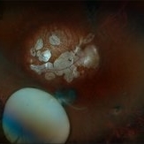
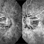
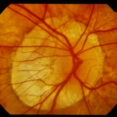
38 year old female with Epiretinal Membrane (ERM) over macula, post laser barrage for multiple symptomatic Horse-shoe Tears (HSTs) and Lattice Degenerations (seen on wide-field image). Posterior pole revealed tilted disc with peripapillary atrophy. There is thick opaque epiretinal membrane obscuring the underlying superior arcade vessels and causing foveal ectopia with distortion of perimacular vasculature. Patient was planned for Right Eye pars plana vitrectomy for ERM peeling.
Photographer: Dr. Shraddha Raj Shrivastava
Imaging device: Nidek Mirante SLO/OCT (Confocal scanning/Spectral domain OCT
Condition/keywords: BARRAGE LASER, ectopic fovea, epiretinal membrane (ERM), horseshoe tear, lattice degeneration, vitreomacular traction (VMT)
-
 ERMageddon - Wrinkle in the Space-time Fabric of Macula
Oct 29 2025 by SHRADDHA RAJ SHRIVASTAVA
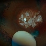
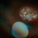
38 year old female with Epiretinal Membrane (ERM) over macula, post laser barrage for multiple symptomatic Horse-shoe Tears (HSTs) and Lattice Degenerations. Posterior pole revealed tilted disc with peripapillary atrophy. There is thick opaque epiretinal membrane obscuring the underlying superior arcade vessels and causing foveal ectopia with distortion of perimacular vasculature. Patient was planned for Right Eye pars plana vitrectomy for ERM peeling.
Photographer: Dr. Shraddha Raj Shrivastava
Imaging device: Nidek Mirante SLO/OCT (Confocal scanning/Spectral domain OCT
Condition/keywords: ectopic fovea, epiretinal membrane (ERM), ERM, horseshoe tear, vitreomacular traction (VMT)
-
 Atrophy
Jun 4 2025 by Paulina Araujo
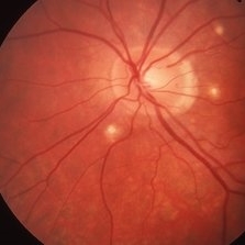
The fundus photograph captures the central 55 degrees of the right eye, revealing alpha and beta peripapillary atrophy.
Photographer: Paulina D.Araujo Martínez, Asociación para Evitar la Ceguera en México I.A.P., Hospital Dr Luis Sánchez Bulnes.
Condition/keywords: atrophy
-
 Multimodal Imaging in CHRPE
Mar 6 2025 by Gerardo - Montante Montelongo, MD
Fundus photograph of an 83-year-old male with a history of Diabetes, smoking, cataract surgery on the right eye in 2022, and open-angle glaucoma. Asymptomatic. Indirect ophthalmoscopy revealed 80% excavation, peripapillary atrophy, and a hyperpigmented perifoveal lesion with 35% atrophy, 10% drusen, and 5.1 mm diameter, corresponding to a CHRPE. At multimodal imaging, FFA shows hypoautofluorescence of the lesion, OCT shows preservation of internal retinal layers, atrophy of external retinal layer, with an RPE disruption, and posterior shadowing. USG shows a flat hyperechoic lesion 5.1 mm in diameter and 1.32 mm in thickness, solid and with high internal reflectance.
Photographer: Gerardo Montante-Montelongo, MD, Mexican Institute of Ophthalmology
Imaging device: Clarus 700
Condition/keywords: congenital hypertrophy of the retinal pigment epithelium (CHRPE), multimodal imaging
-
 Subluxation of the Lens
Dec 12 2024 by Kimberly Wakester
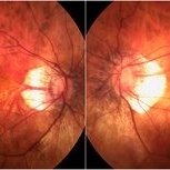
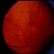
Ultra-wide field fundus photos of an 53-year-old man with a Subluxation of the Lens in the posterior vitreous cavity of the right eye after a trauma that happened many years ago. Patient remains stable with no adverse reaction to the lens at this time. No surgical intervention is recommended at this time. Patient also has myopic degeneration and lattice degeneration that will require patient to have follow up care.
Photographer: Kimberly Wakester, COA
Imaging device: Optos California
Condition/keywords: lattice degeneration, myopic degeneration, peripapillary atrophy, posterior staphyloma, Subluxation of the Lens
-
 Subluxation of the Lens
Dec 12 2024 by Kimberly Wakester
Ultra-wide field fundus photos of an 53-year-old man with a Subluxation of the Lens in the posterior vitreous cavity of the right eye after a trauma that happened many years ago. Patient remains stable with no adverse reaction to the lens at this time. No surgical intervention is recommended at this time. Patient also has myopic degeneration and lattice degeneration that will require patient to have follow up care.
Photographer: Kimberly Wakester, COA
Imaging device: Optos California
Condition/keywords: lattice degeneration, myopic degeneration, peripapillary atrophy, posterior staphyloma, Subluxation of the Lens
-
 Peripapillary Atrophy
Sep 21 2023 by Ben Serar
Fundus photograph showing peripapillary atrophy.
Condition/keywords: peripapillary atrophy
-
 Fluorescein Angiography in High Myopia
Dec 7 2019 by Anfisa Ayalon, MD
Fluorescein angiography pictures of a 55-year-old woman with high myopia.
Photographer: Anfisa Ayalon, MD., Meir Medical Center, Kfar Saba, Israel.
Condition/keywords: fluorescein angiogram (FA), high myopia, peripapillary atrophy
-
 High Myopia
Dec 7 2019 by Anfisa Ayalon, MD
Fundus photograph of a 55-year-old woman with high myopia.
Photographer: Anfisa Ayalon,MD., Meir Medical Center, Kfar Saba, Israel.
Condition/keywords: high myopia, myopia, peripapillary atrophy
-
 Histoplasmosis
Mar 27 2019 by Gary R. Cook, MD, FACS
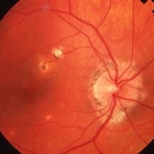
24-year-old white female with presumed ocular histoplasmosis (POHS) demonstrating minimal peripapillary atrophy but 3 atrophic histo spots around the optic nerve of her left eye; patient was asymptomatic; V.A.= 20/20.
Imaging device: Topcon VT-50
Condition/keywords: atrophic spot, ocular histoplasmosis syndrome (OHS), presumed ocular histoplasmosis syndrome (POHS)
-
 Histoplasmosis
Mar 27 2019 by Gary R. Cook, MD, FACS
24-year-old white female with presumed ocular histoplasmosis (POHS) demonstrating some peripapillary atrophy and multiple atrophic histo spots around the optic nerve of her right eye; the patient was asymptomatic; V.A.= 20/20.
Imaging device: Topcon VT-50
Condition/keywords: atrophic spot, ocular histoplasmosis syndrome (OHS), peripapillary atrophy, presumed ocular histoplasmosis syndrome (POHS)
-
 Histoplasmosis and Old Disciform Macular Scar
Mar 27 2019 by Gary R. Cook, MD, FACS
Left eye of a 59-year-old white male with an old, inactive, disciform macular scar secondary to presumed ocular histoplasmosis (POHS); V.A.= counting fingers at 3 feet.
Imaging device: Topcon VT-50
Condition/keywords: central disciform scar, disciform scar, peripapillary atrophy, presumed ocular histoplasmosis syndrome (POHS)
-
 Histoplasmosis and Subfoveal Neovascular Membrane
Mar 27 2019 by Gary R. Cook, MD, FACS
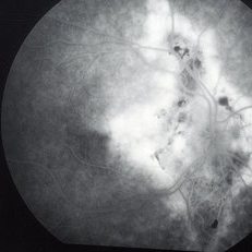
Late-phase fluorescein angiogram image of the right eye of a 59-year-old white male with ocular histoplasmosis and a subfoveal neovascular membrane showing late leakage and diffusion of dye from the membrane; V.A.= 20/80+2.
Imaging device: Topcon VT-50
Condition/keywords: FA late phase, fluorescein angiogram (FA), ocular histoplasmosis syndrome (OHS), peripapillary atrophy, presumed ocular histoplasmosis syndrome (POHS), subfoveal neovascular membrane
-
 Histoplasmosis and Subfoveal Neovascular Membrane
Mar 27 2019 by Gary R. Cook, MD, FACS
Mid-phase (20.4 seconds) fluorescein angiogram image of the right eye of 59-year-old white male with ocular histoplasmosis and a well-defined subfoveal CNVM OD; V.A.= 20/80+2
Imaging device: Topcon VT-50
Condition/keywords: FA mid phase, fluorescein angiogram (FA), ocular histoplasmosis syndrome (OHS), peripapillary atrophy, presumed ocular histoplasmosis syndrome (POHS), subfoveal choroidal neovascularization, subfoveal neovascular membrane
-
 Histoplasmosis with Choroidal Neovascularization
Mar 27 2019 by Gary R. Cook, MD, FACS
59-year-old white male with presumed ocular histoplasmosis (POHS) and a choroidal neovascular membrane (CNVM) along the temporal margins of the peripapillary atrophy; V.A.= 20/80+2.
Imaging device: Topcon VT-50
Condition/keywords: choroidal neovascular membrane (CNVM), peripapillary atrophy, presumed ocular histoplasmosis syndrome (POHS)
-
 Histo and Subfoveal Neovascular Membrane
Mar 27 2019 by Gary R. Cook, MD, FACS
41-year-old white female with a large subfoveal CNVM, subretinal fluid, and hemorrhage secondary to presumed ocular histoplasmosis (POHS) OS; V.A.= 20/400.
Imaging device: Topcon VT-50
Condition/keywords: hemorrhage, peripapillary atrophy, presumed ocular histoplasmosis syndrome (POHS), subfoveal choroidal neovascularization, subfoveal neovascular membrane
-
 Ocular Histoplasmosis
Mar 27 2019 by Gary R. Cook, MD, FACS
Fellow eye (OD) of a 41-year-old white female with ocular histoplasmosis showing peripapillary atrophy and several atrophic histo spots OD; no CNVM present; V.A.= 20/20.
Imaging device: Topcon VT-50
Condition/keywords: atrophic spot, histoplasmosis, peripapillary atrophy, presumed ocular histoplasmosis syndrome (POHS)
-
 Peripapillary Atrophy With High Myopia
Feb 4 2015 by H. Michael Lambert, MD
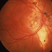
Peripapillary atrophy and central macular degeneration seen in high myopia.
Condition/keywords: high myopia, peripapillary atrophy
-
 Peripapillary Atrophy With High Myopia
Feb 4 2015 by H. Michael Lambert, MD
Peripapillary atrophy and central macular degeneration seen in high myopia.
Condition/keywords: high myopia, peripapillary atrophy
-
 Myopic Nerve Head
Jan 30 2015 by H. Michael Lambert, MD
Myopic nerve heard with peripapillary atrophy.
Condition/keywords: myopia, myopic nerve
-
 Myopic Nerve Head
Jan 30 2015 by H. Michael Lambert, MD
Myopic nerve heard with peripapillary atrophy.
Condition/keywords: myopia, myopic nerve
-
 Myopic Nerve Head
Jan 30 2015 by H. Michael Lambert, MD
Myopic nerve heard with peripapillary atrophy.
Condition/keywords: myopia, myopic nerve
-
 Presumed Ocular Histoplasmosis Syndrome
Jan 5 2015 by H. Michael Lambert, MD
OS with subtle central lesion and peripapillary atrophy. Presumed Ocular Histoplasmosis Syndrome.
Condition/keywords: presumed ocular histoplasmosis syndrome (POHS)
 Loading…