Search results (173 results)
-
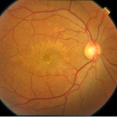
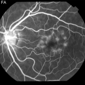
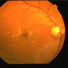
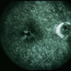
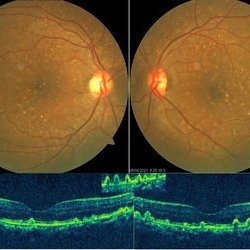 Cuticular Drusen
Jun 13 2021 by Priya Rasipuram Chandrasekaran, MBBS, DO, DNB, FRCS
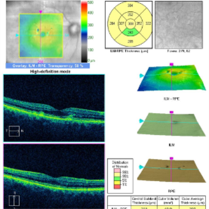
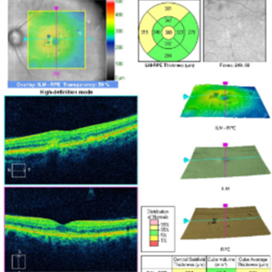
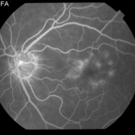
This is the fundus photo showing numerous yellow, small, hard drusen distributed throughout the retina. The corresponding OCT shows numerous elevated lesions underneath the RPE causing RPE elevations and arranged in a saw-tooth manner. Macular complications include acquired vitelliform lesion, choroidal neovascular membrane and geographic atrophy which are common after 60 years of age. It is usually associated with mutations in complement factor H. Basal laminar drusen, diffuse drusen and early adult onset grouped drusen are other alternative names. The differential diagnosis includes autosomal dominant drusen, pattern macular dystrophy, Sorsby macular drusen, mitochondrial macular dystrophy and so on.
Condition/keywords: cuticular drusen
-
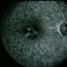
 Macular Pattern Dystrophy Associated with MELAS
Dec 19 2019 by Olivia Rainey
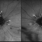
Bilateral wide field fundus autofluorescence images of a 54-year-old female with macular pattern dystrophy associated with MELAS. The patient is positive for m.3243A>G in MT-TL1. She had stroke in her 40s, hearing loss in her 30s, and has early onset diabetes. MyRetinaTracker shows VUS in RP1L1. Mutation in RP1L1 have been describe in other families with occult macular dystrophy. Farnsworth D15 is showing mild tritan abnormality, which is most commonly seen with acquired maculopathies. 12/17/19 patient's Optos and OCT show mild progression of atrophy.
Photographer: Olivia Rainey
Imaging device: Optos California
Condition/keywords: advanced geographic atrophy, bilateral, fundus autofluorescence (FAF), MELAS, Optos, pattern macular dystrophy, wide angle imaging
-
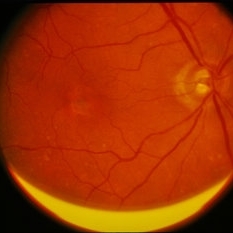
 Macular Pattern Dystrophy Associated with MELAS
Dec 19 2019 by Olivia Rainey
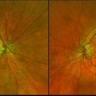
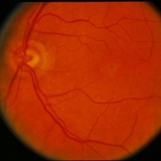
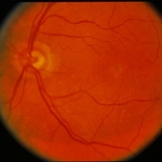
Bilateral wide field pseudocolor images of a 54-year-old female with macular pattern dystrophy associated with MELAS. The patient is positive for m.3243A>G in MT-TL1. She had stroke in her 40s, hearing loss in her 30s, and has early onset diabetes. MyRetinaTracker shows VUS in RP1L1. Mutation in RP1L1 have been describe in other families with occult macular dystrophy. Farnsworth D15 is showing mild tritan abnormality, which is most commonly seen with acquired maculopathies. 12/17/19 patient's Optos and OCT show mild progression of atrophy.
Photographer: Olivia Rainey
Imaging device: Optos California
Condition/keywords: advanced geographic atrophy, bilateral, fundus photograph, MELAS, Optos, pattern macular dystrophy, pseudocolor, wide angle imaging
-
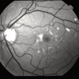
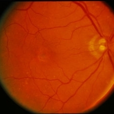
 Reticular Pattern Dystrophy
Oct 15 2017 by Sivakami A Pai, MS, DNB, FRCS ( UK), PhD
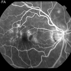
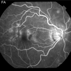
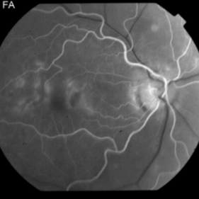
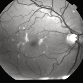
Left eye fluorescence image of 40-year-old Asian gentlemen, who presented with metamorphosia , vision 1.0 in both eye.
Photographer: Dr sivakami A Pai
Condition/keywords: pattern macular dystrophy
-
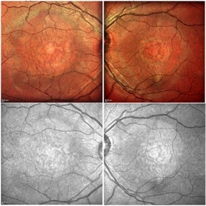 Pattern Dystrophy
Apr 20 2016 by Hashim Ali Khan, OD, FAAO
Color and NIR images of a 32-year-old woman with pattern dystrophy.
Imaging device: Spectralis Multicolor imaging
Condition/keywords: butterfly dystrophy, pattern dystrophy, pattern macular dystrophy
-
 Pattern Dystrophy
Sep 5 2015 by Ali Tavallali, MD, FASRS
A 44-year-old male, with VA of 20/70 both eyes.
Photographer: Neda Shaibani
Condition/keywords: pattern macular dystrophy
-
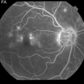
 Pattern Dystrophy Simulating Fundus Flavimaculatus #2
Nov 27 2014 by Thomas A. Ciulla, MD, MBA, FASRS
Note the subretinal hyper-reflective thickening corresponding to the flecks noted clinically.
Condition/keywords: pattern dystrophy, pattern macular dystrophy
-
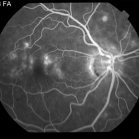
 Pattern Dystrophy Simulating Fundus Flavimaculatus #2
Nov 27 2014 by Thomas A. Ciulla, MD, MBA, FASRS
Note the subretinal hyper-reflective thickening corresponding to the flecks.
Condition/keywords: pattern dystrophy, pattern macular dystrophy
-
 Pattern Dystrophy Simulating Fundus Flavimaculatus #2
Nov 27 2014 by Thomas A. Ciulla, MD, MBA, FASRS
A 54-year-old woman without symptoms was referred to the retina service for assessment of maculopathy. Her visual acuities were 20/25 right and 20/20 left. She was noted to have flecks in each macula. Angiography showed a somewhat dark choroid and staining of the flecks.
Photographer: Charlotte Harris
Condition/keywords: fundus flavimaculatus, pattern dystrophy, pattern macular dystrophy
-
 Pattern Dystrophy Simulating Fundus Flavimaculatus #2
Nov 27 2014 by Thomas A. Ciulla, MD, MBA, FASRS
A 54 year old woman without symptoms was referred to the retina service for assessment of maculopathy. Her visual acuities were 20/25 right and 20/20 left. She was noted to have flecks in each macula. Angiography showed a somewhat dark choroid and staining of the flecks.
Photographer: Charlotte Harris
Condition/keywords: fundus flavimaculatus, pattern dystrophy, pattern macular dystrophy
-
 Pattern Dystrophy Simulating Fundus Flavimaculatus #2
Nov 27 2014 by Thomas A. Ciulla, MD, MBA, FASRS
A 54-year-old woman without symptoms was referred to the retina service for assessment of maculopathy. Her visual acuities were 20/25 right and 20/20 left. She was noted to have flecks in each macula. Angiography showed a somewhat dark choroid and staining of the flecks.
Photographer: Charlotte Harris
Condition/keywords: fundus flavimaculatus, pattern dystrophy, pattern macular dystrophy
-
 Age-Related Macular Degeneration With Pattern Dystrophy Appearance
Feb 21 2014 by David Callanan, MD
71-year-old male with age-related macular degeneration with pattern dystrophy appearance.
Condition/keywords: age-related macular degeneration (AMD), pattern macular dystrophy
-
 Age-Related Macular Degeneration With Pattern Dystrophy Appearance
Feb 21 2014 by David Callanan, MD
71-year-old male with age-related macular degeneration with pattern dystrophy appearance.
Condition/keywords: age-related macular degeneration (AMD), pattern macular dystrophy
-
 Age-Related Macular Degeneration With Pattern Dystrophy Appearance
Feb 21 2014 by David Callanan, MD
71-year-old male with age-related macular degeneration with pattern dystrophy appearance.
Condition/keywords: age-related macular degeneration (AMD), pattern macular dystrophy
-
 Age-Related Macular Degeneration With Pattern Dystrophy Appearance
Feb 21 2014 by David Callanan, MD
71-year-old male with age-related macular degeneration with pattern dystrophy appearance.
Condition/keywords: age-related macular degeneration (AMD), pattern macular dystrophy
-
 Age-Related Macular Degeneration With Pattern Dystrophy Appearance
Feb 21 2014 by David Callanan, MD
71-year-old male with age-related macular degeneration with pattern dystrophy appearance.
Condition/keywords: age-related macular degeneration (AMD), pattern macular dystrophy
-
 Age-Related Macular Degeneration With Pattern Dystrophy Appearance
Feb 21 2014 by David Callanan, MD
71-year-old male with age-related macular degeneration with pattern dystrophy appearance.
Condition/keywords: age-related macular degeneration (AMD), pattern macular dystrophy
 Loading…