Search results (37 results)
-
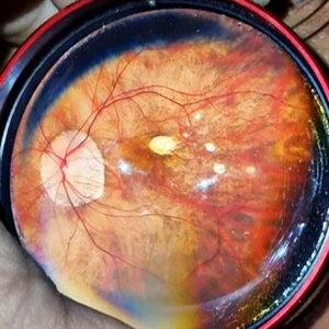
 Myopic CNVM
Jan 31 2025 by Thirumalesh Mochi Basavaraj, MD
Widefield image of a 26 year-old male patient with pathologic myopia with history of central scotoma with a sub macular bleed.
Photographer: Puttaswamy N K
Imaging device: Optos Daytona
Condition/keywords: myopic choroidal neovascularization (CNV), Myopic CNVM, pathologic myopia
-
 Pathological Myopia
Sep 25 2024 by DR Rohit Gupta
Fundus photograph of a 28 year-old male having high myopia on fundus examination Degenerative changes are seen in retina suggestive of pathological myopia.
Photographer: Dr Rohit gupta
Imaging device: Samsung S21
Condition/keywords: choroidal degeneration, degeneration of optic disc, lacquer cracks, myopia, Myopia macular degeneration CNVM foster fuch spot, pathologic myopia, staphyloma
-
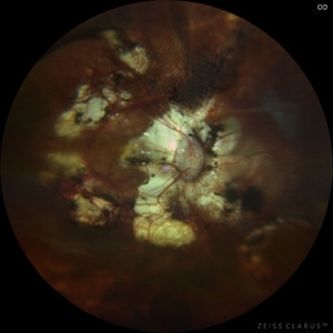 Pathological Myopia with posterior pole retinal detachment & new open break
Jul 31 2023 by Harsh Vardhan Singh, MS
45-year female with redetachment & new break
Photographer: Dr Harsh Vardhan Singh, AIIMS, Guwahati
Imaging device: Zeiss Clarus 700
Condition/keywords: pathologic myopia, posterior staphyloma, retinal break, rrd
-
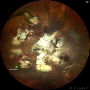 Pathological Myopia with posterior pole retinal detachment & new open break
Jul 31 2023 by Harsh Vardhan Singh, MS
45-year female with redetachment & new break
Photographer: Dr Harsh Vardhan Singh, AIIMS, Guwahati
Imaging device: Zeiss Clarus 700
Condition/keywords: pathologic myopia, posterior staphyloma, retinal break, rrd
-
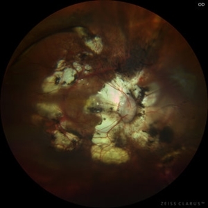 Pathological Myopia with posterior pole retinal detachment
Jul 31 2023 by Harsh Vardhan Singh, MS
45-year female with right eye re-detachment with pathological myopia & posterior pole RRD with open break
Photographer: Dr Harsh Vardhan Singh, AIIMS, Guwahati
Imaging device: Zeiss clarus 700
Condition/keywords: pathologic myopia, posterior pole lesion, posterior staphyloma, rrd
-
 Degenerative Myopia
Apr 12 2023 by Ahmed Abbas Hashmi, OD
Right eye Fundus photograph of a 61-year-old female with pathological myopia.
Condition/keywords: chorioretinal atrophy, high myopia, pathologic myopia
-
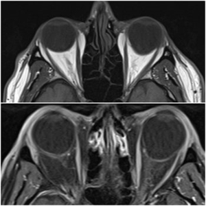 Staphyloma in Pathologic Myopia
Feb 7 2020 by Jonathan C. Tsui, MD
A 51-year-old presents with a six-month history of OS vision loss found to be CF at 3'. Fundus exam demonstrated pathologic myopia OS>OD with tilted discs. MRI Orbit Axial T1 images demonstrate significant compatible findings of staphylomas OS>OD.
Condition/keywords: pathologic myopia, staphyloma
-
 High Myopia
Apr 2 2019 by Gary R. Cook, MD, FACS
61-year-old white female with -7.75D myopia; OS; V.A. = 20/30-1
Imaging device: Topcon VT-50
Condition/keywords: high myopia, pathologic myopia
-
 High Myopia with CNVM
Apr 2 2019 by Gary R. Cook, MD, FACS
61-year-old patient with -8.25D myopia and a type 1 CNVM OD; V.A. = 20/200
Imaging device: Topcon VT-50
Condition/keywords: choroidal neovascular membrane (CNVM), high myopia, pathologic myopia, subretinal neovascular membrane
-
 Bilateral CNV in High Myopia
Apr 2 2019 by Gary R. Cook, MD, FACS
Left eye of a 60-year-old white female with -9D myopia and bilateral visible (Type 1) CNV; V.A. = 20/30.
Imaging device: Topcon VT-50
Condition/keywords: choroidal neovascular membrane (CNVM), choroidal neovascularization (CNV), high myopia, myopic degeneration, myopic fundus, pathologic myopia
-
 Bilateral CNV in High Myopia
Apr 2 2019 by Gary R. Cook, MD, FACS
Right eye of a 60-year-old white female with -9D myopia, myopic maculopathy, and visible (Type 1) CNV; V.A. = 20/40.
Imaging device: Topcon VT-50
Condition/keywords: choroidal neovascular membrane (CNVM), choroidal neovascularization (CNV), high myopia, myopic degeneration, myopic fundus, pathologic myopia
-
 CNVM Due to Pathologic Myopia
Apr 2 2019 by Gary R. Cook, MD, FACS
Fluorescein angiogram frame of the left eye of the 55-year-old Asian male with -9.50D myopia OS with a subfoveal CNVM and hemorrhage secondary to high myopia; V.A.= 20/200.
Imaging device: Topcon VT-50
Condition/keywords: choroidal neovascular membrane (CNVM), high myopia, pathologic myopia, retinal hemorrhage, subfoveal choroidal neovascularization
-
 CNVM due to Pathologic Myopia
Apr 2 2019 by Gary R. Cook, MD, FACS
55-year-old Asian male with -9.50D myopia with a visible (Type I) CNVM and thin hemorrhage in the macula: V.A.= 20/200
Imaging device: Topcon VT-50
Condition/keywords: choroidal neovascular membrane (CNVM), high myopia, pathologic myopia, retinal hemorrhage
-
 High Myopia
Apr 2 2019 by Gary R. Cook, MD, FACS
51-year-old white female with -7.00D myopia with a myopic conus on temporal aspect of the optic nerve and focal choroiretinal atrophy in the macula OS; V.A. = 20/25-1
Imaging device: Topcon VT-50
Condition/keywords: high myopia, myopic degeneration, myopic fundus, pathologic myopia
-
 High Myopia
Apr 2 2019 by Gary R. Cook, MD, FACS
51-year-old white female with -6.25D myopia OD with a myopic conus on the inferotemporal aspect of the optic disc and focal myopic chorioretinal atrophy in the macula OD; V.A. = 20/25
Imaging device: Topcon VT-50
Condition/keywords: high myopia, myopic degeneration, myopic fundus, pathologic myopia
-
 Fuch's Spot
Apr 2 2019 by Gary R. Cook, MD, FACS
20-year-old patient with high myopia and a Fuch's spot OD.
Condition/keywords: Fuchs, high myopia, pathologic myopia
-
 Pathologic Myopia
Apr 2 2019 by Gary R. Cook, MD, FACS
Mid-phase fluorescein angiogram image of the left eye of a 51-year-old white male with -25D myopia; V.A. = 20/40
Imaging device: Topcon VT-50
Condition/keywords: FA mid phase, fluorescein angiogram (FA), high myopia, pathologic myopia
-
 High Myopia
Apr 2 2019 by Gary R. Cook, MD, FACS
51-year-old white male with -25 diopters of myopia OS; V.A. = 20/40
Imaging device: Topcon VT-50
Condition/keywords: high myopia, pathologic myopia
-
 Pathologic Myopia
Apr 2 2019 by Gary R. Cook, MD, FACS
Mid-phase fluorescein angiogram image of the right eye of 51-year-old white male with -25D myopia; V.A. = 20/70-1
Imaging device: Topcon VT-50
Condition/keywords: FA mid phase, fluorescein angiogram (FA), high myopia, pathologic myopia
-
 High Myopia
Apr 2 2019 by Gary R. Cook, MD, FACS
51-year-old white male with -25 diopters of myopia OD; V.A.= 20/70
Imaging device: Topcon VT-50
Condition/keywords: high myopia, pathologic myopia
-
 High Myopia
Apr 2 2019 by Gary R. Cook, MD, FACS
17-year-old Vietnamese male with -23D myopia; V.A.= 20/40
Imaging device: Topcon VT-50
Condition/keywords: high myopia, lacquer cracks, pathologic myopia
-
 High Myopia with CNVM
Apr 2 2019 by Gary R. Cook, MD, FACS
75-year-old white female, aphakic without intraocular lens, with pathologic myopia with a CNVM and hemorrhage; V.A.= 20/200.
Imaging device: Topcon VT-50
Condition/keywords: choroidal neovascular membrane (CNVM), high myopia, macular hemorrhage, pathologic myopia
-
 Visible Myopic CNVM
Apr 1 2019 by Gary R. Cook, MD, FACS
70-year-old white male with visible myopic CNVM OS; V.A.= 20/200.
Imaging device: Topcon VT-50
Condition/keywords: high myopia, myopic choroidal neovascularization (CNV), pathologic myopia
-
 Optos Picture With Speculum: Dislocated Natural Lens
Oct 9 2018 by John S. King, MD
55-year-old white female with history of pathologic myopia+, lattice (laser), SB OU (1990s), and dislocated natural lenses OU that had been watched for years. In the fellow eye she developed phacolytic glaucoma and a PPV, PPL was performed. Plan for both eyes are monitoring. I wanted to get a good picture of her lens today with the optos machine, as the other pics had artifact from the lower lid. It worked out well to use a speculum in the left eye. Vision cc is 20/400 J1+ OD and 20/40 J2 OS; aphakic OU; vitreous clear OD; dislocated lens OS (see pic); retinas attached.
Photographer: Maisee Yang
Imaging device: Optos California
Condition/keywords: dislocated crystalline lens, pathologic myopia, scleral buckle, staphyloma
-
 Myopic Degeneration
Jul 3 2018 by Armando L. Oliver, MD
Myopic Degeneration
Photographer: Moises Castro
Imaging device: Optos California
Condition/keywords: pathologic myopia, posterior staphyloma
 Loading…