Search results (70 results)
-
 Foveal Hypoplasia with Vitreomacular Adhesion
Nov 5 2025 by Kevin Card
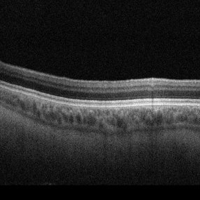
Optical coherence tomography of a 58-year-old female with ocular albinism shows fovea plana/hypoplasia. The overlying vitreomacular adhesion may masquerade as vitreomacular traction.
Imaging device: Zeiss Cirrus 6000
Condition/keywords: fovea plana, hypoplasia, vitreomacular adhesion
-
 Foveal Hypoplasia / Ocular Albinism
Aug 29 2024 by César Adrián Gómez Valdivia, MD
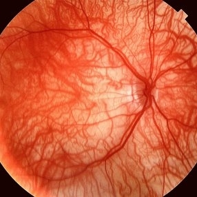
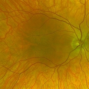
Fundus photograph of a 6-year-old female patient with foveal hypoplasia, ocular albinism and pendular nystagmus. Findings were bilateral. Retinal and choroidal vasculature are exquisitely beautiful.
Photographer: @eyemissu2
Imaging device: TOPCON TRC-50DX
Condition/keywords: foveal hypoplasia, ocular albinism
-
 Foveal Hypoplasia / Ocular Albinism
Aug 29 2024 by César Adrián Gómez Valdivia, MD
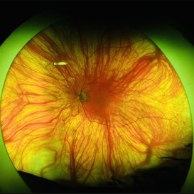
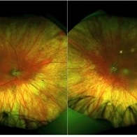
Fundus photograph of a 64-year-old female patient with foveal hypoplasia, ocular albinism and pendular nystagmus. Findings were bilateral. Retinal and choroidal vasculature are exquisitely beautiful.
Photographer: @eyemissu2
Imaging device: California ICG OPTOS
Condition/keywords: foveal hypoplasia, ocular albinism
-
 Ocular Albinism
Aug 27 2024 by Monica Elena Cortizo Brown , MD
9 year old girl with pendular nystagmus, photophobia and a fundus photo that shows clear view of the choroidal vasculature due to the hypopigmentation of retinal pigment epithelium.
Photographer: Mónica Cortizo Brown , Hospital de la Luz, Ciudad de México
Condition/keywords: nystagmus, ocular albinism, oculocutaneous albinism, photophobia
-
 Foveal Hypoplasia / Ocular Albinism
Aug 25 2024 by César Adrián Gómez Valdivia, MD
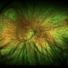
Fundus photograph of a 6-year-old female patient with foveal hypoplasia, ocular albinism and pendular nystagmus. Findings were bilateral. Retinal and Choroidal vasculature are exquisitely beautiful.
Photographer: @eyemissu2
Imaging device: TOPCON TRC-50DX
Condition/keywords: albinism, foveal hypoplasia, ocular albinism
-
 Foveal Hypoplasia / Ocular Albinism
Aug 25 2024 by César Adrián Gómez Valdivia, MD
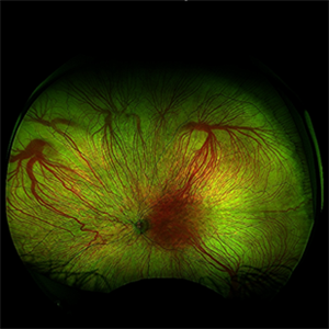
Fundus photograph of a 6-year-old female patient with foveal hypoplasia, ocular albinism and pendular nystagmus. Findings were bilateral. Retinal and choroidal vasculature are exquisitely beautiful.
Photographer: @eyemissu2
Imaging device: TOPCON TRC-50DX
Condition/keywords: Albinism, foveal hypoplasia, ocular albinism, vascula
-
 Albinism
Feb 6 2024 by Thirumalesh Mochi Basavaraj, MD
12 year old child with ocular albinism showing the underlying choroidal vasculature.
Photographer: Puttaswamy
Condition/keywords: ocular albinism
-
 Albinotic Fundus
Jan 24 2024 by Poornachandra B, MS, FVRS
Fundus photo of a 30 year old male with Ocular albinism. Hypopigmented fundus with very evident choroidal vessels.
Photographer: Dr Poornachandra B
Condition/keywords: ocular albinism
-
 Ocular Albinism
Sep 20 2023 by Karen Flores Guevara
Fundus photograph of a 4 year-old children with ocular albinism type 1. The genetic test showed a GPR143 pathogenic variant.
Photographer: Kitty Carolina Franco-Ramírez, Asociación para Evitar la Ceguera en México
Condition/keywords: ocular albinism
-
 Ocular Albinism
Sep 12 2023 by Ben Serar
Fundus photograph of LE showing hypopigmented fundus with clear visualisation of choroidal vasculature, with foveal hypoplasia, in a case of Ocular Albinism.
Condition/keywords: ocular albinism, oculocutaneous albinism
-
 Ocular Albinism
Sep 12 2023 by Ben Serar
Fundus photograph of RE showing hypopigmented fundus with clear visualisation of choroidal vasculature, with foveal hypoplasia, in a case of Ocular Albinism.
Condition/keywords: ocular albinism, oculocutaneous albinism
-
 Ocular albinism
Aug 18 2023 by Dr.Anushri Godbole
41 years old male came to opd with chief complaints of diminution of vision of both eyes since childhood. BCVA RE- FC 2M, LE- 6/60. On examination, patient had BE nystagmus. Fundus was tessellated with prominent choroidal blood vessels and Foveolar hypoplasia. Diagnosis of BE ocular albinism was made..
Condition/keywords: ocular albinism
-
 Ocular albinism
Aug 18 2023 by Dr.Anushri Godbole
41 years old male came to opd with chief complaints of diminution of vision of both eyes since childhood. BCVA RE- FC 2M, LE- 6/60. On examination, Patient had BE nystagmus. Fundus was tessellated with prominent choroidal blood vessels and Foveolar hypoplasia. Diagnosis of BE ocular albinism was made..
Condition/keywords: ocular albinism
-
 Ocular albinism
Aug 14 2023 by Erick Quiroz
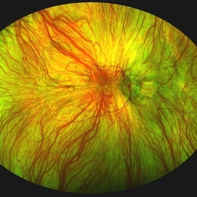
Ultra wide field fundus photograph of a 12 year old woman with ocular albinism.
Photographer: Erick Quiroz-Gonzalez, Hospital Militar Especialidades Oftalmologicas, Mexico City
Imaging device: Optos
Condition/keywords: albinism, ocular albinism
-
 Foveal hypoplasia - "thumb print" sign
Jul 14 2023 by Pedro S Tetelbom, MD
Right eye fundus photograph of a 26-year-old female with ocular albinism and foveal hypoplasia. "Thumb print" sign can be seen due the reflectance pattern of the Henle Fiber Layer.
Photographer: Johnny Ferrell, Jones Eye Institute, University of Arkansas for Medical Sciences
Imaging device: Optos Silverstone
Condition/keywords: foveal hypoplasia
-
 Ocular Albinism
Apr 10 2023 by William Jacob Anderson, MD
Fundus photograph of a 21-year-old girl with ocular albinism. Clinical exam was significant for decreased visual acuity and nystagmus. OCT macula demonstrates foveal hypoplasia
Photographer: William J Anderson, MD, Saint Louis University
Imaging device: Heidelberg Spectralis OCT
Condition/keywords: Albinism, foveal hypoplasia, ocular albinism
-
 Oculocutaneous Albinism
Jan 22 2023 by Pietro Dechichi
Fundus photograph of an 6-year-old girl with oculocutaneous albinism. Pacient´s nystagmus made it difficult to perform the exame. Foveal hypoplasia and evident choroidal vessels can be seen in the retinography
Photographer: Pietro Dechichi
Imaging device: Optos California
Condition/keywords: childhood, foveal hypoplasia, ocular albinism
-
 Oculocutaneous Albinism
Jan 22 2023 by Pietro Dechichi
Fundus photograph of a 6-year-old girl with oculocutaneous albinism. Patient's nystagmus made it difficult to perform the exam. Foveal hypoplasia and evident choroidal vessels can be seen in the retinography
Photographer: Pietro Dechichi
Imaging device: Optos California
Condition/keywords: childhood, foveal hypoplasia, ocular albinism
-
 Ocular Albinism
Dec 11 2021 by Luis Daniel Gutierrez, MD
Optos images of 6-year-old female patient with type 1 oculocutaneous albinism.
Photographer: Luis Daniel Gutierrez García, Hospital Fundación Nuestra Señora de la Luz, Ciudad de México.
Imaging device: Optos
Condition/keywords: Albinism, ocular albinism, Optos, ultra-wide field imaging
-
 Ocular Albinism
Aug 14 2021 by Aditya S Kelkar, MS, FRCS, FASRS,FRCOphth
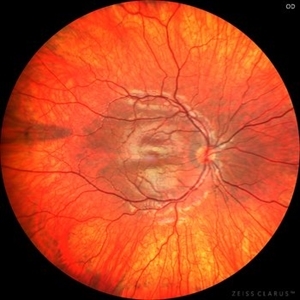
Fundus photograph of the left eye of a 26-year-old young man with ocular albinism.
Photographer: Devesh Kumar Dahariya, National Institute of Ophthalmology, Pune, India.
Imaging device: Zeiss Clarus 500
Condition/keywords: ocular albinism
-
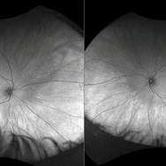
 Albinism with Rhegmatogenous Retinal Detachment
Mar 26 2019 by Gary R. Cook, MD, FACS
Left eye of patient with ocular albinism with rhegmatogenous retinal detachment inferiorly.
Imaging device: Topcon VT-50
Condition/keywords: albinism
-
 Albinism
Mar 26 2019 by Gary R. Cook, MD, FACS
Right eye of patient with ocular albinism.
Imaging device: Topcon VT-50
Condition/keywords: albinism
-
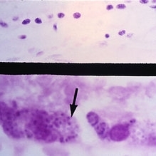 Slide 9-3
Feb 25 2019 by Lancaster Course in Ophthalmology
Tyrosinase-positive oculocutaneous albinism. Macular area is shown with normal-appearing retinal pigment epithelium (between asterisks) except for the lack of pigment (upper view). At very high power, fine melanin pigment granules may be seen (arrow, lower view).
Condition/keywords: melanin granules, ocular albinism, retinal pigment epithelium
-
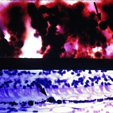 Slide 9-2
Feb 25 2019 by Lancaster Course in Ophthalmology
X-linked ocular albinism. Iris pigment epithelium (upper) and retinal pigment epithelium (lower) showing giant pigment granules (arrows) in the eye of an affected male.
Condition/keywords: ocular albinism, pigment epithelium, retinal pigment epithelium
 Loading…