Search results (13 results)
-
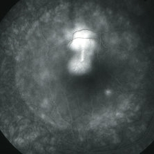
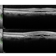
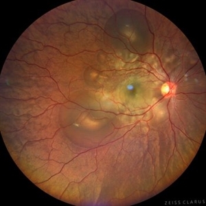 Central Serous Chorioretinopathy in Pregnancy (OD)
Apr 28 2024 by Vishal Agrawal, MD, FRCS,FACS,FASRS
30-year female with sudden loss of vision came for examination. She was in her first trimester of pregnancy. Examination revealed bilateral bullous NSD with subretinal fibrin s/o CSR.
Photographer: Dr Ayushi
Imaging device: Clarus 700
Condition/keywords: Central Serous Chorioretinopathy (CSR), neurosensory detachment of retina, pregnancy
-
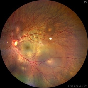 Central Serous Chorioretinopathy in Pregnancy (OS)
Apr 28 2024 by Vishal Agrawal, MD, FRCS,FACS,FASRS
30-year female with sudden loss of vision came for examination. She was in her first trimester of pregnancy. Examination revealed bilateral bullous NSD with subretinal fibrin s/o CSR.
Photographer: Dr Ayushi
Imaging device: Clarus 700
Condition/keywords: Central Serous Chorioretinopathy (CSR), neurosensory detachment of retina, pregnancy
-
 Suspicious Choroidal Nevus S/P Laser Photocoagulation
Apr 2 2019 by Gary R. Cook, MD, FACS
62-year-old white male with suspicious choroidal nevus OD with NSRD involving the macula; immediately S/P delimiting photocoagulation; V.A. = 20/200
Imaging device: Topcon VT-50
Condition/keywords: choroidal nevus, laser photocoagulation, neurosensory detachment of retina
-
 Choroidal Nevus with NSRD
Apr 2 2019 by Gary R. Cook, MD, FACS
62-year-old white male with suspicious choroidal nevus OD with neurosensory retinal detachment involving the macula; VA=20/200
Imaging device: Topcon VT-50
Condition/keywords: choroidal nevus, neurosensory detachment of retina
-
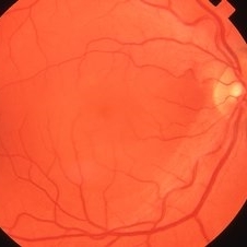
 CSR with large RPED
Mar 26 2019 by Gary R. Cook, MD, FACS
Mid-phase FA showing large RPED inferonasal to optic disc with overlying cruciate pigment figures (black lines) and neurosensory macular detachment OD.
Imaging device: Topcon VT-50
Condition/keywords: central serous retinopathy (CSR), neurosensory detachment of retina, retinal pigment epithelium (RPE) detachment
-
 Bilateral Central Serous Retinopathy
Mar 26 2019 by Gary R. Cook, MD, FACS
Late-phase frame of FA of 37-year-old white male with acute CSR OD showing pooling of dye beneath the small central RPED centrally, a smokestack-type leak from the RPE defect just above it, and mild late pooling of dye outlining the large neurosensory macular detachment; VA = 20/80-1.
Imaging device: Topcon VT-50
Condition/keywords: central serous retinopathy (CSR), FA late phase, FA late phase leakage, neurosensory detachment of retina
-
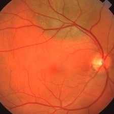
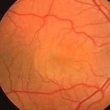
 Central Serous Retinopathy
Mar 26 2019 by Gary R. Cook, MD, FACS
43-year-old white male with acute CSR OD showing a large neurosensory macular detachment in his right eye.
Imaging device: Topcon VT-50
Condition/keywords: central serous retinopathy (CSR), neurosensory detachment of retina
-
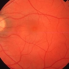
 Bilateral Central Serous Retinopathy
Mar 26 2019 by Gary R. Cook, MD, FACS
Right eye of a 37-year-old white male with a history of bilateral CSR showing a 2 DD NSRD centrally in his symptomatic OD; VA = 20/20-2.
Imaging device: Topcon VT-50
Condition/keywords: central serous retinopathy (CSR), neurosensory detachment of retina
-
 Central Serous Retinopathy
Mar 26 2019 by Gary R. Cook, MD, FACS
32-year-old white female with acute CSR with 1.5DD serous detachment in the macula OS; VA = 20/30-2.
Imaging device: Topcon VT-50
Condition/keywords: central serous retinopathy (CSR), neurosensory detachment of retina
-
 Central Serous Chorioretinopathy-OCT
Jun 22 2018 by Mitzy E Torres Soriano, MD
OCT showing typical subfoveal neurosensory detachment and PEDs in CSR.
Condition/keywords: central serous chorioretinopathy (CSCR), central serous retinopathy (CSR), neurosensory detachment of retina, optical coherence tomography (OCT), retinal pigment epithelium (RPE) detachment
-
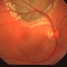
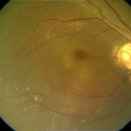 Disc Pit With Maculopathy
Jun 3 2014 by Neha Goel, MS DNB FRCS (Glasg)
Fundus photograph of the right eye of a 28-year-old male.
Photographer: Neha Goel
Imaging device: Zeiss Visucam
Condition/keywords: congenital optic nerve pit, neurosensory detachment of retina
-
---thumb.jpg/image-square;max$300,300.ImageHandler) Myopic Traction Maculopathy
Myopic Traction Maculopathy
Mar 23 2014 by Hamid Ahmadieh, MD
OCT image of the right eye of a 55-year-old woman with marked visual reduction due to myopic traction maculopathy manifesting as foveoschisis and neurosensory retinal detachment.
Photographer: Solmaz Shahmohammad , Negah Eye Center , Tehran
Imaging device: Specteralis
Condition/keywords: myopic foveoschisis, myopic traction maculopathy, neurosensory detachment of retina, optical coherence tomography (OCT)
-
 Retinoschisis Detachment
Nov 9 2012 by Norman Byer
Combined retinoschisis detachment, so-called schisis detachment, in a 47-year-old woman. The large outer layer hole in the center has a posterior yellow border which represents the position of the outer layer. Please observe superior to the hole the dark convexity of the scleral indentation. Just below the hole at the middle of the slide and going to the left the yellow zone comes to lie right against the inner layer and a fluid filled cavity lies deep to the outer layer. At this point, therefore, there is a true neurosensory detachment of the retina. On the right side of the hole, the yellow line slants up and to the right and lies close to the pigment epithelium. On the right side of the photograph, the original schisis cavity can be seen separating the yellow line of the outer layer above from the inner retinal layer below. The mechanism of this detachment is that some of the fluid from the schisis cavity passes through the outer layer hole and detaches the outer layer. This lesion has not been treated and has remained exactly the same for 13 years. A similar symmetrical "schisis-detachment" is present in the fellow eye.
Condition/keywords: neurosensory detachment of retina, outer layer hole, pigment epithelium, retinoschisis, schisis detachment, scleral indentation
 Loading…