Search results (41 results)
-
 Giant Retinal Tear with Multiple Retinal Breaks
Apr 21 2025 by Hrishikesh Naik, MS
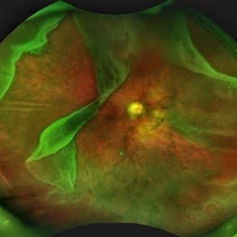
A 28 year old high myope with retinal detachment associated with a supero-temporal giant retinal tear in addition to multiple peripheral horseshoe tears and an additional supero-nasal retinal tear.
Photographer: Hrishikesh Naik
Imaging device: Optos Daytona
Condition/keywords: giant retinal tear, High Myopia, horseshoe tear, retinal break, retinal detachment
-
 Choroidal Melanoma Masquerading as PEHCR
Mar 3 2025 by Tejaswita Verma
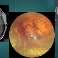
A 65 year old diabetic male presented with large nasal retinal mass giving the appearance of organized dehaemoglobinized subretinal hemorrhage with breakthrough vitreous hemorrhage , with 6/6P vision. Enucleation specimen showed histopathology confirmed choroidal melanoma.
Photographer: DR. TEJASWITA VERMA
Imaging device: MIRANTE
Condition/keywords: vitreous hemorrhage
-
 Choroidal Melanoma Masquerading as Subretinal Hemorrhage With Breakthrough VH
Jan 23 2025 by Tejaswita Verma
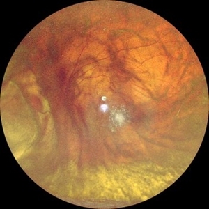
A 65 year old diabetic male presented with large nasal retinal mass giving the appearance of organized dehaemoglobinized subretinal hemorrhage with breakthrough vitreous hemorrhage , with 6/6P vision. Enucleation specimen showed histopathology confirmed choroidal melanoma.
Photographer: DR. TEJASWITA VERMA
Imaging device: MIRANTE
-
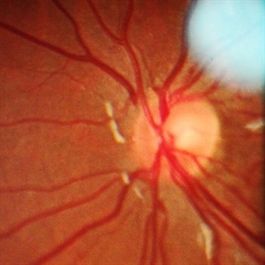
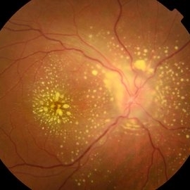
 Morning Glory Disc Anomaly
Feb 12 2024 by NIDHI PANWAR, MD FNB FICO
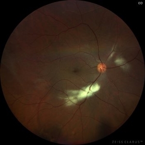
Fundus photograph of 43 year old male, hypertensive on medication, came for routine check up, and has been diagnosed to have poor vision left eye since childhood, denies any history of trauma. Vision left eye 6/18, Anterior segment normal, Fundus left eye shows excavated ,funnel-shaped optic nerve head, with central tuft of glial tissue obscuring the cup . The retinal vessels were seen emanating from the edge of disc in radial manner. In addition, the sectoral nasal retina shows localized area of hyperpigmented bony spicules like lesions. However, no history of nyctalopia or any other neurological disorder could be obtained.
Photographer: Nidhi Panwar, NMC Royal hospital, Sharjah , UAE
Imaging device: OPTOMAP
Condition/keywords: Morning Glory Anomaly, optic disc excavation
-
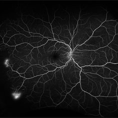
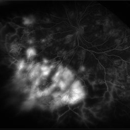
 Proliferative Diabetic Retinopathy with Severe Ischemia
Nov 30 2023 by Gabriel Costa Andrade, PhD
Ultra-widefield fluorescein angiography of the right eye of a 47 year old woman with diabetes mellitus showing macular and nasal retinal capillary dropout and neovascularization of the disc and temporal vascular arcades.
Photographer: Gabriel Andrade
Imaging device: Optos California
Condition/keywords: Diabetic Retinopathy
-
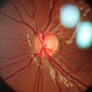
 Retinal detachment
Nov 23 2023 by Anand Temkar
LE color photo montage of a 50 years old male with supero-nasal retinal detachment (with break) and we can see horseshoe tear temporally with sub-retinal fluid.
Photographer: Dr.Anand Temkar- Retina Foundation, Ahmedabad
Imaging device: Mirante
Condition/keywords: RD, retinal break
-
 Isolated myelinated nerve fiber layers
Mar 5 2023 by Niloofar Piri, MD
Fundus photograph of the right eye demonstrating patches of isolated myelinated nerve fiber layers along inferior arcade as well as nasal retina
Photographer: Sean Kelso, Saint Louis University
Condition/keywords: myelinated nerve fiber layer, myelinated nerve fibers
-
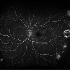
 Proliferative Sickle Cell Retinopathy
Feb 1 2023 by Olivia Rainey
Ultra-widefield fluorescein angiography of a 25-year old male with Proliferative Sickle Cell Retinopathy affecting his right eye. Patient stated that he was born with Sickle disease (SC), and has yearly eye exams. He noted no vision concerns over the last year but has typically experienced sickle attacks about 1-2 per year. The physician noted that the fluorescein obtained showed peripheral nonperfusion affecting the patient's nasal and temporal retina as well as neovascularization affecting his left eye more than his right. He recommended pan retinal photocoagulation in his left eye for his temporal and nasal retina, as as well as his right eye following.
Photographer: Olivia Rainey, OCT-C, COA
Imaging device: Optos California
Condition/keywords: early phase, fluorescein angiogram (FA), fluorescein leakage, neovascularization (NV), non-perfusion, proliferative retinopathy, right eye, sickle cell retinopathy, ultra-wide field imaging, ultra-widefield image
-
 Proliferative Sickle Cell Retinopathy
Feb 1 2023 by Olivia Rainey
Ultra-widefield fluorescein angiography of a 25-year old male with Proliferative Sickle Cell Retinopathy affecting his left eye. Patient stated that he was born with Sickle disease (SC), and has yearly eye exams. He noted no vision concerns over the last year but has typically experienced sickle attacks about 1-2 per year. The physician noted that the fluorescein obtained showed peripheral nonperfusion affecting the patient's nasal and temporal retina as well as neovascularization affecting his left eye more than his right. He recommended pan retinal photocoagulation in his left eye for his temporal and nasal retina, as as well as his right eye following.
Photographer: Olivia Rainey, OCT-C, COA
Imaging device: Optos California
Condition/keywords: early phase, fluorescein angiogram (FA), fluorescein leakage, left eye, neovascularization (NV), proliferative retinopathy, sickle cell retinopathy, ultra-wide field imaging, ultra-widefield image
-
 Methotrexate Bubble following Intravitreal Injection for PVR
Sep 21 2022 by Zach Seim
Ultra-widefield fundus photograph of an 81 year old female with a Methotrexate bubble following an Intravitreal Injection for Proliferative Vitreoretinopathy. Patient has been presenting to the office for two week interval Methotrexate injections in her left eye. The image was taken prior to her eighth injection which revealed a residual Methotrexate bubble in her inferior retinal image. This patient was seeing "lots" of floaters, as well as having visual acuity of cc20/400 cc20/200 PH.
Photographer: Zach Seim
Imaging device: OPTOS California
Condition/keywords: bubble, fundus photograph, fundus photography, intravitreal injection, left eye, methotrexate, nasal retina, Optos, proliferative vitreoretinopathy (PVR), pseudocolor, ultra-wide field imaging
-
 Retinal Blood Vessels in Retinochoroidal (RC) Coloboma
May 4 2021 by Priya Rasipuram Chandrasekaran, MBBS, DO, DNB, FRCS
This is the fundus photo of a 10-year-old girl showing RC coloboma along the infero nasal retina and involving the disc. This belongs to grade 4 of Ida Mann’s classification and grade 5 of Lingam Gopal’s classification of RC coloboma. The optic disc has no cup and BV for superior fundus emanates from superior part of optic disc and that for inferior fundus in the colobomatous area from multiple points. The blood vessels are discontinuous and are cork screw shaped.
Condition/keywords: chorioretinal coloboma
-
 Purtscher's Retinopathy
Mar 22 2021 by Marco Antonio Sauza
First OCT made after 3 weeks after chest and legs injury, with subretinal liquid/fluid, and a disorder of the nasal retina layers.
Photographer: Marco Sauza, hospital Ángeles, México
Imaging device: Spectralis
Condition/keywords: Purtscher's retinopathy
-
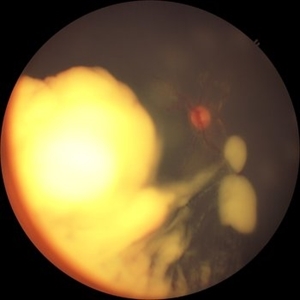 Retinoblastoma
Jan 17 2020 by Paulina Ramirez Neria, MD
Fundus photograph of a 4-year-old male with no familiar ophthalmic history. Call for consultation for leucocoria, the opening opportunity normal segment, normal optical disc, the retinal tumor mass is that involves the nasal retina major than 3mm, with diffuse vitreous seeding.
Photographer: DRA. PAULINA RAMIREZ NERIA, INSTITUTO MEXICANO DE OFTALMOLOGIA ( IMO IAP ) QUERETARO, MEXICO
Imaging device: 3NETHRA
Condition/keywords: retinoblastoma
-
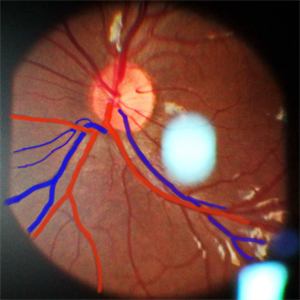
 Disc Edge Veins of Kraupa
Sep 15 2019 by Hashim Ali Khan, OD, FAAO
Color fundus with overlay drawing of 12-year-old girl with superonasal disc edge veins of Kraupa; a rare exit anomaly. The superonasal retina is drained through the venous trunk exiting at the edge of optic disc.
Condition/keywords: disc edge veins of Kraupa, vascular exit anomalies
-
 Disc Edge Veins of Kraupa
Aug 25 2019 by Hashim Ali Khan, OD, FAAO
Color fundus with overlay drawing of 10-year-old girl with inferior disc edge veins of Kraupa; a rare exit anomaly. The inferonasal retina is drained through the venous trunk exiting at the edge of optic disc.
Condition/keywords: disc edge veins of Kraupa, vascular exit anomalies
-
 Disc Edge Veins of Kraupa
Aug 24 2019 by Hashim Ali Khan, OD, FAAO
Red free and color fundus images of 10-year-old girl with inferior disc edge veins of Kraupa; a rare exit anomaly. The inferonasal retina is drained through the venous trunk exiting at the edge of optic disc.
Condition/keywords: disc edge veins of Kraupa, vascular exit anomalies
-
 Disc edge Veins of Kraupa
Aug 24 2019 by Hashim Ali Khan, OD, FAAO
Red free and color fundus images of 10-years-old girl with inferior disc edge veins of Kraupa; a rare exit anomaly. The inferonasal retina is drained through the venous trunk exiting at the edge of optic disc.
Condition/keywords: disc edge veins of Kraupa, vascular exit anomalies
-
 Proliferative Diabetic Retinopathy with Active Neovascularization
Jul 30 2019 by Olivia Rainey
Ultra-wide field fluorescein angiogram of a 36-year-old male with proliferative diabetic retinopathy with active neovascularization affecting his left eye. Patient presented with seeing flashing lights and trouble seeing to drive at night. His vision was sc20/50-1 PH20/40-2 in the left eye. There are suspicious vessels within the inferonasal retina of the patient's left eye. Labs ordered and are negative for sickle cell.
Photographer: Olivia Rainey
Imaging device: Optos
Condition/keywords: diabetes, diabetic macular edema, fluorescein angiogram (FA), fluorescein leakage, ischemia, late phase, left eye, neovascularization (NV), Optos, proliferative diabetic retinopathy (PDR), ultra-wide field imaging
-
 Uveitis Posterior
Jul 19 2019 by JEFFERSON R SOUSA, Tecg.º (Biomedical Systems Technology)
A 23-year-old male patient attended the clinic with low vision of the right eye. In the evaluation it presented important fundoscopical alterations like retinal exudations in the posterior pole and nasal retina, aspects of macular star. It was proven that it was a posterior uveitis.
Photographer: JEFFERSON R SOUSA - Study Center and Ophthalmological Research Dr. Andre M V Gomes, Institute Dr. Suel Abujamra São Paulo-Brazil
Imaging device: Topcon TRC-50 DX, Imaginet 4.0, angle de 50 graus. Flash 50w-s
Condition/keywords: uveitis
-
 Eales Disease
Apr 3 2019 by Paola Brito, MD
8-year-old girl with positive Matoux test. She received laser in nasal retina. Peripheral vein occlusion, ischemic areas and neovascularization.
Photographer: Paola Brito, Hospital de la Luz, Mexico
Imaging device: retcam
Condition/keywords: Eales disease
-
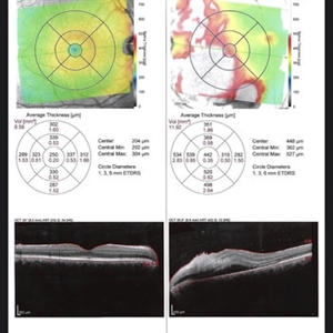
 Alport's Syndrome
Aug 29 2018 by Abhishek Das, MBBS, MS
OCT of a 54-year-old woman diagnosed to have Alport's syndrome. OCT shows temporal thinning of retina with nasal retina preserved.
Photographer: Abhishek Das, The Eye Foundation,Coimbatore,India
Imaging device: Optovue
Condition/keywords: Alports disease
-
 Retina Dialysis Associated Retinal Detachment
Aug 22 2018 by Luis J Haddock, MD
Optos color fundus photography showing nasal macula-on retinal detachment associated to a superonasal chronic retinal dialysis. This is a 19-year-old male who presented to glaucoma for elevated IOP. On dilated fundus exam retinal detachment was noted. Extended DFE showed nasal macula-on retinal detachment associated to a nasal retinal dialysis with peripheral vitreous contraction. The patient reported remote history of BB gun injury to his left eye at 5-years-old.
Imaging device: Optos California
Condition/keywords: blunt trauma, retinal dialysis
-
 Buckle intrusion with Retinal detachment
Feb 8 2018 by Manish Nagpal, MD, FRCS (UK), FASRS
Patient operated on 10 years back for a scleral buckling surgery presented with decreased vision and had a superonasal retinal detachment along with intrusion of the scleral buckle.
Photographer: Mehul Prajapati
Condition/keywords: acute retinal detachment, retinal break, scleral buckle
-
 Self-Applied Retinal Detachment
Sep 24 2017 by Ivonne Jocelyn Rivera Alvarado
40-year-old female, asymptomatic, without history of trauma. VA 20/20. No comorbilities. No ophthalmologic surgeries. It was a incidental finding. It can be observed a large RPE hypertrophy at the nasal retinal zone that borders the optic nerve with a line of demarcation that corresponds to a self applied retinal detachment.
Photographer: Ivonne Jocelyn Rivera Alvarado, Tec de Monterrey, Mexico
Condition/keywords: retinal pigment epithelium (RPE) hypertrophy
 Loading…