Search results (102 results)
-
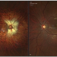
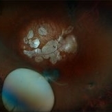 Subluxation of the Lens
Dec 12 2024 by Kimberly Wakester
Ultra-wide field fundus photos of an 53-year-old man with a Subluxation of the Lens in the posterior vitreous cavity of the right eye after a trauma that happened many years ago. Patient remains stable with no adverse reaction to the lens at this time. No surgical intervention is recommended at this time. Patient also has myopic degeneration and lattice degeneration that will require patient to have follow up care.
Photographer: Kimberly Wakester, COA
Imaging device: Optos California
Condition/keywords: lattice degeneration, myopic degeneration, peripapillary atrophy, posterior staphyloma, Subluxation of the Lens
-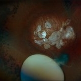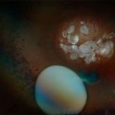
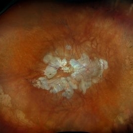 Myopic Degeneration
Dec 9 2024 by Virginia Gebhart
67 year old female with myopic degeneration. Posterior staphylomas are stable. VA limited by extensive chorioretinal atrophy. BCVA 20/160 (ecc)
Photographer: Virginia Gebhart, Retina Consultants of Carolina
Imaging device: Optos California
Condition/keywords: chorioretinal atrophy, myopic degeneration, staphyloma
-
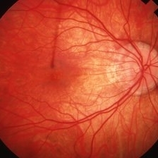
 Right Myelinated Nerve Fibre, LE Normal
May 6 2024 by Anupama Janardhanan
Fundus photograph of a 24 year old male patient with Anisometropia and Right Eye High Myopia associated with Diffuse circumpapillary myelinated Nerve Fibre and Left Eye normal fundus.
Photographer: Dr. Anupama Janardhanan, Aravind Eye hospital, Tirunelveli, India
Imaging device: Heidelberg Spectralis
Condition/keywords: anisometropia, myelinated nerve fibers, myopic degeneration
-
 Diffuse Chorioretinal Atrophy
Feb 21 2024 by Virginia Gebhart
61 year male with myopic degeneration and diffuse chorioretinal atrophy. BCVA 20/200.
Photographer: Virginia Gebhart
Imaging device: Topcon TRC 50DX
Condition/keywords: chorioretinal atrophy, myopic degeneration
-
 Macular Dystrophy vs Myopic Degeneration
Dec 22 2023 by Virginia Gebhart
35 year old female with myopic degeneration (-18.00 OU). BCVA 20/100 OU. RPE atrophy present in both eyes, but no significant chorioretinal atrophy. OCT not consistent with degenerative myopia due to dome shape appearance rather than posterior bowing. Possible macular dystrophy over degeneration. Will observe
Photographer: Virginia Gebhart
Imaging device: Topcon
Condition/keywords: Macular Dystrophy, myopic degeneration
-
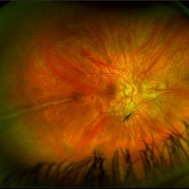
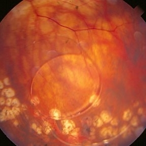
 Pathological Myopia
Oct 18 2023 by Anand Temkar
LE widefield CF montage of a 24 year old male with pathological myopia showing multiple lattice degenerations in periphery along with holes.
Photographer: Dr.Anand Temkar- Retina Foundation, Ahmedabad
Imaging device: Mirante
Condition/keywords: holes, lattice degeneration, myopic degeneration, myopic eye
-
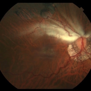
 Prominent Long Ciliary Nerve
Jan 25 2022 by Kachelle Brown
Ultra-wide field photograph of a 48-year-old female with a prominent long ciliary nerve. Patient presented asymptomatic, and was referred for a macula on retinal detachment. Patient was diagnosed with high myopia and a posterior vitreous detachment, and the physician discussed increased risk of floaters, myopic degeneration and retinal detachment associated with high myopia. -24.00 prior to cataract surgery OU per patient.
Photographer: Kachelle Brown
Imaging device: Optos California
Condition/keywords: fundus photograph, high myopia, long ciliary nerve, optos, right eye, ultra-widefield image
-
 PRP Marks
Apr 26 2021 by Priya Rasipuram Chandrasekaran, MBBS, DO, DNB, FRCS
This is the fundus photo montage of both eyes of a patient showing pan retinal photocoagulation marks. Theses marks can be confused with gyrate atrophy, cobble stone degeneration and myopic degeneration.
Condition/keywords: pan-retinal photocoagulation (PRP)
-
 Bilateral CNV in High Myopia
Apr 2 2019 by Gary R. Cook, MD, FACS
Left eye of a 60-year-old white female with -9D myopia and bilateral visible (Type 1) CNV; V.A. = 20/30.
Imaging device: Topcon VT-50
Condition/keywords: choroidal neovascular membrane (CNVM), choroidal neovascularization (CNV), high myopia, myopic degeneration, myopic fundus, pathologic myopia
-
 Bilateral CNV in High Myopia
Apr 2 2019 by Gary R. Cook, MD, FACS
Right eye of a 60-year-old white female with -9D myopia, myopic maculopathy, and visible (Type 1) CNV; V.A. = 20/40.
Imaging device: Topcon VT-50
Condition/keywords: choroidal neovascular membrane (CNVM), choroidal neovascularization (CNV), high myopia, myopic degeneration, myopic fundus, pathologic myopia
-
 High Myopia
Apr 2 2019 by Gary R. Cook, MD, FACS
51-year-old white female with -7.00D myopia with a myopic conus on temporal aspect of the optic nerve and focal choroiretinal atrophy in the macula OS; V.A. = 20/25-1
Imaging device: Topcon VT-50
Condition/keywords: high myopia, myopic degeneration, myopic fundus, pathologic myopia
-
 High Myopia
Apr 2 2019 by Gary R. Cook, MD, FACS
51-year-old white female with -6.25D myopia OD with a myopic conus on the inferotemporal aspect of the optic disc and focal myopic chorioretinal atrophy in the macula OD; V.A. = 20/25
Imaging device: Topcon VT-50
Condition/keywords: high myopia, myopic degeneration, myopic fundus, pathologic myopia
-
 Macular Hemorrhage
Apr 1 2019 by Gary R. Cook, MD, FACS
29-year-old white female with -12.25D high myopia OS and fresh macular hemorrhage; V.A.= 20/25.
Imaging device: Topcon VT-50
Condition/keywords: high myopia, lacquer cracks, macular hemorrhage, myopic degeneration
-
 High Myopia
Apr 1 2019 by Gary R. Cook, MD, FACS
29-year-old white female with -10D high myopia OD; V.A.= 20/20.
Imaging device: Topcon VT-50
Condition/keywords: high myopia, myopic degeneration, myopic fundus
-
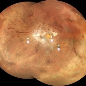
 Bilateral RD and Final Myopic Maculopahty Stage
Aug 11 2018 by Matias Iglicki, MD
Middle age male with myopic macular degeneration. This patient have bilateral vitrectomy plus sclera buckle due to a bilateral RD, here you can see the post op. Retinas have been re attached right eye with silicon oil tamponade, left eye c3f8 tamponade plus LASER and buckle indentation.
Photographer: matias iglicki MD certifed Teacher of Ophthalmology. University of Buenos Aires
Imaging device: DAYTONA
Condition/keywords: myopic degeneration, post-op
-
 Myopic Degeneration
Jul 3 2018 by Armando L. Oliver, MD
Myopic Degeneration
Photographer: Moises Castro
Imaging device: Optos California
Condition/keywords: pathologic myopia, posterior staphyloma
-
 Myopic Degeneration
Jul 3 2018 by Armando L. Oliver, MD
Myopic Degeneration
Photographer: Moises Castro
Imaging device: Optos California
Condition/keywords: pathologic myopia, posterior staphyloma
-
 Myopic Degeneration
Jul 3 2018 by Armando L. Oliver, MD
Late views IVFA.
Photographer: Moises Castro
Imaging device: Optos California
Condition/keywords: pathologic myopia, posterior staphyloma
-
 Myopic Degeneration
Jul 3 2018 by Armando L. Oliver, MD
Late Views IVFA
Photographer: Moises Castro
Imaging device: Optos California
Condition/keywords: pathologic myopia, posterior staphyloma
-
 Myopic Degeneration
Jul 3 2018 by Armando L. Oliver, MD
FAF
Photographer: Moises Castro
Imaging device: Optos California
Condition/keywords: pathologic myopia, posterior staphyloma
-
 Myopic Degeneration
Jul 3 2018 by Armando L. Oliver, MD
FAF
Photographer: Moises Castro
Imaging device: Optos California
Condition/keywords: pathologic myopia, posterior staphyloma
-
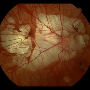
 LIO Dipped in the Vitreo
Aug 29 2016 by JEFFERSON R SOUSA, Tecg.º (Biomedical Systems Technology)
Patient Male, 51-years-old, with treatment with laser photocoagulation in myopic degeneration peripheral. Did FEC. suffered trauma (elbow) and had LIO dipped in the víteo.
Photographer: JEFFERSON R SOUSA - Institute Dr. Suel Abujamra / São Paulo - Brazil
Imaging device: Topcon TRC-50VT, Film, Kodak Ektachrome 160 - ASA 100 / 35mm, field of 35 degrees. Flash 100.
Condition/keywords: lens, myopic degeneration
-
 ERM / Myelinated NFL
Jun 10 2016 by John S. King, MD
High myope who dev ERM post-RD repair.
Condition/keywords: epiretinal membrane (ERM), myelinated nerve fibers, myopic degeneration
 Loading…