Search results (561 results)
-
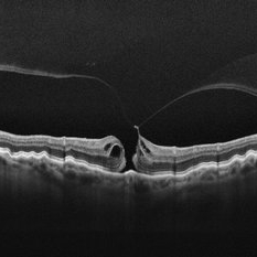
 Internal Limiting Membrane Peeling for Macular Hole Repair
Dec 10 2025 by Ahmad B. Tarabishy, MD
Intraoperative images of a patient undergoing internal limiting membrane peeling for a macular hole.
Photographer: Kristine Lawn
Imaging device: Alcon NGENUITY 3D Visualization System
Condition/keywords: internal limiting membrane (ILM) peeling, Macular hole, Macular surgery
-
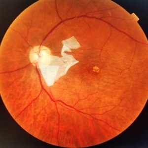
 Retinal Detachment with Macular Hole
Nov 10 2025 by Korey Starkey
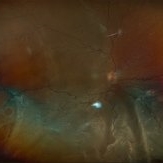
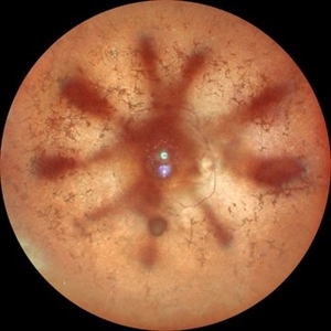
A 58 year-old female presented with re-detached retina through macular hole. Planned for surgical intervention.
Photographer: Korey Starkey
Imaging device: Optos
Condition/keywords: gas bubble, intermediate uveitis, macular hole, pars plana vitrectomy (PPV), retinal detachment, scleral buckle, traction detachment
-
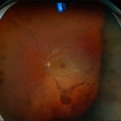
 Total Retinal Detachment
Sep 30 2025 by Kimberly Wakester
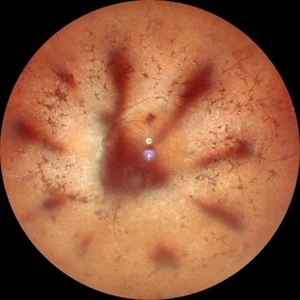
Optomap RGB of a 70-year-old woman with a total retinal detachment in the right eye. Exam confirms a chronic appearing retinal detachment with bare LP vision. Thorough scleral depressed exam was performed, revealing IT retinoschisis with large outer and inner holes as the likely causative break. Additionally, there is a full thickness macular hole. Surgery was recommended. Patient is to continue follow up care post operatively.
Photographer: Kimberly Wakester, COA, OCT-C
Imaging device: Optos California
Condition/keywords: macular hole, Retinoschisis, total retinal detachment
-
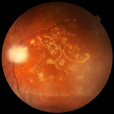
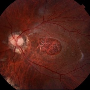
 Macular Hole Due to Proliferative Diabetic Retinopathy
Aug 13 2025 by Ricardo Leitão Guerra
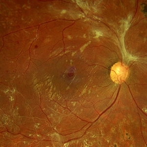
A macular hole formation after anti-VEGF injection prior to vitrectomy for tractional retinal detachment in a patient presenting proliferative diabetic retinopathy.
Photographer: Ricardo Leitão Guerra
Imaging device: ZEISS CLARUS 700
Condition/keywords: macular hole, proliferative diabetic retinopathy (PDR)
-
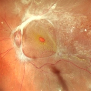
 Giant Persistent Macular Hole
Jul 5 2025 by César Adrián Gómez Valdivia, MD
Giant Persistent Macular Hole found in a 48YO male patient 1 year after vitrectomy.
Photographer: @eyemissu2
Imaging device: TOPCON TRC-50DX
Condition/keywords: macular hole
-
 Double Trouble
Jun 28 2025 by Tejaswita Verma
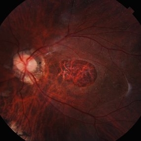
Fundus image of a 60 year old diabetic female with double macular holes with 6/60 vision status post LE PPV+gas 4 months ago. Other eye also had an unoperated large macular hole. Known case of glaucoma
Photographer: Dr. Tejaswita Verma
Imaging device: MIRANTE
Condition/keywords: macular hole
-
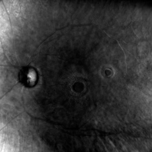 Double Trouble
Jun 28 2025 by Tejaswita Verma
Retro image of aa double macular hole in a 60 yr old diabetic female status post PPV + gas 4 months ago. Vision was 6/60 in LE.
Photographer: Dr. Tejaswita Verma
Imaging device: MIRANTE
Condition/keywords: macular hole, retro mode
-
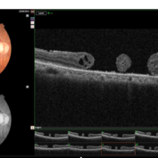 Double Macular Holes
Jun 26 2025 by Moazzam Parvez
OCT image of a 62 year old man after a blunt trauma by a tennis ball with a vision of CF 3 mt in the right eye.
Photographer: Moazzam Parvez , Netralayam , Kolkata
Imaging device: Topcon Maestro 2
Condition/keywords: double, traumatic macular hole
-
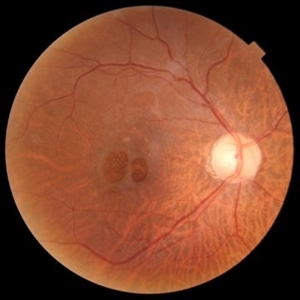 Two Suns in the Macular Sky
Jun 26 2025 by Moazzam Parvez
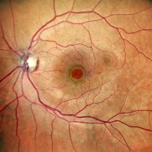
Fundus photograph of a 62 year old gentleman presenting with double adjacent full thickness macular holes in the right eye maintaining a vision of CF 3 mts.
Photographer: Moazzam Parvez ,Netralayam , Kolkata
Imaging device: Topcon Maestro 2
Condition/keywords: double, Macular hole, traumatic macular hole
-
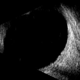 Macular Hole
May 9 2025 by Gustavo Uriel Fonseca Aguirre
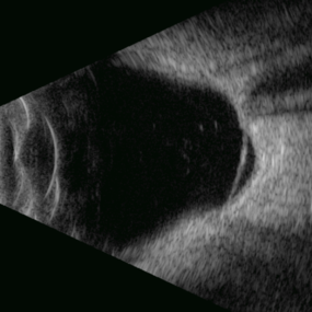
This B-mode longitudinal ultrasound scan demonstrates a full-thickness macular hole, appearing as a well-defined hypoechoic defect in the retinal surface with elevated edges.
Photographer: Gustavo U. Fonseca Aguirre, Hospital Conde de Valenciana, Ciudad de México
Condition/keywords: full thickness macular hole, macular hole
-
 Retinitis Pigmentosa with Macular Hole with Posterior Subcapsular Cataract
Apr 28 2025 by Malvika Singh
Fundus photograph of the left eye of a 31 year old with retinitis pigmentosa, showing the shadow of posterior subcapsular cataract over the fundus.
Photographer: Dr Malvika Singh, Retina Foundation, Ahmedabad, India
Imaging device: Mirante SLO/OCT
Condition/keywords: posterior subcapsular cataract, retinitis pigmentosa
-
 Retinitis Pigmentosa with Macular Hole with Posterior Subcapsular Cataract
Apr 28 2025 by Malvika Singh
Fundus photograph of the right eye of a 31 year old with retinitis pigmentosa with a macular hole, showing the shadow of posterior subcapsular cataract over the fundus.
Photographer: Dr Malvika Singh, Retina Foundation, Ahmedabad, India
Imaging device: Mirante SLO/OCT
Condition/keywords: macular hole, posterior subcapsular cataract, retinitis pigmentosa
-
 Retinal Detachment Associated With a Posterior Staphyloma
Apr 9 2025 by Gustavo Uriel Fonseca Aguirre
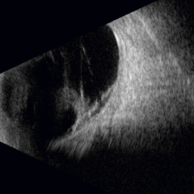
B-mode axial ultrasound scan of a highly myopic eye shows a posterior staphyloma with an associated macular hole-induced retinal detachment.
Photographer: Gustavo U. Fonseca Aguirre, Hospital Conde de Valenciana, Ciudad de México
Condition/keywords: high myopia, posterior staphyloma, rhegmatogenous retinal detachment
-
 Macular Hole in Tractional Retinal Detachment
Apr 1 2025 by Gustavo Uriel Fonseca Aguirre
B-scan findings in a diabetic patient reveal vitreous hemorrhage, blood-soaked hyaloids, and tractional retinal detachment with an associated macular hole in the posterior pole.
Photographer: Gustavo U. Fonseca Aguirre, Hospital Conde de Valenciana, Ciudad de México
Condition/keywords: macular hole, tractional retinal detachment
-
 Stage 2 Macular Hole From VMT
Mar 21 2025 by Drew Mitchell
HD 1 line 100x OCT showcasing a full thickness macular hole caused by vitreomacular traction on fovea. Choroidal folds can also be seen on scan.
Photographer: Drew Mitchell OCT-C
Imaging device: Zeiss Cirrus 6000
Condition/keywords: Choroidal Folds, FTMH, macular hole, OCT, PVD
-
 Myopic Traction Maculopathy
Mar 17 2025 by Drew Mitchell
HD 1 line 100x 9 mm scan of a right eye with MTM at stage 3c. Macular Schisis Detachment.
Photographer: Drew Mitchell OCT-C
Imaging device: Zeiss Cirrus 5000
Condition/keywords: full thickness macular hole, Macular hole, myopic foveoschisis, myopic macular schisis, myopic traction maculopathy, PVD
-
 AMG on Papillomacular Bundle
Mar 16 2025 by PUJA NEGI
Patient came to our OPD with history of AMG done for macular hole . On examination we found that the AMG had displaced over the papillomacular bundle from the hole.
Photographer: DR Nuzhat
Condition/keywords: amniotic membrane graft, macular hole
-
 Emulsified Silicon Oil in Macular Hole- Hyperoleon in Hole
Mar 8 2025 by PUJA NEGI
Fundus photograph showing emulsified silicon oil bubbles in macular hole, hyperoleon in hole.
Photographer: Dr Vaidehi Jethwa
Condition/keywords: Inverse hypopyon, macular hole
-
Macular Hole Surgery: Inverse Flaps
Jan 31 2025 by Thirumalesh Mochi Basavaraj, MD
This video demonstrates, PVD induction , followed by ILM peeling in multiple flower petal flap technique.
Condition/keywords: ILM flaps, ILM peel, induction
-
 Dislocated Lens
Jan 30 2025 by Kimberly Wakester
Fundus photograph of a 37-year-old man with an anteriorly dislocated lens in the left eye. The natural lens has displaced anteriorly in the AC secondary to trauma to the eye. There is also a Macular hole present with vitreous hemorrhage. Patient was recommended to proceed with lensectomy, iris repair and MH repair in the left eye.
Photographer: Kimberly Wakester, COA
Imaging device: Topcon TRC-50DX
Condition/keywords: dislocated lens, iridodialysis
-
 Macular Hole
Jan 30 2025 by Kimberly Wakester
Fundus photograph of a 37-year-old man with an anteriorly dislocated lens in the left eye. The natural lens has displaced anteriorly in the AC secondary to trauma to the eye. There is also a Macular hole present with vitreous hemorrhage. Patient was recommended to proceed with lensectomy, iris repair and MH repair in the left eye.
Photographer: Kimberly Wakester, COA
Imaging device: Optos California
Condition/keywords: dislocated lens, macular hole, vitreous hemorrhage
-
 Polyploidal Choroidal Vasculopathy
Dec 27 2024 by Tejaswita Verma
Fundus image of a 74 year old woman with CF1mt vision in right eye showing large PED in a case of PCV. There was associated full thickness macular hole in the same eye.
Photographer: DR. TEJASWITA VERMA
Imaging device: MIRANTE
Condition/keywords: PED, polypoidal choroidal vasculopathy (PCV)
-
 Giant Persistent Macular Hole
Dec 6 2024 by César Adrián Gómez Valdivia, MD
Giant Persistent Macular Hole found in a 48 year-old male patient one year after vitrectomy.
Photographer: @eyemissu2
Imaging device: TOPCON TRC-50DX
Condition/keywords: macular, macular hole
-
 Traumatic Macular Hole pre and post repair
Nov 25 2024 by Shobhit Chawla, M.S.
31 year-old male reported with h/o of blunt trauma over right eye ,from cricket ball. On examination DVA RE 6/60,LE 6/18,ant segment BE :WNL,FUNDUS RE:Sub retinal hemorrhage at macula with chroidal tear,LE :WNL. Undwer went 25G vitrectomy+sub retinal TPA+C3F8(RE).Post op 1 month DVA RE:6/24 ,ANT SEGMENT:WNL,FUNDUS:resolved sub retinal haem with traumatic macular hole. Under went repeat vit+autologous retinal transplant +SOI RE.POST SOR AFTER4monthsV/A :6/18 RE
Photographer: Ranjit Ray
Imaging device: Clarus 500
Condition/keywords: Macular hole, retinal graft, subretinal hemorrhage
-
 Combined Retinal Detachment With Macular Hole
Sep 28 2024 by Tejaswita Verma
Fundus image of the LE of a 67 year old diabetic, hypertensive female with CF 3metres vision showing combined RD with FTMH, in a pseudophakic eye. She was lost to follow up status post 2 anti VEGF injections received 8 months back due to typhoid fever.
Photographer: DR. TEJASWITA VERMA
Imaging device: MIRANTE
Condition/keywords: full thickness macular hole, proliferative diabetic retinopathy (PDR), tractional retinal detachment
 Loading…