Search results (655 results)
-
 Starstruck by Stargardt
Nov 17 2025 by SHRADDHA RAJ SHRIVASTAVA
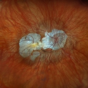
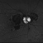
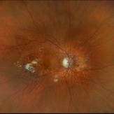
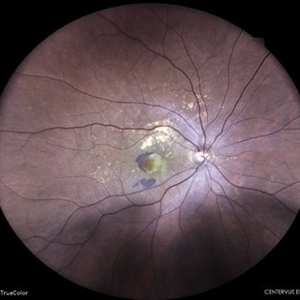
Left eye G-FAF image of a 26 year old patient diagnosed with Stargardt Disease, showing hyperautofluorescent flecks of increased lipofuscin accumulation and dark areas of hypoautofluorescence representing retinal pigment epithelium (RPE) atrophy.
Photographer: Dr. Shraddha Raj Shrivastava
Imaging device: Nidek Mirante SLO/OCT (Confocal scanning/Spectral domain OCT)
Condition/keywords: fleck dystrophy, fundus autofluorescence (FAF), hereditary macular dystrophy, heredomacular degeneration, lipofuscin, Stargardt Disease
-
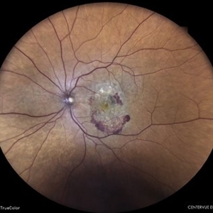
 Dry AMD, Advanced Atrophic with Subfoveal Involvement
Oct 3 2025 by Kimberly Wakester
Optomap RGB of an 76-year-old woman with Dry AMD, Advanced Atrophic with Subfoveal Involvement in the right eye. Longstanding, more likely due to myopic/choroidal atrophy. End-stage disease, would not recommend aggressive intervention. Patient is to continue follow up care and repeat OCT/imaging as directed per doctor. Just a fun mention about the image, if you look at the optic nerve and the vessels it appears to look like an eye.
Photographer: Kimberly Wakester, COA, OCT-C
Imaging device: Optos California
Condition/keywords: Advanced Atrophic with Subfoveal Involvement, dry age-related macular degeneration (dry AMD), Myopic Degeneration
-
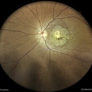
 Optic Disc Drusen
Aug 20 2025 by Drew Mitchell
Fundus Autofluorescence photo of an 86 year old woman with neovascular AMD with active CNV and optic disc drusen.
Photographer: Drew Mitchell OCT-C
Imaging device: Optos California
Condition/keywords: fundus autofluorescence (FAF), neovascular age-related macular degeneration (AMD), optic disc drusen, OPTOS
-
 Subretinal Neovascular Membrane
Aug 15 2025 by Akansha Sharma
Color fundus photograph of a 40 year old male with subretinal neovascular membrane.
Photographer: DR. AKANSHA SHARMA
Condition/keywords: choroidal neovascular membrane (CNVM), CNVM, SRNVM, subretinal neovascularization (SRNV), wet age-related macular degeneration (wet AMD)
-
 Subretinal Neovascular Membrane
Aug 15 2025 by Akansha Sharma
Color fundus photograph of a 40 year old male with subretinal neovascular membrane.
Photographer: DR. AKANSHA SHARMA
Condition/keywords: choroidal neovascular membrane (CNVM), CNVM, Myopic CNVM, SRNVM, subretinal neovascularization (SRNV), Wet age related macular degeneration
-
 Neovascular AMD with Active CNV
May 22 2025 by Kimberly Wakester
Optomap RGB of an 82-year-old man with Neovascular AMD with Active CNV and Dry AMD in the right eye. There is advanced atrophic changes without subfoveal involvement located temporally to the fovea. Patient is to continue follow up care with dilated exam, repeat OCT, and treatment of intravitreal injection of Vabysmo every 5 weeks at this time.
Photographer: Kimberly Wakester, COA, OCT-C
Imaging device: Optos California
Condition/keywords: advanced geographic atrophy, dry age-related macular degeneration (dry AMD), neovascular age-related macular degeneration (AMD)
-
 Retromode Image of Dry AMD
May 13 2025 by Anupama Kiran Kumar
A retromode pseudo3D image of a case of dry AMD(NIDEK Mirante ). Retromode deviated left depicts depressed (pseudo concave) medium to large drusen. Here the drusenoid deposits appear to have a spectacular "moon surface" like appearance.
Photographer: Mr Pratap
Imaging device: Mirante SLO/OCT (Nidek Co., Gamagori, Japan)
Condition/keywords: AMD, dry age-related macular degeneration (dry AMD), retro mode
-
 Toxic Maculopathy (Elmiron)
Apr 9 2025 by Virginia Gebhart
79 year old male with toxic maculopathy from long term use of Elmiron (15+ yrs.) On exam there is stippled RPE changes, pigment clumping, and subretinal deposits. BCVA 20/100 | 20/40.
Photographer: Virginia Gebhart, Retina Consultants of Carolina
Imaging device: Optos California
Condition/keywords: autofluorescence imaging, cystoid macular degeneration, Elmiron Toxicity, Toxic Maculopathy
-
 Subretinal Fibrosis
Jan 14 2025 by Kimberly Wakester
Fundus photograph of an 86-year-old woman with the end stage of Age-related Macular Degeneration in the left eye. Patient went unseen for 3-4 years prior to establishing care at our practice. Due to the significant amount of subretinal fibrosis, treatment was not recommended due to limited visual recovery. Patient was advised of monocular vision and the importance of follow up care.
Photographer: Kimberly Wakester, COA
Imaging device: Optos California
Condition/keywords: AMD, subretinal fibrosis
-
 New Subretinal Hemorrhage in AMD
Jan 8 2025 by Drew Mitchell
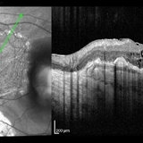
HD 1 line 100x OCT scan of a New Subretinal Hemorrhage in a established patient with AMD.
Photographer: Drew Mitchell, OCT-C
Imaging device: Zeiss Cirrus 6000
Condition/keywords: age-related macular degeneration (AMD), OCT, subretinal hemorrhage
-
OCT Video Imaging of Left Eye Age Related Macular Degeneration
Jan 6 2025 by Kavitha Duraipandi, MD DNB FICO FRCS
Left eye OCT macula shows various biomarkers like PED, sub retinal fluid, sub retinal hyper reflective material and hyper reflective foci suggestive of wet age-related macular degeneration.
Condition/keywords: OCT biomarkers, wet age-related macular degeneration (wet AMD)
-
 Macular Degeneration
Dec 3 2024 by Sarah D Kang
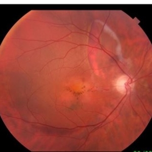
Fundus photograph of an 85-year-old female patient with macular degeneration observed for retinal clearance before cataract surgery.
Condition/keywords: floaters, macular degeneration
-
 Macular Degeneration
Dec 3 2024 by Sarah D Kang
Fundus photograph of an 85-year-old female patient with macular degeneration observed for retinal clearance before cataract surgery.
Condition/keywords: floaters, macular degeneration
-
 Peripheral Exudative Hemorrhagic Chorioretinopathy
Nov 19 2024 by Toolie Winters
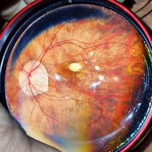
Ultra-wide field fundus photograph of an 85-year-old woman with Peripheral Exudative Hemorrhagic Chorioretinopathy (PECHR) affecting the right eye. Patient presented with a blind spot centrally in the right eye which she first noticed 4 months prior to this image being taken. The patient states that in the month prior to this image, she began noticing bright lights flash across her vision 4-5x/day which last about 15 seconds. The flashes are either black with a blue ring around them or yellow, and their frequency has increased over time. The patient's vision at the time of this appointment was Dcc20/100+1 PHNI. This photo also shows diffuse hemorrhage, lipid, and an eccentric disciform lesion.
Photographer: Toolie Winters
Imaging device: Optos California
Condition/keywords: fundus photography, neovascular age-related macular degeneration (AMD), Optos, OPTOS CALIFORNIA, peripheral exudative hemorrhagic chorioretinopathy (PEHCR), pseudocolor, ultra-wide field imaging, wet age-related macular degeneration (wet AMD)
-
 Large Subretinal Bleed in Case of Wet ARMD
Sep 28 2024 by Anjana Mirajkar, MS Ophthalmology
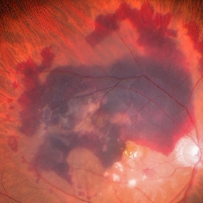
An intra operative image showing large sub retinal hemorrhage involving the macular area and along the superior arcade with exudation at the macular area in case of wet ARMD.
Photographer: Dr. Anjana Mirajkar -Retina Foundation, Ahmedabad
Condition/keywords: polypoidal choroidal vasculopathy (PCV), subretinal hemorrhage, wet age-related macular degeneration (wet AMD)
-
 Pathological Myopia
Sep 25 2024 by DR Rohit Gupta
Fundus photograph of a 28 year-old male having high myopia on fundus examination Degenerative changes are seen in retina suggestive of pathological myopia.
Photographer: Dr Rohit gupta
Imaging device: Samsung S21
Condition/keywords: choroidal degeneration, degeneration of optic disc, lacquer cracks, myopia, Myopia macular degeneration CNVM foster fuch spot, pathologic myopia, staphyloma
-
 Look With Your Heart
Sep 20 2024 by Virginia Gebhart
FA of 65 year old male with exudative AMD superior to a chorioretinal defect in the nasal macula. FA shows classic CNV with late leakage. Treated with IVA, will consider PDT if no improvement.
Photographer: Virginia Gebhart, Retina Consultants of Carolina
Imaging device: Optos California
Condition/keywords: choroidal neovascularization (CNV), exudative age-related macular degeneration, FA early phase
-
 Large PED with RPE-rip
Aug 21 2024 by Amirfarbod Yazdanyar, MD , PhD
Fundus photograph of a 85 year-old male with AMD who developed very large PED with RPE-rip. BCVA is 20/70
Photographer: Justin Cocilo , Retina Group of New England
Condition/keywords: large pigment epithelial detachment, RPE-rip, wet age-related macular degeneration (wet AMD)
-
 RPE-Transplantation
Jul 25 2024 by Gabriel Costa Andrade, PhD
Postoperative period of RPE-transplantation in a patient with neovascular AMD after RPE tear.
Photographer: Gabriel Andrade
Condition/keywords: neovascular age-related macular degeneration (AMD), pars plana vitrectomy (PPV), wet age-related macular degeneration (wet AMD)
-
 Neovascular AMD with Polypoidal Choroidal Vasculopathy
Jun 11 2024 by Gregg T. Kokame, MD, MMM, FASRS
Structure OCT from the OCT angiography shows definition of the PCV complex.
Photographer: Jaclyn Pisano
Condition/keywords: branching vascular network (BVN), OCTA, polypoidal choroidal vasculopathy (PCV), wet age-related macular degeneration (wet AMD)
-
 Subretinal Neovascular Membrane
Jun 5 2024 by Akansha Sharma
Color fundus photograph of a 49 year old female with subretinal bleed suggestive of subretinal neovascular membrane.
Photographer: Dr. Akansha Sharma, Bharati Eye Hospital
Condition/keywords: choroidal neovascular membrane (CNVM), CNVM, SRNVM, subretinal neovascularization (SRNV), wet age-related macular degeneration (wet AMD)
-
 Subretinal Neovascular Membrane
Jun 5 2024 by Akansha Sharma
Color fundus photograph of a 94 year old female with subretinal bleed suggestive of subretinal neovascular membrane.
Photographer: Dr. Akansha Sharma, Bharati Eye Hospital
Condition/keywords: choroidal neovascular membrane (CNVM), CNVM, SRNVM, subretinal neovascularization (SRNV), wet age-related macular degeneration (wet AMD)
-
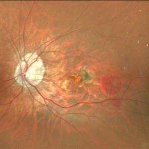
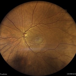
 Hereditary Macular Degeneration
May 27 2024 by Akansha Sharma
Color fundus photograph of a 42 year old female with hereditary macular degeneration.
Photographer: Dr. Akansha Sharma, Bharati Eye Hospital
Condition/keywords: HMD, macular degeneration
-
 Peripheral Retinal Degeneration (L-ORD)
Apr 17 2024 by Virginia Gebhart
92 year old female with bilateral patchy, sharply demarcated circular areas of chorioretinal atrophy with hyperpigmented margins in the mid to far periphery. Labs showed normal plasma ornithine levels ruling out generalized gyrate atrophy. Also intermediate uveitis and CMD/CME. FTA-ABS, Quant gold, and HLA-A29 labs all negative.
Photographer: Virginia Gebhart
Imaging device: Optos California
Condition/keywords: cystoid macular degeneration, cystoid macular edema (CME), FA, Fluorescein angiography, peripheral retinal degeneration
-
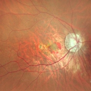
 Subretinal Neovascular Membrane with PED
Apr 17 2024 by Akansha Sharma
Color fundus photograph of a 72 year old male with ped along with subretinal bleed around it.
Photographer: Dr. Akansha Sharma, Bharati Eye Hospital
Condition/keywords: CNVM, PED, SRNVM, subretinal neovascularization (SRNV), wet age-related macular degeneration (wet AMD)
 Loading…