Search results (46 results)
-
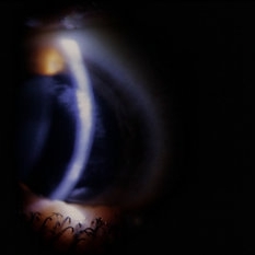
 Dendritic Epithelial Keratitis
Jun 24 2025 by Arkaprava Ray
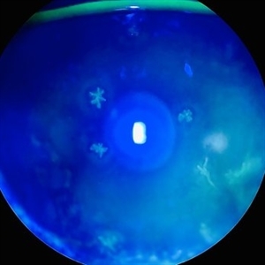
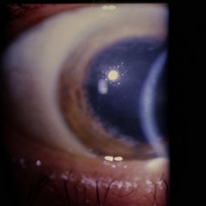
Slit-lamp photograph of a 46-year-old male showing dendritic epithelial lesion of herpes simplex keratitis, stained with 2% fluorescein sodium and viewed under cobalt blue filter.
Photographer: Arkaprava Ray, Trilochan Netralaya, Sambalpur, India
Condition/keywords: dendritic keratitis, HSV KERATITIS, keratitis
-
 Vitreous Bands
Apr 28 2025 by Gustavo Uriel Fonseca Aguirre
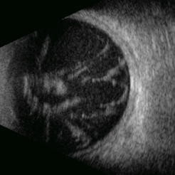
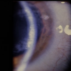
This B-mode transversal ultrasound scan shows condensed vitreous bands with anterior traction toward the cornea, accompanied by vitreous cellularity, in a patient with corneal perforation secondary to bacterial keratitis. The findings indicate severe intraocular inflammation with potential vitreous involvement.
Photographer: Gustavo U. Fonseca Aguirre, Hospital Conde de Valenciana, Ciudad de México
Condition/keywords: keratitis, vitreous bands
-
 Necrotizing Scleritis
Apr 17 2025 by Gustavo Uriel Fonseca Aguirre
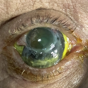
The clinical photograph shows necrotizing scleritis with perilimbal involvement, featuring marked scleral thinning and violaceous episcleral injection in the inferior quadrant. Focal uveal prolapse is visible at the area of maximal scleral necrosis, accompanied by peripheral ulcerative keratitis. Fluorescein staining residue is observed on the ocular surface. Associated findings include mild conjunctival chemosis and dilated episcleral vessels.
Photographer: Gustavo U. Fonseca Aguirre, Hospital Conde de Valenciana, Ciudad de México
Condition/keywords: necrotizing scleritis
-
 Herpetic Corneal Ulcer
Sep 24 2024 by DR Rohit Gupta
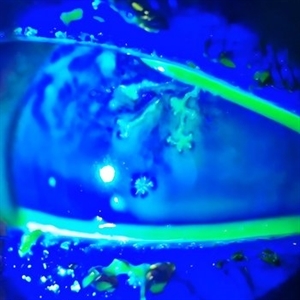
Slit lamp photograph of 32 year old male presented with herpetic corneal ulcer on staining with fluorescein dye under cobalt blue filted dendrits can be seen.
Photographer: Dr Rohit gupta
Imaging device: Samsung S21
Condition/keywords: corneal ulcer, dendritic keratitis, herpes dendrite, Herpes simplex infection, Herpes zoster, staining
-
 VZV Keratitis: Slit lamp Photo
Dec 8 2021 by Wen Hu
Slit lamp photograph of a 29-year-old man with VZV keratitis, who later developed choroidopathy.
Condition/keywords: varicella zoster virus (VZV), Varicella Zoster Virus Keratitis
-
 VZV Choroidopathy
Dec 8 2021 by Wen Hu
Optos fundus photograph of a 29-year-old man with VZV choroidopathy that developed after an episode of keratitis and anterior uveitis.
Imaging device: Optos
Condition/keywords: varicella zoster virus (VZV), Varicella Zoster Virus choroidopathy
-
 VZV Choroidopathy OCT
Dec 8 2021 by Wen Hu
OCT macula of a 29-year-old man with VZV choroidopathy that developed after an episode of keratitis and anterior uveitis.
Imaging device: Heidelberg
Condition/keywords: varicella zoster virus (VZV), Varicella Zoster Virus choroidopathy
-
 VZV Choroidopathy FA
Dec 8 2021 by Wen Hu
Late frame fluorescein angiogram of a 29-year-old man with VZV choroidopathy that developed after an episode of keratitis and anterior uveitis.
Imaging device: Optos fluorescein angiogram
Condition/keywords: varicella zoster virus (VZV), Varicella Zoster Virus choroidopathy
-
 VZV Choroidopathy ICG
Dec 8 2021 by Wen Hu
Indocyanine green image of a 29-year-old man with VZV choroidopathy that developed after an episode of keratitis and anterior uveitis.
Imaging device: Optos indocyanine green
Condition/keywords: varicella zoster virus (VZV), Varicella Zoster Virus choroidopathy
-
 VZV Choroidopathy Fundus Photo
Dec 8 2021 by Wen Hu
Zeiss fundus photograph of a 29-year-old man with VZV choroidopathy that developed after an episode of keratitis and anterior uveitis.
Imaging device: Zeiss
Condition/keywords: varicella zoster virus (VZV), Varicella Zoster Virus choroidopathy
-
 VZV Choroidopathy Early FA
Dec 8 2021 by Wen Hu
Early frame fluorescein angiogram of a 29-year-old man with VZV choroidopathy that developed after an episode of keratitis and anterior uveitis.
Imaging device: Optos fluorescein angiogram
Condition/keywords: varicella zoster virus (VZV), Varicella Zoster Virus choroidopathy
-
 Mooren Ulcer
May 18 2020 by McGill University Health Centre
A Mooren ulcer is a rapidly progressive ulcerative keratitis that first affects the periphery of the cornea before spreading circumferentially toward its center. Mooren ulcer is a diagnosis of exclusion: it can only be diagnosed after ruling out infectious and systemic causes. In this enucleation specimen, the periphery of the cornea is circumferentially ulcerated (arrow) and the cornea is thinning. There is also scarring of the corneoscleral junction.
Condition/keywords: Mooren's ulcer, ulcerative keratitis
-
 Slide 6-60
Mar 20 2019 by Lancaster Course in Ophthalmology
Retinoblastoma has extended out of the right eye into the orbit, causing proptosis and exposure keratitis of the eye.
Condition/keywords: keratitis, proptosis, retinoblastoma
-
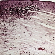 Slide 7-36
Feb 25 2019 by Lancaster Course in Ophthalmology
Herpes simplex keratitis is characterized by irregularity of the epithelium, patchy loss of Bowman's membrane, infiltration by lymphocytes and plasma cells, and vascularization of the stroma.
Condition/keywords: Bowman's membrane, epithelium, Herpes, lymphocytes, plasma cells, stroma
-
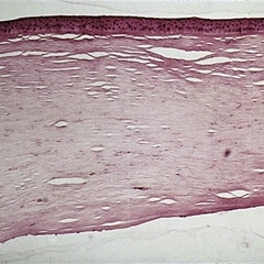 Slide 7-35
Feb 25 2019 by Lancaster Course in Ophthalmology
In interstitial keratitis, the only pathology noted is the presence of blood vessels in the deep stroma.
Condition/keywords: interstitial keratitis, stroma
-
 Slide 3-11
Feb 20 2019 by Lancaster Course in Ophthalmology
Higher-power view of hyphae and spores of Candida albicans in Candida keratitis ( x160).
Condition/keywords: endophthalmitis, hyphae, keratitis, spores
-
 Disseminated Chorioretinitis With Unknown Etiology
Apr 5 2018 by Kim Barrett
Ultra-wide field fluorescein angiogram of a 31-year-old female with intermittent pain in her left eye. Her condition has been managed in Liberia until recently when she moved to the United States. She suffers from multiple modalities including central retinal artery occlusion, posterior synechiae of the iris, interstitial keratitis, disseminated chorioretinitis, as well as HIV. An infectious cause is high on the differential in light of her HIV status. DDx: hypertensive crisis, an embolism (? IV drug use), coagulopathy, trauma, infectious. Blood work was normal. Her current vision is 20/30 right eye and 20/400 left eye.
Photographer: Kim Barrett, COA
Imaging device: Optos
Condition/keywords: central retinal artery occlusion (CRAO), chorioretinal scar, ciliary artery sparring, disseminated chorioretinitis, HIV, left eye, optic atrophy, staining
-
 Herpetic Keratitis
Feb 8 2018 by Claire Kiernan, MD
Slit lamp photograph of a 29-year-old female with herpetic keratitis complicated by deep corneal neovascularization and lipid keratopathy, shown here following Argon laser sectioning and subconjunctival bevacizumab with marked reduction of neovascularization and lipid keratopathy.
Photographer: Steve Crow, University of Tennessee Hamilton Eye Institute, Memphis, TN
Condition/keywords: Herpes simplex infection, keratitis
-
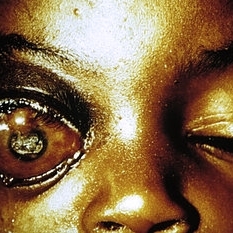
 Congenital Syphilis
Feb 20 2015 by H. Michael Lambert, MD
Interstitial keratitis in syphilis.
Condition/keywords: congenital, cornea, interstitial and deep keratitis, syphilis
-
 Congenital Syphilis
Feb 20 2015 by H. Michael Lambert, MD
Interstitial keratitis in syphilis.
Condition/keywords: congenital, cornea, interstitial and deep keratitis, syphilis
-
 Congenital Syphilis
Feb 20 2015 by H. Michael Lambert, MD
Interstitial keratitis in syphilis.
Condition/keywords: congenital, cornea, interstitial and deep keratitis, syphilis
-
 Wegener's Disease
Feb 20 2015 by H. Michael Lambert, MD
Thinning and vascularization of the peripheral cornea, a sequelae of peripheral ulcerative keratitis and scleritis.
Condition/keywords: corneal furrow, Wegener's granulomatosis
-
 Scleritis
Jul 16 2014 by John S. King, MD
Keratoscleritis with suprachoroidal hemorrhage in an elderly male with history of cataract surgery.
Photographer: URMC
Condition/keywords: keratitis, scleritis, suprachoroidal hemorrhage
-
 Scleritis
Jul 16 2014 by John S. King, MD
Keratoscleritis with suprachoroidal hemorrhage in an elderly male with history of cataract surgery.
Photographer: URMC
Condition/keywords: keratitis, scleritis, suprachoroidal hemorrhage
-
 Scleritis
Jul 16 2014 by John S. King, MD
Keratoscleritis with suprachoroidal hemorrhage in an elderly male with history of cataract surgery.
Photographer: URMC
Condition/keywords: keratitis, scleritis, suprachoroidal hemorrhage
 Loading…