Search results (111 results)
-
 Kissing Choroidals
Dec 18 2025 by Talhah - Zubair, MD
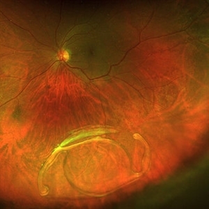
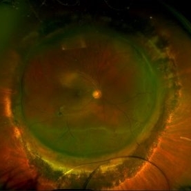
72 year old woman developed suprachoroidal hemorrhage during Yamane scleral intraocular lens fixation. At clinic follow up they were found to be appositional. Suprachoroidal tissue plasminogen activator was injected in clinic and an emergent choroidal cutdown drainage was performed the following day with subsequent resolution of apposition. Appositional choroidals were managed urgently to avoid retinal adhesion/membrane formation.
Condition/keywords: appositional choroidals, kissing choroidals, suprachoroidal hemorrhage
-
 Kissing Choroidals
Dec 18 2025 by Talhah - Zubair, MD
72 year old woman developed suprachoroidal hemorrhage during Yamane scleral intraocular lens fixation. At clinic follow up they were found to be appositional. Suprachoroidal tissue plasminogen activator was injected in clinic and an emergent choroidal cutdown drainage was performed the following day with subsequent resolution of apposition. Appositional choroidals were managed urgently to avoid retinal adhesion/membrane formation.
Condition/keywords: appositional choroidals, kissing choroidals, suprachoroidal hemorrhage
-
 Posterior Dislocated Intraocular Lens
Oct 23 2025 by Aditya S Kelkar, MS, FRCS, FASRS,FRCOphth
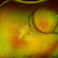
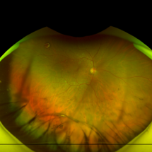
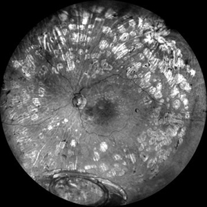
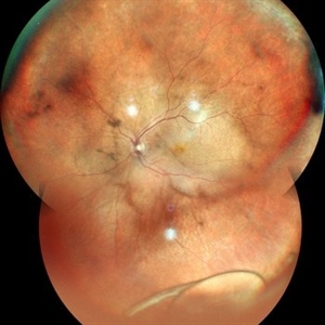
Fundus photograph of an 53-year-old man with a posteriorly dislocated intraocular lens near the posterior pole.
Photographer: Dr Tejal Rao, National Institute of Ophthalmology, Pune, India
Imaging device: Optos Daytona
Condition/keywords: dislocated intraocular lens (IOL), IOL drop
-
 Dislocated ACIOL
Oct 23 2025 by KANWALJEET HARJOT MADAN, M.S. (Ophthalmology); FAICO (Vitreous - Retina)
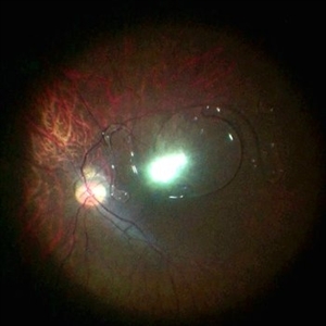
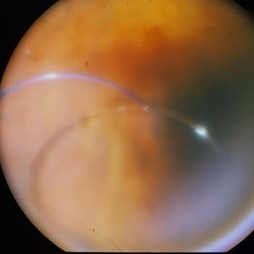
This is intraoperative image of a young male who presented with sudden diminution of vision in RE after Trauma. Fundus exam revealed presence of dislocated anterior chamber IOL in Vitreous Cavity.
Photographer: Dr. Kanwaljeet Harjot Madan, Thind Eye Hospital, Jalandhar City (Punjab). INDIA.
Imaging device: Zeiss Fundus Camera
Condition/keywords: dislocated anterior chamber intraocular lens (ACIOL)
-
 Dislocated IOL
Sep 20 2025 by JORGE SOBERANES
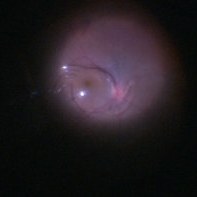
Fundus photograph of a 65-year-old man with a history of cataract surgery one year ago and bad vision since that.
Photographer: Dr. Jorge Soberanes, APEC, Universidad Nacional Autónoma México
Condition/keywords: dislocated lens, intraocular lens dislocation
-
 Dislocated Cataractous Lens
Jun 19 2025 by Mrinali Gupta, MD, FASRS
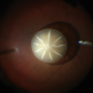
Intraoperative image of a chronically dislocated cataractous lens. The patient underwent pars plana vitrectomy, lensectomy, and placement of an anterior chamber intraocular lens, with improvement in vision from Count Fingers to 20/20 without correction.
Photographer: Mrinali Gupta, MD
Imaging device: Intraoperative surgical video (Zeiss Lumera scope, Resight lens)
Condition/keywords: dislocated crystalline lens
-
 Dislocated Intraocular Lens
Jun 4 2025 by Aditya S Kelkar, MS, FRCS, FASRS,FRCOphth
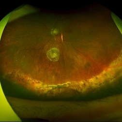
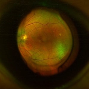
Fundus photograph of a 79-year-old man with a posteriorly dislocated intraocular lens in the inferior quadrant.
Photographer: Optom Chandrakanta Bhandare, National Institute of Ophthalmology, Pune
Imaging device: Optos Daytona
Condition/keywords: dislocated intraocular lens (IOL)
-
 Dislocated IOL
Apr 23 2025 by Anjana Mirajkar, MS Ophthalmology
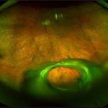
A widefield imaging of the right eye of a 55 year old male showing dislocated IOL inferiorly.
Photographer: Dr. Anjana Mirajkar- HV desai eye hospital ,Pune
Imaging device: Optos
Condition/keywords: dislocated intraocular lens (IOL)
-
Eye of the Hurricane
Apr 9 2025 by Gustavo Uriel Fonseca Aguirre
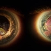
Ultrasound biomicroscopy of a post-operative eye (status post trabeculectomy and phacoemulsification) reveals a patent ostium on the right side, along with an intraocular lens in position. A hyphema is observed displaying small convection currents, creating a circular motion pattern due to the temperature gradient between the iris and cornea. Notably, the blood flow can be seen circulating toward the trabeculectomy site.
Condition/keywords: hyphema, trabeculectomy
-
 Eye of the Hurricane
Apr 8 2025 by Gustavo Uriel Fonseca Aguirre
Ultrasound biomicroscopy of a post-operative eye (status post trabeculectomy and phacoemulsification) reveals a patent ostium on the right side, along with an intraocular lens in position. A hyphema is observed displaying small convection currents, creating a circular motion pattern due to the temperature gradient between the iris and cornea. Notably, the blood flow can be seen circulating toward the trabeculectomy site.
Photographer: Gustavo U. Fonseca Aguirre, Hospital Conde de Valenciana, Ciudad de México
Condition/keywords: Hyphema, trabeculectomy
-
 PCIOL Opacification
Mar 31 2025 by DR Rohit Gupta
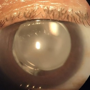
A pseudophakic patient visiting after 6 months of cataract surgery. On slit lamp examination a complete hazy white PCIOL was seen, which is a rare complication after cataract surgery.
Photographer: Dr Rohit gupta
Imaging device: Samsung S21
Condition/keywords: posterior chamber intraocular lens (PCIOL)
-
 Lacteocrumenasia
Mar 11 2025 by Gustavo Uriel Fonseca Aguirre
A 75-year-old female with a history of cataract surgery with intraocular lens implantation 20 years ago presented with progressive visual loss. On slit lamp examination, opaque material was found in the capsular bag behind the intraocular lens. Ultrasound biomicroscopy revealed hyperechoic material contained in the temporal-posterior sector of the capsular bag corresponding to lacteocrumenasia.
Photographer: Gustavo U. Fonseca Aguirre, Hospital Conde de Valenciana, Ciudad de México
Condition/keywords: Lacteocrumenasia, ultrasound biomicroscopy
-
 Dislocated Intraocular Lens
Nov 15 2024 by Tejaswita Verma
Fundus image of a spontaneously posteriorly dislocated IOL 10 years following surgery. Other eye had a subluxated opacified IOL.
Photographer: DR. TEJASWITA VERMA
Imaging device: MIRANTE
Condition/keywords: dislocated intraocular lens (IOL)
-
 What Lies Beneath
Jun 14 2024 by SHISHIR VERGHESE, MS, FVRS, FAICO (Retina)
Grey color fundus photograph of the left eye of a 78 year old gentleman who has undergone pars plana vitrectomy for proliferative diabetic retinopathy, shows dislocated intraocular lens bag complex lying on the inferior retina
Photographer: SHISHIR VERGHESE
Condition/keywords: dislocated intraocular lens (IOL), dislocated IOL, proliferative diabetic retinopathy (PDR)
-
 Dislocated IOL
Jun 4 2024 by Marlee Curnutt
Slit lamp photo of a 64 year old woman presenting with worsening vision and depth perception after trauma induced by a dog, which dislocated her IOL. The patient's IOL haptic was seen in the AC, and almost abutting cornea. Patient's vision upon presentation was DCC CF@1 feet. Patient was counseled and underwent an IOL exchange.
Photographer: Marlee Curnutt, COA
Imaging device: Galaxy A42
Condition/keywords: dislocated intraocular lens (IOL), haptic, IOL, right eye, slit lamp photo, slit lamp photography, trauma
-
 Dislocated IOL
May 24 2024 by Anjana Mirajkar, MS Ophthalmology
Intra-operative photo of a dislocated IOL in the left eye.
Photographer: Dr. Anjana Mirajkar -Retina Foundation, Ahmedabad
Condition/keywords: dislocated IOL, dislocated posterior chamber intraocular lens (PCIOL)
-
 Posteriorly Dislocated Intraocular Lens
Feb 22 2024 by Nikhil K Bommakanti, MD
A woman in her seventies with a history of retinal detachment repair presented with poor vision and was found to have a posteriorly dislocated intraocular lens. She subsequently underwent pars plana vitrectomy, intraocular lens extraction, and scleral-fixated intraocular lens implantation.
Condition/keywords: dislocated intraocular lens (IOL), dislocated posterior chamber intraocular lens (PCIOL)
-
 Dislocated Iol With Hypotony Maculopathy and Hemorrhagic Choroidal
Feb 9 2024 by Sandra R Montezuma, MD
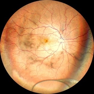
28 year old year-old male with history of congenital cataract of the right eye, s/p cataract extraction in 1999, s/p lens implant in 2011, presented with a dislocated IOL, hypotony, retina folds, hypotony maculopathy and hemorrhagic nasal choroidal after unsuccessful surgery to attempt remove the dislocated lens.
Photographer: Scott Baker, University of Minnesota
Condition/keywords: choroidals, dislocated posterior chamber intraocular lens (PCIOL), hypotony maculopathy, retina folds
-
 Dislocated IOL
Jan 28 2024 by Anjana Mirajkar, MS Ophthalmology
A widefield image of a 60 year old female with dislocated IOL inferiorly.
Photographer: Dr. Anjana Mirajkar -Retina Foundation, Ahmedabad
Imaging device: Mirante-Nidek
Condition/keywords: dislocated intraocular lens (IOL)
-
 Antero-Posterior Glance
Nov 5 2023 by rahul saradge
image through the principle axis with visibility of all structure in pathway .
Photographer: Optom Rahul , Isha Netralaya
Condition/keywords: optical, Optos, posterior chamber intraocular lens (PCIOL)
-
 Dislocated IOL
Oct 12 2023 by Virginia Gebhart
Fundus photo of an 83-year-old man with a 3 piece dislocated IOL. Surgery performed, PPV/removal of nonmagnetic FB/secondary Akreos. Eye is stable, vision limited due to grade 3 VH
Photographer: Virginia Gebhart, Retina Consultants of Carolina
Imaging device: Optos
Condition/keywords: dislocated intraocular lens (IOL), dislocated lens
-
 Dropped IOL
Sep 12 2023 by Ben Serar
Fundus photograph showing dropped 3-piece intraocular lens.
Condition/keywords: dropped IOL
-
 Scleral Buckling IOL Drop
Aug 6 2023 by Dr.Sheetal Divate
A 27 year old female with an old history of trauma and operated with scleral buckling and cataract surgery in the past came recently with complaints of DOV . Findings noted where IOL drop, inferior retinal detachment and old scleral buckle indent.
Photographer: Dr.Sheetal Divate
Imaging device: Optos Advance
Condition/keywords: dislocated intraocular lens (IOL), Retinal Detachment, scleral buckle
-
 Rhegmatogenous retinal detachment with dislocated IOL in a Morning Glory anomaly
Jul 27 2023 by Gustavo Aguirre-Suarez
Fundus photograph of a 13-year-old male with a history of congenital cataract surgery in his right eye in 2019. The patient presents with sudden visual loss. Upon examination, a dislocated IOL is observed in the posterior segment, accompanied by a rhegmatogenous retinal detachment featuring peripheral retinal tears and horseshoe breaks. Additionally, a morning glory disc anomaly is also present in this patient.
Photographer: Gustavo Aguirre-Suarez
Imaging device: Mirante, NIDEK
Condition/keywords: dislocated posterior chamber intraocular lens (PCIOL), Morning Glory Anomaly, rhegmatogenous retinal detachment
-
 Intraocular lens luxated to the vitreous cavity
Jun 24 2023 by Mariam Cernichiaro-Espinosa, MD
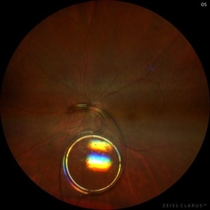
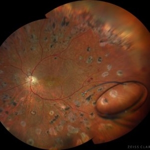
Three-piece intraocular lens luxated to the vitreous cavity in a patient with photocoagulated diabetic retinopathy after blunt trauma.
Photographer: Mariam Cernichiaro-Espinosa, Asociación para Evitar la Ceguera en México, I.A.P. Mexico City, Mexico.
Imaging device: Zeiss Clarus
Condition/keywords: diabetic retinopathy, intraocular lense in vitreous, lens luxation
 Loading…