Search results (73 results)
-
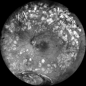
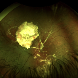
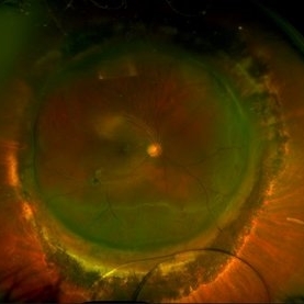
 Vasoproliferative Tumor
Aug 29 2024 by César Adrián Gómez Valdivia, MD
Inferior retinal vasoproliferative tumor found in a 66 year-old female patient. Asymptomatic.
Photographer: @eyemissu2
Imaging device: California ICG OPTOS
Condition/keywords: Vasoproliferative Tumor
-
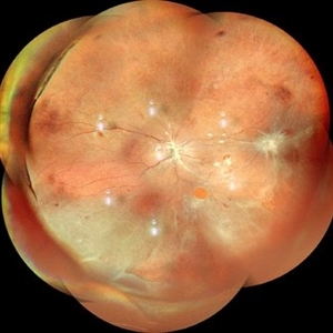
 What Lies Beneath
Jun 14 2024 by SHISHIR VERGHESE, MS, FVRS, FAICO (Retina)
Grey color fundus photograph of the left eye of a 78 year old gentleman who has undergone pars plana vitrectomy for proliferative diabetic retinopathy, shows dislocated intraocular lens bag complex lying on the inferior retina
Photographer: SHISHIR VERGHESE
Condition/keywords: dislocated intraocular lens (IOL), dislocated IOL, proliferative diabetic retinopathy (PDR)
-
 Retinal Detachment
Apr 28 2024 by Anjana Mirajkar, MS Ophthalmology
A montage of a 40 year old male showing multiple breaks with inferior retinal detachment with peripheral traction in a silicon filled eye.
Photographer: Dr. Anjana Mirajkar -Retina Foundation, Ahmedabad
Imaging device: Mirante-Nidek
Condition/keywords: Retinal detachment under Silicon Oil
-
 Status Post Prophylactic Barrage Laser for Inferior Retinal Detachment
Apr 9 2024 by Akansha Sharma
Color fundus photograph of a 19 year old male with an inferior retinal detachment holding well years after prophylactic barrage laser in a one eyed patient.
Photographer: Dr. Akansha Sharma, Bharati Eye Hospital
Condition/keywords: BARRAGE LASER, RD
-
 Serpiginous Choroidopathy
Mar 21 2024 by Ogugua Ndubuisi Okonkwo, MD, FRCS (Edin), FASRS
This is a right eye widefield fundus photograph of a 13-year-old male with a peripapillary ring of fibrotic scar that extends subretinally in finger-like projects along the vascular arcades and into the macula, with an extension of the scarring into the inferior retina, where it appears as a pigmented mottling.
Photographer: Zainab Ogunsanu, Eye Foundation Hospital & Eye Foundation Retina Institute, Lagos
Imaging device: ZEISS CLARUS 700
Condition/keywords: serpiginous like choroiditis
-
 Dislocated Crystalline Lens
Mar 19 2024 by Annaka Gooding
Ultra Wide field fundus photography of a 70 year old male who presented to clinic with a sudden increase of vision due to dropped crystalline lens secondary to severely dense cataract. Patient reported seeing a full black circle in his inferior visual field. Patient's visual acuity at time of visit was 20/100 with a +5.00 diopter lens. The physician recommended surgical intervention, and discussed surgery for PPV/PPL/IOL implantation with an ACIOL.
Photographer: Annaka Gooding, CPO
Imaging device: Optos California RGB
Condition/keywords: dislocated crystalline lens, fundus photography, inferior retina, OPTOS CALIFORNIA RGB, Right Eye, Ultra-wide field retinal imaging
-
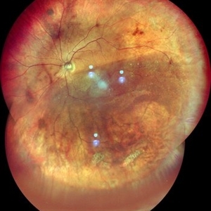
 Central Serous Retinopathy
Mar 19 2024 by Corey Grant
Ultra Wide-Field Fundus Autofluorescence Imaging of a 37 year old female with Central Serous Retinopathy affecting her right eye. Patient Visual Acuity was 20/20 in both eyes. Patient reported black spots in her vision onset three years ago, with associating flashes of light. Patient reports history of cortisone back injections a few years ago and denies Flonase use. The physician stated that there is hyperautofluorescence in the area of gutter of Sub-Retinal Fluid which likely happened from CSR.
Photographer: Corey Grant, OSC
Imaging device: OPTOS CALIFORNIA RGB
Condition/keywords: Central Serous Chorioretinopathy (CSR), central serous retinopathy (CSR), fundus autofluorescence (FAF), Guttering, hyperautofluorescence, inferior retina, OPTOS, Retina, Right Eye, subretinal fluid, ULTRA WIDE FIELD
-
 Retinal Detachment After Retinoblastoma Treatment
Mar 10 2024 by Alexandre Grandinetti, MD, PhD
Inferior retinal detachment occurring 6 years after treatment with intraarterial chemotherapy and laser in an 8-year-old boy.
Photographer: Corina Szrek
Condition/keywords: pediatric, retinoblastoma
-
 Choroidal Mass
Mar 4 2024 by ANKIT JAIN
Left eye color photo montage of 39 year old female with sub retinal mass in nasal quadrant with hemorrhages and subretinal fluid with inferior retinal detachment.
Photographer: Dr Ankit Jain
Imaging device: MIRANTE
Condition/keywords: choroidal mass
-
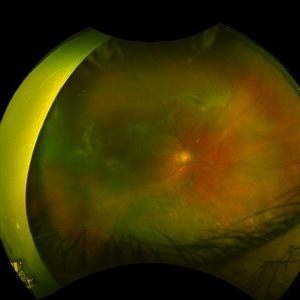
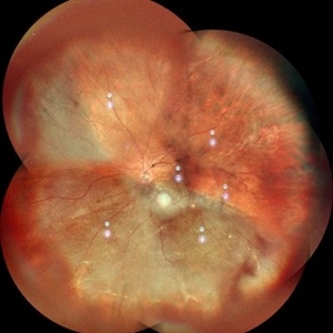 Retinal Detachment
Nov 3 2023 by Anjana Mirajkar, MS Ophthalmology
A widefield image (montage) of OS of a 55 year old female case of inferior retinal detachment with macula off.
Photographer: Dr. Anjana Mirajkar -Retina Foundation, Ahmedabad
Imaging device: Mirante-Nidek
Condition/keywords: inferior retinal detachment, Retinal Detachment
-
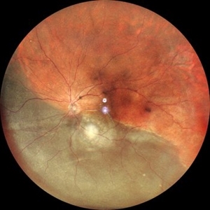 Retinal Detachment
Nov 3 2023 by Anjana Mirajkar, MS Ophthalmology
A widefield image of OS of a 55 year old female case of inferior retinal detachment with macula off.
Photographer: Dr. Anjana Mirajkar -Retina Foundation, Ahmedabad
Imaging device: Mirante-Nidek
Condition/keywords: inferior retinal detachment, Retinal Detachment
-
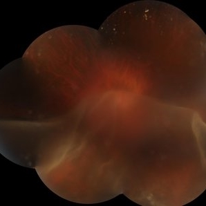 SUNSET THROUGH A VEIL
Oct 12 2023 by Deepti A Kulkarni, M.B.B.S., D.N.B., F.V.R.
HIGH MYOPE WITH A CORRECTION OF -24D. SUDDEN VISION LOSS FOLLOWING TRAUMA. SUPERIOR RETINA FOLDED ON TO THE INFERIOR RETINA MASKING THE DISC IN A TRANSLUCENT VEIL.
Photographer: DEEPTI KULKARNI, DR ANIL KULKARNI EYE HOSPITAL, MIRAJ, INDIA
Imaging device: TOPCON
Condition/keywords: GIANT RETINAL TEAR
-
 Choroidal Melanoma
Sep 7 2023 by Annaka Gooding
Ultra-Widefield pseudo-color and autofluorescence imaging of a 59 year old male with Choroidal Melanoma affecting his left eye. Patient reported floaters OS for months prior to examination as well as 1-2 weeks of "tunnel vision". Patient denies personal history of cancer. Patient's vision at time of examination was CF@5FT. Due to the Tumor size, the patient has developed a serous retina detachment in their inferior retina
Photographer: Annaka Gooding
Imaging device: Optos California
Condition/keywords: autofluorescence imaging, choroidal tumor, fundus photography, OPTOS CALIFORNIA, serous retinal detachment
-
 Scleral Buckling IOL Drop
Aug 6 2023 by Dr.Sheetal Divate
A 27 year old female with an old history of trauma and operated with scleral buckling and cataract surgery in the past came recently with complaints of DOV . Findings noted where IOL drop, inferior retinal detachment and old scleral buckle indent.
Photographer: Dr.Sheetal Divate
Imaging device: Optos Advance
Condition/keywords: dislocated intraocular lens (IOL), Retinal Detachment, scleral buckle
-
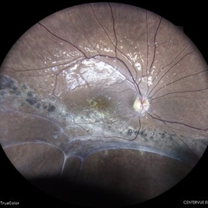
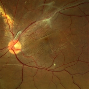
 Inferior retinal detachment with lattice and holes
May 31 2023 by Aditya S Kelkar, MS, FRCS, FASRS,FRCOphth
Importance of dilated retina check up before Lasik surgery can't be better demonstrated...patient totally asymptomatic came for Lasik opinion and has inferior retinal detachment with lattice and holes, sparing the macula
Photographer: Dr. Sahil Wagh , National Institute of Opthalmology, Pune , India
Imaging device: Zeiss Clarus 500
Condition/keywords: inferior retinal detachment
-
 Retinal detachment
Apr 12 2023 by Ahmed Abbas Hashmi, OD
Color fundus photograph of the left eye of a 30-year-old man with asymptomatic inferior retinal detachment with pigmented demarcation line. Macula and Disc healthy.
Photographer: Ahmed Abbas Hashmi
Imaging device: Topcon TRC-NW8F
Condition/keywords: Pigmentary demarcation line, Retinal Detachment
-
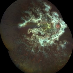
 Displaced & folded macula
Oct 10 2022 by Ricardo Leitão Guerra
Tractional retinal detachment due to sickle cell retinopathy leading to a displaced and folded appearance of the macula in this 36-yo male. Subretinal bands are also noticed crossing the macula towards inferior retinal detachment area.
Photographer: Ricardo Leitão Guerra
Imaging device: Clarus 700 - Zeiss
Condition/keywords: folds, sickle cell retinopathy, subretinal bands, tractional retinal detachment
-
 Methotrexate Bubble following Intravitreal Injection for PVR
Sep 21 2022 by Zach Seim
Ultra-widefield fundus photograph of an 81 year old female with a Methotrexate bubble following an Intravitreal Injection for Proliferative Vitreoretinopathy. Patient has been presenting to the office for two week interval Methotrexate injections in her left eye. The image was taken prior to her eighth injection which revealed a residual Methotrexate bubble in her inferior retinal image. This patient was seeing "lots" of floaters, as well as having visual acuity of cc20/400 cc20/200 PH.
Photographer: Zach Seim
Imaging device: OPTOS California
Condition/keywords: bubble, fundus photograph, fundus photography, intravitreal injection, left eye, methotrexate, nasal retina, Optos, proliferative vitreoretinopathy (PVR), pseudocolor, ultra-wide field imaging
-
 Recurrent Retinal Detachment with PVR with Subretinal Oil after Retinal Detachment with Silicone Oil
Feb 2 2022 by Manish Nagpal, MD, FRCS (UK), FASRS
Intraoperative photo of inferior retinal contraction due to PVR and presence of subretinal silicone oil globule noted.
Photographer: Manish Nagpal, Retina Foundation, Ahmedabad, Gujarat
Imaging device: Sony PMW -10 MD surgical camera
Condition/keywords: proliferative vitreoretinopathy (PVR), retina, retina surgery, retina surgery complications, silicone oil, subretinal
-
 Retinal Detachment with Proliferative Vitreoretinopathy
Jan 31 2022 by Ahmad B. Tarabishy, MD
Ultra wide-field fundus photograph of a 55-year-old gentleman who had previously underwent laser retinopexy for multiple inferior retinal breaks. He presented with a macula-off retinal detachment from a new temporal break with proliferative vitreoretinopathy with fixed folds noted temporally and superonasally.
Photographer: Megan McLandsborough, Lakeland Eye Clinic
Imaging device: Optos California UWF Camera
Condition/keywords: laser retinopexy, macula off Retinal Detachment, proliferative retinopathy, proliferative vitreoretinopathy (PVR), Retinal Detachment, retinal detachment with retinal defect
-
 Ozurdex Implant Related Tear
Jan 26 2022 by Tracey Grabowski
Ultra wide-field photograph of a 73-year-old female with an Ozurdex implant causing a retinal tear in the inferior retina. Prompt laser was added to prevent a retinal detachment and patient has been doing well since. Patient had no symptoms following the occurrence.
Photographer: Tracey Grabowski
Imaging device: Optos California
Condition/keywords: fundus photograph, inferior retina, optos, ozurdex, Ozurdex implant, retinal tear, treated retinal tear, ULTRA WIDE FIELD
-
 Dexamethasone Implant
Jul 3 2021 by Gerardo Rivera Arroyo
42-year-old male, operated on for vitrectomy plus scleral buckling plus silicone plus dexamethasone implant for inferior retinal detachment with PVR.
Photographer: Rosa Elizabeth Moreno Anda, MD, Hospital Central Militar CDMX
Condition/keywords: dexamethasone implant, retina surgery, vitrectomy
-
 Spontaneous Reattachment of Retinal Detachment
Apr 26 2021 by Priya Rasipuram Chandrasekaran, MBBS, DO, DNB, FRCS
This is the fundus photo montage and red free montage of a 27-year-old male showing pigmentary changes and atrophic changes in the inferior retina involving the macula. This has sharply demarcated margins and a convex border with subretinal bands suggestive of spontaneous reattachement of retinal detachment.
Condition/keywords: re-attached retinal detachment (RRD), spontaneous retinal reattachment
-
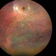
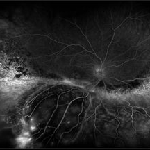 Exudative Retinal Detachment and Branch Retinal Vein Occulsion
Oct 29 2020 by Olivia Rainey
Ultra-widefield fluorescein anigogram of a 51-year-old female with an exudative retinal detachment and branch retinal vein occlusion with retinal neovascularization affecting her right eye. The physician stated that the multiple aneurysmal dilations noted in the inferior periphery are responsible for the exudative RD seen on exam. He is considering Coat's vs FEVR given family history of aneurysms/congenital heart pathology per patient. He encouraged the patient to control their blood pressure, cholesterol, blood sugar, and co-morbidities which may have promoted the BRVO. He recommended antiVEGF injections to control the vascular leakage. Given the severe presentation and imminent threat to her vision, he recommended Eylea as first line therapy.
Photographer: Olivia Rainey, OCT-C, COA
Imaging device: Optos California
Condition/keywords: branch retinal vein occlusion (BRVO), chronic retinal detachment, fluorescein angiogram (FA), fluorescein leakage, inferior retina, inferior retinal detachment, Optos, ultra-wide field imaging
-
 Inferior Retinal Detachment with Lattice
Sep 30 2020 by Sham Talati, DOMS
A patient of inferior retinal detachment with lattice inferiorly.
Photographer: Dr. Sham Talati,Retina Foundation,Ahmedabad
Imaging device: Nidek Mirante
Condition/keywords: lattice degeneration
 Loading…