Search results (169 results)
-
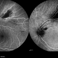
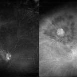
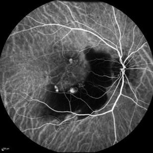
Central Serous Chorioretinopathy
Apr 15 2025 by Filip Kecer
FA&ICG late phase of a young woman with CSCR
Photographer: Filip Kecer, Oftalmocentrum Betliarska, Bratislava, Slovakia
Imaging device: Spectralis, Heidelberg Engineering
Condition/keywords: central serous chorioretinopathy (CSCR), Central Serous Chorioretinopathy (CSR), FA late phase, indocyanine green (ICG) angiography
-
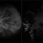
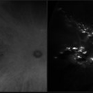
 Choroidal Hemangioma 4 Ways
Mar 13 2025 by Virginia Gebhart
Color fundus, FAF, late FA, late ICG of 64 year old male with choroidal hemangioma. Early hyperfluorescence with late leakage on FA, early hypercyanescence with late washout (25 min) on ICG.
Photographer: Virginia Gebhart, Retina Consultants of Carolina
Imaging device: Optos California
Condition/keywords: autofluorescence imaging, choroidal hemangioma, FA late phase, Fluorescein angiography, hemangioma, indocyanine green (ICG) angiography
-
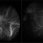
 FA/ICG Choroidal Melanoma
Mar 10 2025 by Virginia Gebhart
Side by Side comparison of late FA/ICG on choroidal melanoma. FA showed early lacy hyperfluorescence with late leakage, ICG showed late Hypocyanescence.
Photographer: Virginia Gebhart, Retina Consultants of Carolina
Imaging device: Optos California
Condition/keywords: FA, Fluorescein angiography, fluorescein leakage, indocyanine green (ICG) angiography
-
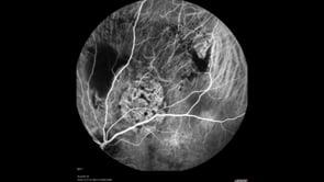
 Indocyanine Green (ICG) of Circumscribed Choroidal Hemangioma (CCH)
Feb 6 2025 by Jack B Margines, MD, MHCI
Peripheral patchy hyperfluorescence is seen on this early image of ICG-A on a 53-year-old asymptomatic with an extramacular circumscribed choroidal hemangioma.
Photographer: W Ryan Miliam, CRA, OCT-C, University of California, Irvine Gavin Herbert Eye Institute
Imaging device: Optos
Condition/keywords: choroidal hemangioma, indocyanine green (ICG) angiography
-
 Choroidal Melanoma 3 Ways
Jan 16 2025 by Virginia Gebhart
RGB/FA/ICG of 76 year old female with a new choroidal melanoma. Pt scheduled for plaque radiation. BCVA 20/400
Photographer: Virginia Gebhart, Retina Consultants of Carolina
Imaging device: Optos California
Condition/keywords: fluorescein angiogram (FA), indocyanine green (ICG) angiography, OPTOS CALIFORNIA RGB
-
 Toxoplasmosis
Dec 5 2024 by Tejaswita Verma
26 year old male with 6/18 vision , anterior chamber reaction, vitritis and retinitis lesion along the superotemporal arcade with full thickness involvement on OCT . FFA showing hypofluorescence with surrounding hyperfluorescence characterstic of toxoplasma retinitis . ICGA shows hypocyanescence.
Photographer: DR. TEJASWITA VERMA
Imaging device: MIRANTE
Condition/keywords: Fundus Fluorescein Angiography, indocyanine green (ICG) angiography, toxoplasmosis
-
 Fluorescein and Indocyanine Green Angiography in Right Eye in Case of Choroidal Hemangioma
Nov 29 2024 by Anand Temkar
Right eye Fluorescein and Indocyanine green angiography of a 42 year old male in case of Choroidal hemangioma. Choroidal hemangioma have a unique pattern of circulation where the large blood vessels produce a “COARSE VASCULAR PATTERN.” Fluorescein angiography of circumscribed choroidal hemangiomas typically reveals very early hyperfluorescence of larger-caliber choroidal blood vessels either before or simultaneously with the initial filling of the retinal arterioles. Indocyanine green angiography typically shows filling of the intralesional vascular channels, intense hypercyanescence of the lesion by the intermediate frames (peaks around 3-4 minutes) and late washout of the central portion of the lesion.
Photographer: Dr.Anand Temkar- Retina Foundation, Ahmedabad
Imaging device: Mirante
Condition/keywords: Choroidal Hemangioma, FLUORESCEIN ANGIOGRAPHY, indocyanine green (ICG) angiography
-
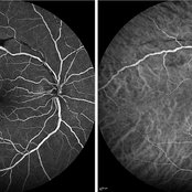 Early FA/ICG at 1 Minute of Atypical ANCA Associated Retinal Vasculitis
Nov 13 2024 by Deepak Sambhara, MD
Fluorescein and Indocyanine Green Angiography of a 49-year-old male with high ANA titer, atypical ANCA positivity, who presented to clinic with 1 month of vision loss. Exam revealed anterior chamber cell, mild vitreous cell, sclerotic vessels along arterioles. Early FA/ICG at 1 minute demonstrates absent arteriole fill.
Photographer: Killian Roberts, Micaela Hertz; Eye Clinic of Wisconsin
Imaging device: Heidelberg Spectralis
Condition/keywords: A-ANCA, autoimmune vasculitis, fluorescein angiogram (FA), indocyanine green (ICG) angiography, retinal vasculitis
-
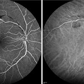 Late FA/ICG at 4 Minutes of Atypical ANCA Associated Retinal Vasculitis
Nov 13 2024 by Deepak Sambhara, MD
Fluorescein and Indocyanine Green Angiography of a 49-year-old male with high ANA titer, atypical ANCA positivity, who presented to clinic with 1 month of vision loss. Exam revealed anterior chamber cell, mild vitreous cell, sclerotic vessels along arterioles. Late FA/ICG at 4 minutes demonstrates absent arteriole fill with venular periphlebitis.
Photographer: Killian Roberts, Micaela Hertz; Eye Clinic of Wisconsin
Imaging device: Heidelberg Spectralis
Condition/keywords: A-ANCA, autoimmune vasculitis, fluorescein angiogram (FA), indocyanine green (ICG) angiography, retinal vasculitis
-
 Birdshot Retinochoroiditis
Aug 8 2024 by Virginia Gebhart
ICG angiogram of 45 year old male with Birdshot Retinochoroiditis. Has been improving on Humira and Methotrexate.
Photographer: Virginia Gebhart
Imaging device: Optos California
Condition/keywords: birdshot, indocyanine green (ICG) angiography
-
 Choroidal Hemangioma
Jul 30 2024 by Korey Starkey
ICG image of a77 year-old female with choroidal hemangioma. The physician states the hypercyanesence in the right eye is consistent with hemangioma but no typical late washout observed. He also notes high internal reflectivity make hemangioma possible. Patients vision at time of imaging VA OD: sc20/200 PH20/60-1; plan to follow patient at 6 month intervals at this time.
Photographer: Korey Starkey
Imaging device: Optos
Condition/keywords: Choroidal Hemangioma, Fluorescein angiography, indocyanine green (ICG) angiography, Optos
-
 Posterior Uveitis
Jul 5 2024 by Zach Seim
FA/ICG OS of a 39 year old female with Posterior Uveitis. VA at time of photos was Dsc 20/20-1.
Photographer: Zach Seim
Imaging device: Optos California
Condition/keywords: FA, indocyanine green (ICG) angiography, Optos, OPTOS CALIFORNIA, posterior uveitis
-
 Adult Onset Coats' Disease
Jul 5 2024 by Zach Seim
FA/ICG of a 64 year old female with Adult Onset Coats' Disease. VA DCC CF@3 feet upon presentation. Therapy options discussed extensively.
Photographer: Zach Seim
Imaging device: Optos California
Condition/keywords: Coats' disease, FA, indocyanine green (ICG) angiography, Optos, OPTOS CALIFORNIA
-
 Polypoidal Choroidal Vasculopathy
Jul 20 2023 by Gregg T. Kokame, MD, MMM, FASRS
64 Year Old Male, with Polypoidal Choroidal Vasculopathy. Pre-op and Post-op PDT/Vabysmo Injection
Photographer: Jaclyn Pisano
Imaging device: Heidelberg Spectralis
Condition/keywords: FA late phase, indocyanine green (ICG) angiography, OCT, PDT, polypoidal choroidal vasculopathy (PCV), subretinal, subretinal fluid
-
 FA/ICG of Retinal Vasoproliferative Tumor
Apr 26 2023 by Kelli Nyenhuis
FA/ICG of a 64-year-old woman referred to our office to rule out choroidal melanoma.
Photographer: Kelli Nyenhuis, COA
Imaging device: Optos California
Condition/keywords: fluorescein angiogram (FA), indocyanine green (ICG) angiography, tumor
-
 Peripapillary Hypopigmentation
Feb 9 2023 by Harold Rodriguez
Peripapillary Hypopigmentation
Photographer: Harold Rodriguez OCT-C
Condition/keywords: fluorescein angiogram (FA), indocyanine green (ICG) angiography, peripapillary
-
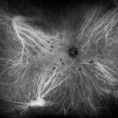
 Acute Central Retinal Artery Occlusion
Jul 27 2022 by Becca Harris
Ultra widefield FA/ICG of a 24 year old female with an acute central retinal artery occlusion affecting the right eye. Patient presented with extreme headaches following DAVF surgery the previous day. Patient has Factor VIII deficiency and had a cerebral venous thrombosis 9 years ago and lost vision in the right eye at that time. Patient has history of optic sheath fenestration OU and craniotomy. On initial evaluation, she had a CRAO as well as diffuse choroidal nonperfusion noted on optos FA. Suspect nonperfusion to third and sixth nerve leading to palsy. Occlusion of vasculature in the setting of recent endovascular embolization of fistulas in the CNS. Discussed diagnosis and poor prognosis with parents and patient. Patient had no light perception at the time of her initial appointment.
Photographer: Becca Harris
Imaging device: Optos California
Condition/keywords: Choroidal non-perfusion, fluorescein angiogram (FA), indocyanine green (ICG) angiography, non-perfusion, Optos, Right Eye, ultra-wide field imaging
-
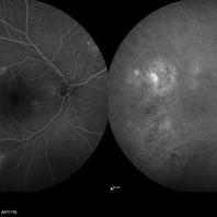 Central Serous Chorioretinopathy
Jan 25 2022 by Olivia Rainey
Late phase widefield fluorescein angiography of a 60-year-old male with Central Serous Chorioretinopathy. Chronic history of CSR followed with observation without treatment prior to presenting at our office. The physician noted significant findings on exam and imaging with multifocal areas of inactive and active changes OD. FA shows superotemporal macular leakage, subtle inferonasal macular leakage and staining as well as multifocal hypercyanescence on ICG. Fortunately foveal sparing and thus observation is recommended at this time OD.
Photographer: Olivia Rainey, OCT-C, COA
Imaging device: Heidelberg Spectralis
Condition/keywords: 55-degrees, central serous chorioretinopathy (CSCR), central serous retinopathy (CSR), chronic central serous chorioretinopathy (CSCR), fluorescein angiogram (FA), fluorescein leakage, heidelberg spectralis, indocyanine green (ICG) angiography, late phase
-
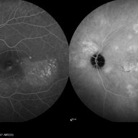 Central Serous Chorioretinopathy
Jan 25 2022 by Olivia Rainey
Late phase widefield fluorescein angiography of a 60-year-old male with Central Serous Chorioretinopathy. Chronic history of CSR followed with observation without treatment prior to presenting at our office. The physician noted subfoveal subretinal fluid with recent visual decline. FA shows multifocal leakage and ICG shows hypercyanescence. OCTA, ICG, and FA consistent with CSR, and without concern for CNVM thus will observe without anti-VEGF at this time. PDT therapy recommended.
Photographer: Olivia Rainey, OCT-C, COA
Imaging device: Heidelberg Spectralis
Condition/keywords: 55-degrees, central serous chorioretinopathy (CSCR), central serous retinopathy (CSR), chronic central serous chorioretinopathy (CSCR), fluorescein angiogram (FA), heidelberg spectralis, indocyanine green (ICG) angiography, left eye
-
 Sub-Macular Hemorrhage Indocyanine Green Angiography
Jul 5 2021 by Fang Helen Mi
Indocyanine green angiography revealed three polypoidal choroidal vasculopathy lesions, and the patient underwent photodynamic therapy the following week. His vision improved to 20/30.
Condition/keywords: indocyanine green (ICG) angiography, polypoidal choroidal vasculopathy (PCV), submacular hemorrhage
-
 Retinal Cavernous Hemangioma
Oct 22 2020 by Olivia Rainey
Ultra-widefield fluorescein and ICG angiogram of a 31-year-old male presenting with a retinal cavernous hemangioma affecting his left eye. Patient was 18-years-old when he was diagnosed with a retinal cavernous hemangioma. He has had a few episodes of vitreous hemorrhages since then. His vision was 20/20-1 in both eyes.
Photographer: Becca Harris
Imaging device: Optos California
Condition/keywords: cavernous hemangioma of the retina, fluorescein angiogram (FA), indocyanine green (ICG) angiography, late phase, left eye, Optos, ultra-wide field imaging
-
 Peripheral Exudative Hemorrhagic Chorioretinopathy
Oct 7 2020 by Olivia Rainey
Fluorecein and ICG angiography of a 80-year-old male with peripheral exudative hemorrhagic chorioretinopathy affecting his right eye. Patient noted only mild floaters for a couple weeks OD on 10/5/2020. The physician strongly suspects that the lesion is subretinal blood (likely from PEHCR) rather than choroidal melanoma. There is blocking on FA and ICG with a definite lack of intrinsic vessels within the mass lesion. He will monitor closely, as the patient is monocular with a history of multiple surgeries (which the family believes PPV for "scar tissue") OS mostly in 2013. His family also reports remembering possibly being told there was a "small mass" in the left eye at one point in their surgical course. The physician believes that it's possible this was a bleed related to PEHCR as it typically exists as a bilateral condition.
Photographer: Olivia Rainey, OCT-C, COA
Imaging device: Optos California
Condition/keywords: fluorescein angiogram (FA), indocyanine green (ICG) angiography, monocular, Optos, peripheral exudative hemorrhagic chorioretinopathy (PEHCR), periphery, subretinal hemorrhage, ultra-wide field imaging
-
 Peripheral Polypoidal Choroidal Vasculopathy Causing PEHCR
Aug 25 2020 by Kshitij Raizada, MS Ophthalmology
A 36-year-old female presented to with complaints of diminution of vision in LE for 3 months. Her BCVA in the RE was 6/6 and CF@1m in the LE. She was a K/C/O polypoidal choroidal vasculopathy (PCV) in the RE and had a history of receiving 2 doses of intravitreal Aflibercept (Eylea) in the RE. On her visit, she had dense Vitreous hemorrhage in the LE. 25G pars plana vitrectomy + intravitreal Aflibercept was planned for her. On clearing the vitreous hemorrhage, the patient was found to have Peripheral Exudative Hemorrhagic Chorioretinopathy (PEHCR). An on-table diagnosis of "PCV causing PEHCR" was made. Endolaser was done to sites suspicious to have underlying polyps. The patient's vision improved to 6/18 in the LE after one week of surgery. One month post-surgery, her BCVA in the LE had improved to 6/9. ICG angiography was done which revealed non-leaking BVN(branching vascular network) and no polyps. The patient has been doing well and has been kept under observation.
Condition/keywords: indocyanine green (ICG) angiography, peripheral exudative hemorrhagic chorioretinopathy (PEHCR), polypoidal choroidal vasculopathy (PCV), video
-
 Tilted Disc Syndrome Complicated with RPE Atrophy and Polypoidal Choroidal Vasculopathy
Jan 20 2020 by Pierre-Henry Gabrielle, MD
Coupled OCT B-scan and ICG angiography of an 81-year-old woman with a tilted disc syndrome complicated with RPE atrophy and polypoidal choroidal vasculopathy.
Photographer: Pierre-Henry Gabrielle, Ophthalmology department, Dijon University Hospital, France.
Imaging device: Heidelberg spectralis
Condition/keywords: atrophic pigment epithelium, indocyanine green (ICG) angiography, optical coherence tomography (OCT), polypoidal choroidal vasculopathy (PCV), tilted disc
 Loading…