Search results (76 results)
-
 Rhegmatogenous Retinal Detachment
Nov 27 2025 by Jacob Adrián Ruíz García
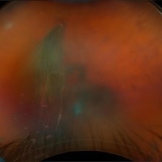
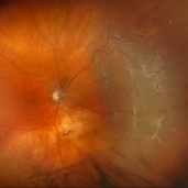
Ultra–widefield fundus image demonstrates an extensive rhegmatogenous retinal detachment (RRD) with several large, irregular retinal tears are visible in the temporal retina, with associated surrounding subretinal fluid. The detachment appears bullous, with fluid extending widely across the mid-periphery and involving much of the posterior pole.
Photographer: Jacob Adrián Ruíz Garcia, Fundación Hospital Nuestra Señora de la Luz I.A.P, México City
Imaging device: Optos California
Condition/keywords: horseshoe tear, Rhegmatogenous retinal detachment, tear
-
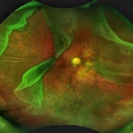
 ERMageddon - Wrinkle in the Space-time Fabric of Macula
Oct 29 2025 by SHRADDHA RAJ SHRIVASTAVA
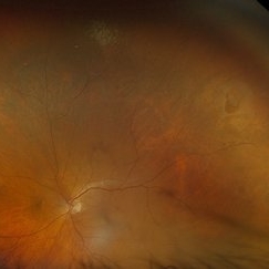
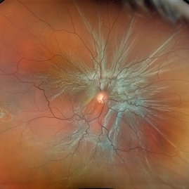
38 year old female with Epiretinal Membrane (ERM) over macula, post laser barrage for multiple symptomatic Horse-shoe Tears (HSTs) and Lattice Degenerations (seen on wide-field image). Posterior pole revealed tilted disc with peripapillary atrophy. There is thick opaque epiretinal membrane obscuring the underlying superior arcade vessels and causing foveal ectopia with distortion of perimacular vasculature. Patient was planned for Right Eye pars plana vitrectomy for ERM peeling.
Photographer: Dr. Shraddha Raj Shrivastava
Imaging device: Nidek Mirante SLO/OCT (Confocal scanning/Spectral domain OCT
Condition/keywords: BARRAGE LASER, ectopic fovea, epiretinal membrane (ERM), horseshoe tear, lattice degeneration, vitreomacular traction (VMT)
-
 ERMageddon - Wrinkle in the Space-time Fabric of Macula
Oct 29 2025 by SHRADDHA RAJ SHRIVASTAVA
38 year old female with Epiretinal Membrane (ERM) over macula, post laser barrage for multiple symptomatic Horse-shoe Tears (HSTs) and Lattice Degenerations. Posterior pole revealed tilted disc with peripapillary atrophy. There is thick opaque epiretinal membrane obscuring the underlying superior arcade vessels and causing foveal ectopia with distortion of perimacular vasculature. Patient was planned for Right Eye pars plana vitrectomy for ERM peeling.
Photographer: Dr. Shraddha Raj Shrivastava
Imaging device: Nidek Mirante SLO/OCT (Confocal scanning/Spectral domain OCT
Condition/keywords: ectopic fovea, epiretinal membrane (ERM), ERM, horseshoe tear, vitreomacular traction (VMT)
-
 Multiple Tear RD
Oct 10 2025 by Angela Rico
67 year-old male with complaint of floaters and veil OD for 2 weeks.
Condition/keywords: horseshoe tear, retinal detachment, Retinal tear
-
 Retinal Tear w/VH
Aug 22 2025 by Virginia Gebhart
69 year old male referred for sudden vision loss. Difficult view secondary to VH. Ultrasound and photos show small break with clotting, heavy amount of layered blood inferior and scattered IRH's. Cryotherapy performed to seal current tear, will give VH time to clear on its own. Pt takes Eliquis twice a day.
Photographer: Virginia Gebhart, Retina Consultants of Carolina
Imaging device: Optos California
Condition/keywords: cryo-retinal tear, horseshoe tear, tear, vitreous hemorrhage
-
 Horseshoe Retinal Tear
Aug 6 2025 by Korey Starkey
80 year-old patient presented with HSRT without detachment in the left eye and macula-off detachment in the right eye. Scheduled patient for prompt surgical repair OD and same day laser retinopexy OS to reduce risk of retinal detachment.
Photographer: Korey Starkey
Imaging device: Optos
Condition/keywords: color fundus photograph, fundus photography, horseshoe tear, Optos
-
 Multiple Retinal Tears
Jul 25 2025 by Virginia Gebhart
60 year old male referred for horseshoe tear of the retina. Scleral depressed exam revealed 7 tears in the same eye. Prophylaxis laser performed to seal all tears.
Photographer: Virginia Gebhart, Retina Consultants of Carolina
Imaging device: Optos California
Condition/keywords: horseshoe tear, multiple retinal tears, operculated tear, Retinal tear
-
 Multiple HST Causing Subtotal RD
Jul 24 2025 by Tejaswita Verma
Fundus image of a 64 year old male with HM vision with pseudophakic bullous RD.
Photographer: Dr. Tejaswita Verma
Imaging device: MIRANTE
Condition/keywords: horseshoe tear, RD
-
 Retinal Detachment with Multiple Horse Shoe Shaped Tears
Jul 14 2025 by Malvika Singh
Fundus photograph of a 46 year old showing a retinal detachment with multiple peripheral horse show shaped tears.
Photographer: Dr Malvika Singh, Retina Foundation, Ahmedabad, India
Imaging device: Mirante SLO/OCT
Condition/keywords: horseshoe tear, retinal detachment
-
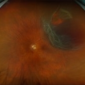
 The Horseshoe Of Havoc
Jun 28 2025 by Tejaswita Verma
Fundus image of a 50 year old male with a very large horseshoe tear causing RRD with macula off, hydration folds.
Photographer: Dr. Tejaswita Verma
Imaging device: MIRANTE
Condition/keywords: horseshoe tear, large break
-
 The Horseshoe Of Havoc
Jun 28 2025 by Tejaswita Verma
Fundus image of a 50 year old male with a very large horseshoe tear causing RRD with macula off, hydration folds.
Photographer: DR. TEJASWITA VERMA
Imaging device: MIRANTE
Condition/keywords: horseshoe tear
-
 A Vessel That Would Not Let Go
May 5 2025 by Malvika Singh
Fundus photograph of a retinal detachment showing a horse shoe shaped tear and a bridging vessel.
Photographer: Dr Tejaswita Verma, Retina Foundation, Ahmedabad, India
Imaging device: Mirante SLO/OCT
Condition/keywords: bridging vessel, horseshoe tear
-
 Horse Shoe Tear With Retinal Detachment
Apr 28 2025 by rohan jain
56 year-old male with idiopathic HST and RRD
Photographer: Dr. ROHAN JAIN
Condition/keywords: horseshoe tear, Retinal Detachment, rrd
-
 Giant Retinal Tear with Multiple Retinal Breaks
Apr 21 2025 by Hrishikesh Naik, MS
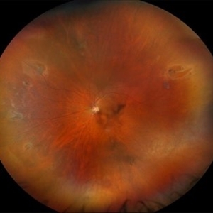
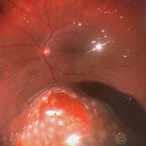
A 28 year old high myope with retinal detachment associated with a supero-temporal giant retinal tear in addition to multiple peripheral horseshoe tears and an additional supero-nasal retinal tear.
Photographer: Hrishikesh Naik
Imaging device: Optos Daytona
Condition/keywords: giant retinal tear, High Myopia, horseshoe tear, retinal break, retinal detachment
-
 Large Horseshoe Tear
Apr 4 2025 by Tejaswita Verma
Fundus photo of a 29 year old myopic male with RE 6/12P vision with large ragged tear on the posterior edge of lattice degeneration
Photographer: DR. TEJASWITA VERMA
Imaging device: MIRANTE
Condition/keywords: horseshoe tear
-
 Hosreshoe Tears on Posterior Pole
Mar 22 2025 by Deepak Bhojwani, MS
A fundus image of an asymptomatic 64 year old male with large horseshoe shaped breaks in inferonasal quadrant on posterior pole, an unusual location for retinal breaks.
Photographer: DR DEEPAK BHOJWANI
Condition/keywords: horseshoe tear, posterior pole break, retinal break
-
 Retinal Detachment (Mac-Off)
Feb 20 2025 by Virginia Gebhart
63 year old male with a mac-off retinal detachment from 4:00 to 1:30 with a single break at 10:00. Pt schedule for PPV/GFE. Guarded prognosis for visual recovery.
Photographer: Virginia Gebhart, Retina Consultants of Carolina
Imaging device: Optos California
Condition/keywords: horseshoe tear, retinal detachment, retinal detachment of the macula
-
 Retinal Detachment with Horseshoe Retinal Tear
Feb 17 2025 by Kimberly Wakester
Optomap RGB image of a 62-year-old woman with a retinal detachment with a horseshoe retinal tear in the left eye. Patient had emergent surgery same day. She is doing well post operatively. Will continue follow up care as directed.
Photographer: Kimberly Wakester, COA
Imaging device: Optos California
Condition/keywords: horseshoe tear, retinal detachment
-
 Retinal Detachment with Single Break
Feb 5 2025 by Virginia Gebhart
61 year old male with mac-off retinal detachment with single horseshoe tear. Macula has been off for several days and has developed associated cystic edema. Visual prognosis guarded. Pt schedule for PPV/Laser/GFE
Photographer: Virginia Gebhart, Retina Consultants of Carolina
Imaging device: Optos California
Condition/keywords: horseshoe tear, PVD, retinal detachment
-
 Retinal Detachment with Multiple Breaks
Feb 3 2025 by Kimberly Wakester
Fundus photograph of a 67-year-old man with a retinal detachment with multiple breaks in the right eye. Patient is doing well s/p PPV and will continued to be observed during PO period.
Photographer: Kimberly Wakester, COA
Imaging device: Optos California
Condition/keywords: horseshoe tear, multiple retinal tears, retinal detachment
-
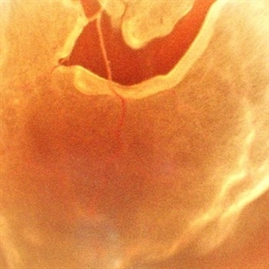
 Pneumatic Retinopexy
Jan 6 2025 by Mateus Queiroz Corrêa, MD
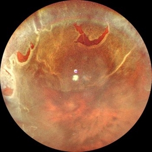
Fundus photograph of a pneumatic retinopexy. The upper photo taken just 30 minutes after C3F8 gas injection shows rhegmatogenous retinal detachment with superior temporal horseshoe tear and gas bubbles resembling fish-eggs. After two days with appropriated head position (botton photo), the retina is attached and laser photocoagulation was performed on the border of the break. A Single great gas bubble was formed.
Photographer: Mateus Queiroz Corrêa, Sorocada Eye Bank Hospital
Imaging device: Optos California
Condition/keywords: pneumatic retinopexy
-
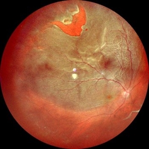
 Horseshoe Tear with Vitreous Hemorrhage
Sep 19 2024 by Virginia Gebhart
New horseshoe tear without detachment in 64 year old male. Vitreous hemorrhage secondary to HST. Prophylactic laser performed to seal tear
Photographer: Virginia Gebhart, Retina Consultants of Carolina
Imaging device: Optos California
Condition/keywords: retinal tear, vitreous hemorrhage
-
 Rhegmatogenous Retinal Detachment
Aug 22 2024 by STEFANY DAVILA
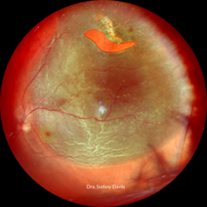
Fundus photograph of a 64-year-old male patient, HIV positive for 20 years, diagnosed with rhegmatogenous retinal detachment in the upper temporal sector from M7 to M1 in the left eye with a horseshoe tear in M10.
Photographer: Stefany Dávila, Instituto Mexicano de Oftalmología, Santiago de Querétaro
Imaging device: MIRANTE NIDEK
Condition/keywords: retina, Retinal Detachment, rhegmatogenous retinal detachment
-
 Twinkle Twinkle
Aug 5 2024 by Virginia Gebhart
65 year old male with mac-off retinal detachment with 360 folds and horseshoe tear.
Photographer: Virginia Gebhart
Imaging device: Optos California
Condition/keywords: macula off retinal detachment, RD, Retinal Detachment
-
 Lasered Horseshoe Tear
Jun 16 2024 by Anjana Mirajkar, MS Ophthalmology
An intra operative image showing a lasered horse shoe tear which is being indented for checking the tear being lasered 360 degree.
Photographer: Dr. Anjana Mirajkar -Retina Foundation, Ahmedabad
 Loading…