Search results (91 results)
-
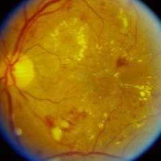
 New Choroidal Melanoma
Jul 16 2025 by Virginia Gebhart
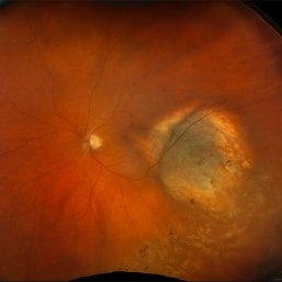
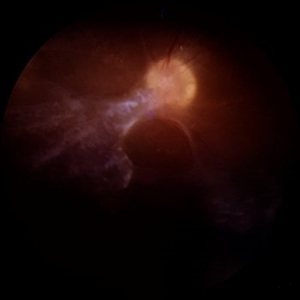
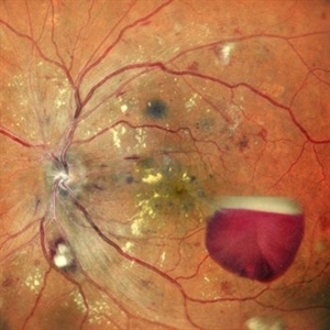
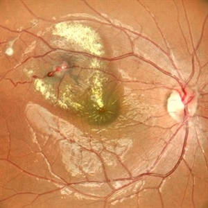
78 year old male with a partially amelanotic dome-shaped lesion with RPE changes, hard exudates, overlying intraretinal fluid and minimal SRF temporally. Exam and ultrasound findings consistent with choroidal melanoma. Pt will be scheduled for brachytherapy pending CT scan results.
Photographer: Virginia Gebhart, Retina Consultants of Carolina
Imaging device: Optos California
Condition/keywords: amelanotic melanoma, choroidal melanoma
-
 Central Retinal Vein Occlusion
Jun 21 2025 by Moazzam Parvez
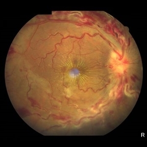
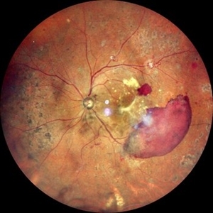
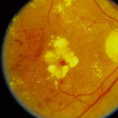
Fundus photograph of a 56 year old male presenting with dilated tortuous vessels with adjoining Hard exudates and macular star.
Photographer: Moazzam Parvez , Netralayam , Kolkata
Imaging device: Topcon Maestro 2
Condition/keywords: CRVO with macular edema, hard exudates, macular star
-
 Diabetic Retinopathy
Jun 4 2025 by Paulina Araujo
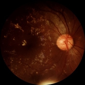
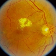
The 55-degree central fundus photograph of the right eye demonstrates numerous hard exudates, dot intraretinal hemorrhages, and microaneurysms.
Photographer: Paulina D.Araujo Martínez, Asociación para Evitar la Ceguera en México I.A.P., Hospital Dr Luis Sánchez Bulnes.
Condition/keywords: diabetic retinopathy
-
 Macular Edema
Jun 4 2025 by Paulina Araujo
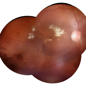
The composite fundus photograph of the right eye demonstrates circinate hard exudates in the thickened macular area, along with flame-shaped intraretinal hemorrhages along the inferior temporal arcade.
Photographer: Paulina D.Araujo Martínez, Asociación para Evitar la Ceguera en México I.A.P., Hospital Dr Luis Sánchez Bulnes.
Condition/keywords: macular edema
-
 Tractional Retinal Detachment
Jun 4 2025 by Paulina Araujo
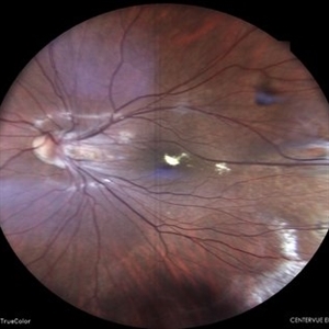
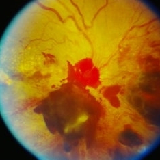
The 55-degree central fundus photograph of the right eye reveals a thickened and opacified hyaloid exerting traction on the optic disc and posterior pole of the retina, along with hard exudates and microaneurysms consistent with advanced proliferative diabetic retinopathy.
Photographer: Paulina D.Araujo Martínez, Asociación para Evitar la Ceguera en México I.A.P., Hospital Dr Luis Sánchez Bulnes.
Condition/keywords: tractional retinal detachment
-
 Coats Disease
May 27 2025 by César Adrián Gómez Valdivia, MD
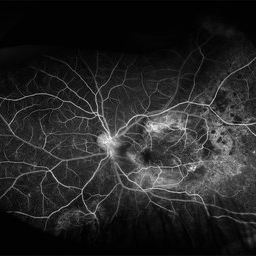
Fluorescein Angiography on an 8 year-old male patient with Coats disease. Vascular leakage causes hard exudates which may be peripheral (near the vascular abnormalities) or midperipheral and central (at the macula. Findings were bilateral.
Photographer: @eyemissu2
Imaging device: California ICG OPTOS
Condition/keywords: Coats disease
-
 Coats Disease
May 27 2025 by César Adrián Gómez Valdivia, MD
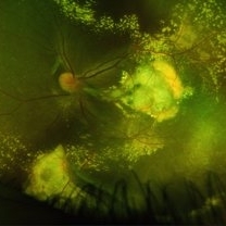
Fundus photograph of an 8 year-old male patient with Coats disease. Vascular leakage causes hard exudates which may be peripheral (near the vascular abnormalities) or midperipheral and central (at the macula). Findings were bilateral.
Photographer: @eyemissu2
Imaging device: California ICG OPTOS
Condition/keywords: Coats disease
-
 Circinate Mark at the Macula — a Lasting Trace of Branch Retinal Vein Occlusion
May 13 2025 by Malvika Singh
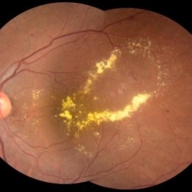
Fundus photograph of a 60 year old with a sclerosed vessel with hard exudates at the macula showing circinate retinopathy after an episode of branch retinal vein occlusion.
Photographer: Dr Malvika Singh, Retina Foundation, Ahmedabad, India
Imaging device: Mirante SLO/OCT
Condition/keywords: branch retinal vein occlusion (BRVO), circinate retinopathy
-
 PDR with NVD
Dec 5 2024 by Tejaswita Verma
Fundus image of a middle aged male with NVD, multiple dot blot and flame shaped hemorrhages, cotton wool spots, hard exudates at the posterior pole in a case of PDR . Vision was 6/9.
Photographer: DR. TEJASWITA VERMA
Imaging device: MIRANTE
Condition/keywords: NEOVASCULARISATION OF DISC, proliferative diabetic retinopathy (PDR)
-
 Severe Exudative Diabetic Retinopathy - Left Eye
Aug 19 2024 by Nishikant J Borse, MS, FMRF, FASRS
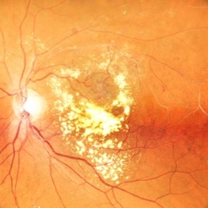
52-year-old diabetic lady presented with diminution of vision for 7 months. She had uncontrolled Diabetes mellitus with an HbA1C of 11.5. On examination she showed Severe Non-Proliferative Diabetic Retinopathy with exudates filling the macular area up to the arcades.
Photographer: Dr Nishikant Borse , Insight eye Clinic , Mumbai
Imaging device: Topcon Triton
Condition/keywords: Diabetic Retinopathy, foveal hard exudates
-
 Severe Exudative Diabetic Retinopathy - Right Eye
Aug 19 2024 by Nishikant J Borse, MS, FMRF, FASRS
52-year-old diabetic lady presented with diminution of vision for 7 months. She had uncontrolled Diabetes mellitus with an HbA1C of 11.5. On examination she showed Severe Non-Proliferative Diabetic Retinopathy with exudates filling the macular area up to the arcades.
Photographer: Dr Nishikant Borse , Insight eye Clinic , Mumbai
Imaging device: Topcon Triton
Condition/keywords: Diabetic Retinopathy, foveal hard exudates
-
 A Fleet of Boat-Shaped Hemorrhages
Aug 1 2024 by James P Dossett, MD
Pseudocolor fundus photograph of the left eye of a 54-year-old diabetic man presenting with bilateral vision loss. Examination revealed 20/200 vision OS with extensive preretinal and vitreous hemorrhage, marked diffuse neovascularization, macular edema and hard exudates.
Photographer: Beth Smith, West Virginia University Eye Institute
Condition/keywords: proliferative diabetic retinopathy (PDR)
-
 Proliferative Diabetic Retinopathy
May 24 2024 by Anjana Mirajkar, MS Ophthalmology
A central photo of a 50 year old male case of PDR showing a sub-hyaloid hemorrhage with cotton wool spots , hard exudates at the fovea with dot and blot hemorrhages.
Photographer: Dr. Anjana Mirajkar -Retina Foundation, Ahmedabad
Imaging device: Mirante-Nidek
Condition/keywords: proliferative diabetic retinopathy (PDR), Sub hyaloid haemorrhage
-
 Proliferative diabetic retinopathy
Apr 28 2024 by Anjana Mirajkar, MS Ophthalmology
A widefield color image of a 60 year old male with type II diabetes showing sub hyaloid hemorrhage with traction at the fovea with hard exudates with venous looping along the supero temporal arcade with NVE inferiorly with surrounding laser marks.
Photographer: Dr. Anjana Mirajkar -Retina Foundation, Ahmedabad
Imaging device: Mirante-Nidek
Condition/keywords: pan-retinal photocoagulation (PRP), proliferative diabetic retinopathy (PDR)
-
 Leaking Aneurysms in Diabetic Retinopathy
Mar 22 2024 by Vaidehi Sathaye
Fundus photograph of LE of a 50 year old female with leaking aneurysms encircled by hard exudates, as a sequelae of Diabetic Retinopathy.
Photographer: Dr. Vaidehi Sathaye
Imaging device: Topcon
Condition/keywords: aneurysm, diabetic retinopathy, hard exudates
-
 Familial Exudative Vitreo-Retinopathy
Jan 30 2024 by Akansha Sharma
Colour fundus photograph of a 19 year old male with both eyes familial exudative vitreo-retinopathy. Left eye shows aggregation of hard exudates over the fovea.
Photographer: Dr. Akansha Sharma, Bharati Eye Hospital
Condition/keywords: familial exudative vitreoretinopathy (FEVR), REGRESSED ROP
-
 Macroaneurysms
Jan 28 2024 by Anjana Mirajkar, MS Ophthalmology
Fundus image in a 20 year old female showing multiple macro aneurysms surrounded with exudation along the supero-temporal arcade and hard exudates at macula suggestive of macular edema.
Photographer: Dr. Anjana Mirajkar -Retina Foundation, Ahmedabad
Imaging device: Mirante-Nidek
Condition/keywords: macroaneurysm
-
 Impending STBRVO
Jan 7 2024 by MEENAL SONI
A middle aged female presented to the OPD with diminution of vision in right eye for past 7 days. Fundus examination findings depict supero-temporal AV crossing changes with macular hard exudates and oedema. On FFA we could clearly visualise the artery compressing the vein with leakage of dye in late phase extending into the macular region. On systemic evaluation the patient was found to be hypertensive with deranged lipid profile. She was advised injection anti VEGF for macular oedema and a physician consult for commencing the treatment for systemic condition. Despite a physician reference patient was not started on anti hypertensives and later presented with frank STBRVO with macular oedema after 3 months.
Photographer: Dr. Meenal Soni, VR fellow ASG eye hospital, Jodhpur (Raj)
Imaging device: Visucam
Condition/keywords: Impending BRVO with macular edema
-
 Von Hippel Lindau with retinal capillary hemangioma
Nov 2 2023 by Marcelo Zas, MD PhD
30-year-old female patient diagnosed with Syndrome VHL (Von Hippel Lindau). Stage II. In the first wide-field retinography of the right eye we can observe the exophytic retinal hemangiomas, rounded, slightly delimited, located in the peripheral retina in the upper and lower temporal quadrants and due to the exudation produced by them, hard exudates are observed in the star hemisphere, affecting the macula.
Photographer: Mariano Cotic MD
Imaging device: Silverstone SS OCT Optos
Condition/keywords: abnormal retinal vessel
-
 Diabetic Retinopathy
Sep 26 2023 by Ben Serar
Fundus photograph of RE showing clumps of hard exudates at the posterior pole, with dot-blot haemorrhages in a case of Diabetic Retinopathy.
Condition/keywords: diabetic retinopathy
-
 Proliferative Diabetic Retinopathy (PDR)
Sep 26 2023 by Ben Serar
Fundus photograph showing pre-retinal haemorrhages obscuring the disc, with surrounding hard exudates, in a case of
Condition/keywords: pre retinal haemorrhage, proliferative diabetic retinopathy (PDR)
-
 Hard exudates
Sep 21 2023 by Ben Serar
Fundus photograph of RE showing hard exudates at the macula, superior to the fovea.
Condition/keywords: hard exudates
-
 Non-proliferative Diabetic Retinopathy (NPDR)
Sep 14 2023 by Ben Serar
Fundus photograph of LE showing dot-blot and flame shaped haemorrhages at the macula, along with hard exudates, in a case of Non-proliferative Diabetic Retinopathy (NPDR).
Condition/keywords: Non-proliferative Diabetic Retinopathy (NPDR)
-
 Hypertensive Retinopathy
Sep 12 2023 by Ben Serar
Fundus photograph of LE showing Disc edema with optic disc pallor, hard exudates with dot-blot haemorrhages at the macula ,along with arteriolar attenuation, in a case of Hypertensive retinopathy.
Condition/keywords: arteriolar attenuation, disc edema, Hard exudates, hypertensive retinopathy
-
 Diabetic Retinopahty
Nov 2 2022 by pedro fernandes souza neto
Fundus photograph of a 40-year-old man with diabetes and hypertension shows hard exudates, difuse intraretinal hemorrhages and splinter hemorrhages.
Photographer: Pedro Fernandes, Universidade Federal da Bahia, Brazil.
Condition/keywords: diabetic mellitus, hypertensive retinopathy, retinopathy
 Loading…