Search results (179 results)
-
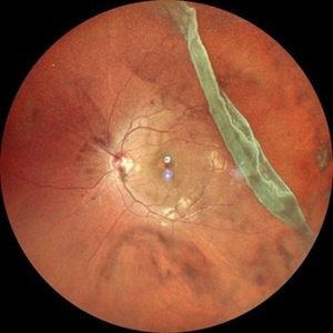
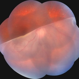
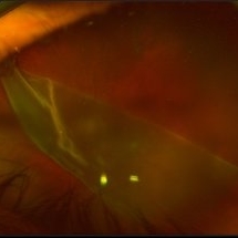
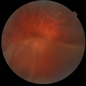
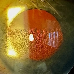 PFO Bubbles
Feb 25 2025 by Parnian Arjmand, MD, MSc, FRCSC, DABO
Post operative day 7 after repair of an RD secondary to a giant retinal tear with temporary PFO tamponade.
Condition/keywords: GRT, PFO
-
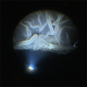
 Giant Retinal Tear with Detachment
Jan 3 2025 by Virginia Gebhart
67 year old male with mac-on RD with giant tear. Pt scheduled for sx
Photographer: Virginia Gebhart
Imaging device: Optos California
Condition/keywords: giant retinal tear, retinal tear with detachment
-
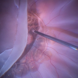
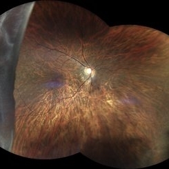
 Giant Retinal Tear with Bare Choroid
Dec 13 2024 by Thirumalesh Mochi Basavaraj, MD
Intra-operative view of a Pediatric Giant Retinal Tear with a view of the Bare Choroid Superiorly.
Photographer: Thirumalesh Mochi Basavaraj
Imaging device: Leica Proveo 8
Condition/keywords: GIANT RETINAL TEAR, Retinal Detachment
-
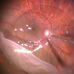
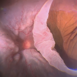
 Intraoperative View of a Giant Retinal Tear
Dec 13 2024 by Thirumalesh Mochi Basavaraj, MD
Intraoperative view of 12 year old child with Giant retinal tear with Retinal detachment.
Photographer: Thirumalesh Mochi Basavaraj
Imaging device: Lumera Proveo 8
Condition/keywords: GIANT RETINAL TEAR, PVR, Retinal Detachment
-
 Giant Tear
Oct 28 2024 by Andreas Paulo Di Luciano Rojas, MD
Giant retinal tear secondary to trauma.
Photographer: Andreas Di-Luciano, MD
Imaging device: Optos
Condition/keywords: giant retinal tear, ocular trauma, proliferative vitreoretinopathy (PVR), retinectomy, Trauma
-
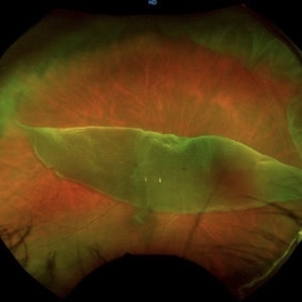
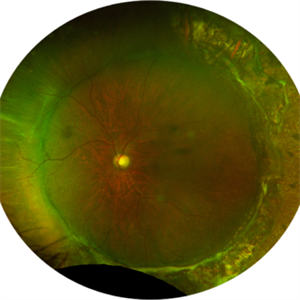
 Giant Retinal Tear
Oct 11 2024 by Anjana Mirajkar, MS Ophthalmology
Fundus photograph montage of LE showing a giant retinal extending from 12 to 4 o clock.
Photographer: Dr. Anjana Mirajkar -Retina Foundation, Ahmedabad
Imaging device: Mirante-Nidek
Condition/keywords: GIANT RETINAL TEAR
-
 Giant Retinal Tear
Oct 11 2024 by Anjana Mirajkar, MS Ophthalmology
Widefield fundus photograph of LE showing giant retinal tear extending from 12 to 4 o clock.
Photographer: Dr. Anjana Mirajkar -Retina Foundation, Ahmedabad
Imaging device: Mirante-Nidek
Condition/keywords: giant retinal tear
-
 Flattening Out the Retina With PFCL
Sep 28 2024 by Anjana Mirajkar, MS Ophthalmology
An intra operative image showing slow injection of PFCL to flatten out the folded retina.
Photographer: Dr. Anjana Mirajkar -Retina Foundation, Ahmedabad
Condition/keywords: GIANT RETINAL TEAR, PFCL
-
 Giant Retinal Tear
Sep 28 2024 by Anjana Mirajkar, MS Ophthalmology
An intra operative image showing slow injection of PFCL on the posterior pole to unfold the posterior margins in case of giant retinal tear.
Photographer: Dr. Anjana Mirajkar -Retina Foundation, Ahmedabad
Condition/keywords: GIANT RETINAL TEAR
-
 Giant Retinal Tear
Sep 28 2024 by Anjana Mirajkar, MS Ophthalmology
An intra-operative still showing giant retinal tear from 6 to 11 clock hour with folded posterior margins.
Photographer: Dr. Anjana Mirajkar -Retina Foundation, Ahmedabad
Condition/keywords: GIANT RETINAL TEAR
-
 Giant Retinal Tear
Sep 28 2024 by Anjana Mirajkar, MS Ophthalmology
An intra operative image of the right eye showing a giant retinal tear with a superior retinal detachment.
Photographer: Dr. Anjana Mirajkar -Retina Foundation, Ahmedabad
Condition/keywords: GIANT RETINAL TEAR
-
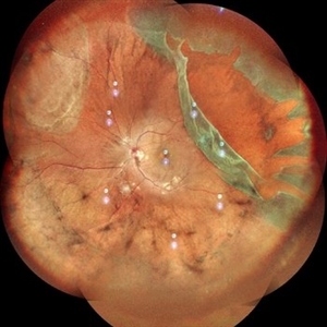
 Retinal Detachment with Giant Retinal Tear
Aug 9 2024 by Aditya S Kelkar, MS, FRCS, FASRS,FRCOphth
Fundus photograph of an 12-year-old boy with a Retinal detachment with Giant retinal tear of acute onset.
Photographer: Sakshi Naik, National Institute of Ophthalmology
Imaging device: Optos Daytona
Condition/keywords: giant retinal tear, pediatric retina, Retina detachment
-
 Giant Retinal Tear
Jul 15 2024 by Arthi Mohankumar , MS,MRCS ED, FICO,FAICO
Fundus montage of a 15 year old boy with Marfans syndrome who presented with defective vision in the right eye.
Photographer: Arthi Mohankumar
Condition/keywords: giant retinal tear, Retinal detachment
-
 Giant Retinal Tear
Jun 24 2024 by Akansha Sharma
Color fundus photograph of a 30 year old female with a giant retinal tear.
Photographer: Dr. Akansha Sharma, Bharati Eye Hospital
Condition/keywords: GIANT RETINAL TEAR, GRT
-
 Giant Retinal Tear With Retinal Fold
Jun 13 2024 by Anand Temkar
Intraoperative still of a 34 year old male showing giant retinal tear with retinal fold.
Photographer: Dr.Anand Temkar- Retina Foundation, Ahmedabad
Condition/keywords: giant retinal tear, GRT, retinal fold
-
 Giant Retinal Tear with Choroidal Detachment
Jun 12 2024 by Anand Temkar
Intra operative still of a 34 year old male showing Giant Retinal Tear with Choroidal Detachment.
Photographer: Dr.Anand Temkar- Retina Foundation, Ahmedabad
Condition/keywords: choroidal detachment, giant retinal tear
-
 Giant Retinal Tear
May 20 2024 by Aysha AlOqab, MB BCh BAO
Fundus photograph of a 40-year-old man who presented with a history of progressive inferior visual field defect in the right eye over 2-3 weeks.
Photographer: Saleh AlDhafiri, King Khaled Eye Specialist Hospital, Riyadh, KSA
Imaging device: Optos
Condition/keywords: giant retinal tear, idiopathic
-
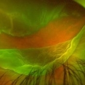
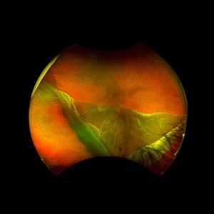
 Revealing the Invisible
Apr 25 2024 by Ricardo Leitão Guerra
In this remarkable wide-field autofluorescence image, we observe a patient with a giant retinal tear that has precipitated both retinal detachment and subsequent folding upon itself. Notably, the image distinctly maps the original anatomical configuration of the blood vessels within the affected retina.
Photographer: Ricardo Leitão Guerra, Leitão Guerra - Oftalmologia.
Imaging device: Clarus 700 - Zeiss Medical technology.
Condition/keywords: fundus autofluorescence (FAF), GIANT RETINAL TEAR
-
 Retinal Detachment with Giant Retinal Tear
Mar 26 2024 by Xitlali Caterina
Ultra-widefield fundus photograph of a 43-year-old male with a Retinal Detachment with Giant Retinal Tear affecting his left eye. Patient presented to the office with count fingers vision at 2 feet. He stated that about 8-9 days ago, he developed a clear curtain/veil and his vision started to get blurry. He also noted that he had floaters and flashes for about 8-9 days as well. The patient had cataract surgery a month prior to his visit. He stated that since his surgery, his vision had been better, but he had an area where he was not able to see well. The physician recommended a complex retinal detachment repair.
Photographer: Xitlali Caterina
Imaging device: OPTOS California RGB
Condition/keywords: fundus photograph, giant retinal tear, left eye, Optos, OPTOS CALIFORNIA, retinal detachment of the macula, retinal detachment with tear, ultra-wide field imaging, ultra-widefield image
-
 Giant Retinal Tear
Feb 20 2024 by Soobien Lee
Optos color fundus photograph of a 40-year-old caucasian male who is a UFC fighter with a total retinal detachment in his right eye secondary to a giant retinal tear from 10 o'clock to 2 o'clock.
Photographer: Trinity Wolf, Elman Retina Group
Imaging device: Optos Ultra-Widefield Imaging
Condition/keywords: giant retinal tear, optos, Retinal Detachment, Retinal tear with detachment, trauma
-
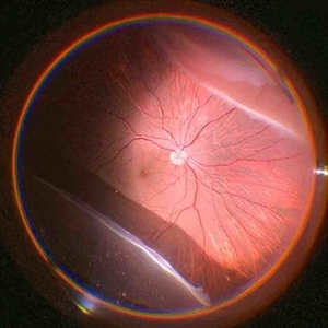
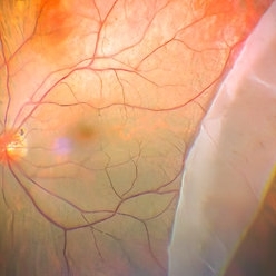
 Giant Retinal Tear
Oct 24 2023 by Ivan J. Suner, MD, MBA
Fundus photograph of 49-year-old man with a giant retinal tear in the right eye.
Photographer: Norelys Alexander Jimenez, Retina Associates of Florida, Tampa, FL
Imaging device: Optos California
Condition/keywords: GIANT RETINAL TEAR
-
 SUNSET THROUGH A VEIL
Oct 12 2023 by Deepti A Kulkarni, M.B.B.S., D.N.B., F.V.R.
HIGH MYOPE WITH A CORRECTION OF -24D. SUDDEN VISION LOSS FOLLOWING TRAUMA. SUPERIOR RETINA FOLDED ON TO THE INFERIOR RETINA MASKING THE DISC IN A TRANSLUCENT VEIL.
Photographer: DEEPTI KULKARNI, DR ANIL KULKARNI EYE HOSPITAL, MIRAJ, INDIA
Imaging device: TOPCON
Condition/keywords: GIANT RETINAL TEAR
-
 SUNSET THROUGH A VEIL
Oct 12 2023 by Deepti A Kulkarni, M.B.B.S., D.N.B., F.V.R.
HIGH MYOPE WITH A CORRECTION OF -24D. SUDDEN VISION LOSS FOLLOWING TRAUMA.
Photographer: DEEPTI KULKARNI, DR ANIL KULKARNI EYE HOSPITAL, MIRAJ, INDIA
Imaging device: TOPCON
Condition/keywords: GIANT RETINAL TEAR
-
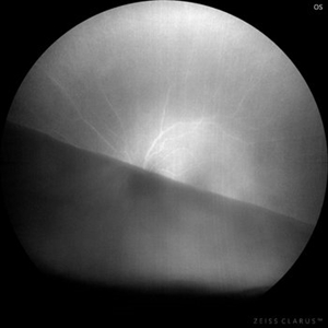
 Retinal Fold
Sep 26 2023 by Mauricio Bayram-Suverza, MD
A 38-year-old man underwent vitrectomy in the left eye due to a giant tear in the upper retina. SF6 gas was used as endotamponade. During the post-surgical check-up, it was identified that the patient developed a full-thickness retinal fold due to retinal slippage during fluid-air exchange. As the fold was away from the macular area, it was decided to observe the patient. Three weeks after the surgery, his best-corrected visual acuity was 20/30.
Photographer: Mauricio Bayram-Suverza, Fundación Hospital Nuestra Señora de la Luz
Imaging device: TRC-50DX
Condition/keywords: giant retinal tear, retina surgery complications, Retinal slippage, vitreoretinal surgery
-
 Giant Retinal Tear After Successful Re-Attachment
Jun 12 2023 by Ethan K Sobol, MD
Appearance of a giant retinal tear eight months after a successful scleral buckle with vitrectomy and silicone oil tamponade, followed by silicone oil removal. Central haziness in the image is due to the development of a cataract.
Condition/keywords: GRT, scleral buckle
 Loading…