Search results (2824 results)
-
 Filigree Networks
Nov 14 2025 by Malvika Singh
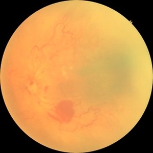
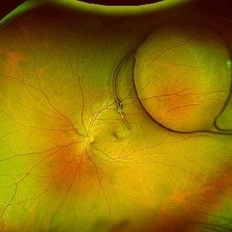
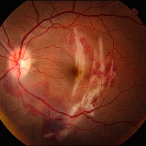
Fundus photograph of a 31 year old male with type 1 diabetes mellitus showing neovascularisation along the superotemporal arcade.
Photographer: Dr Malvika Singh, Retina Foundation, Ahmedabad, India
Imaging device: Mirante SLO/OCT
Condition/keywords: neovascularization (NV), proliferative diabetic retinopathy (PDR)
-
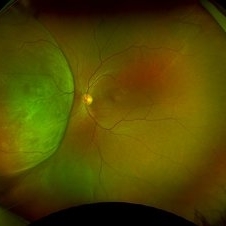
 Unilateral Pigmentary Retinopathy
Nov 9 2025 by Hrishikesh Naik, MS
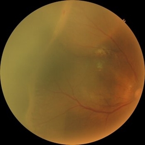
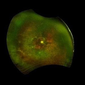
Montage fundus photographs of a 47 year old female presenting with unilateral vision loss in the left eye. Fundoscopy revealed extensive intraretinal pigment clumps, waxy disc pallor, and marked vessel attenuation in the left eye with a normal fundus in the right. Electroretinography showed unilateral reduction in rod and cone function. Unilateral pigmentary retinopathy, an uncommon variant of retinitis pigmentosa (reported incidence ˜ 5%) presents with RP-like changes in one eye, the fellow eye being completely normal. Proposed causes include lyonization and somatic mosaicism. Conditions which mimic RP should be excluded, and any diagnoses should be supported with electrodiagnostic tests and autofluorescence imaging. Management parallels RP, focusing on cataract and macular complications and long-term follow-up to monitor possible bilateral progression.
Imaging device: Zeiss Visucam 224
Condition/keywords: montage, retinitis pigmentosa, unilateral
-
 X-Linked Juvenile Retinoschisis
Nov 5 2025 by Kristen Wagner
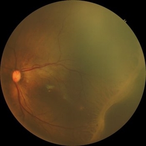
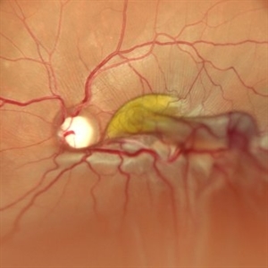
Fundus photograph of a 24 year old male patient who has X-Linked Retinoschisis (XLRS). Findings include Mild to moderate diffuse maculoschisis OD with vitreous veils. Discussed mutations with RS1 gene.
Photographer: Kristen Cross, COT Tennessee Retina
Imaging device: Optos
Condition/keywords: juvenile retinoschisis, maculoschisis, retinoschisis, virreous veils
-
 Multilayer Trauma
Nov 3 2025 by Malvika Singh
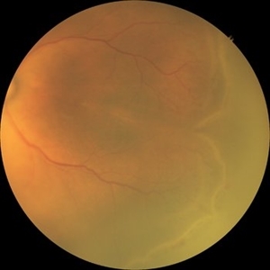
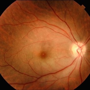
Fundus photograph of a 34 year old following trauma showing a choroidal rupture, a sub RPE and sub retinal bleed.
Photographer: Dr Malvika Singh, Retina Foundation, Ahmedabad, India
Imaging device: Mirante SLO/OCT
Condition/keywords: Choroidal Rupture, subretinal hemorrhage
-
 CRAO
CRAO
Oct 29 2025 by Jeffrey Barker
94 year old female with a CRAO with macular edema.
Photographer: Jeffrey P. Barker, B.S.
Condition/keywords: color fundus photograph, CRAO
-
 Retinopathy of Prematurity
Oct 27 2025 by Anjana Mirajkar, MS Ophthalmology
Fundus photograph of a premature baby showing flat neovascularization with looping of the vessels with bleed in zone 1/2 with plus disease suggestive of A-ROP.
Photographer: Dr. Anjana Mirajkar- HV desai eye hospital ,Pune
Imaging device: Retcam
Condition/keywords: aggressive posterior retinopathy of prematurity (APROP)
-
 Retinopathy of Prematurity
Oct 26 2025 by Anjana Mirajkar, MS Ophthalmology
Fundus photograph of right eye of premature baby showing stage 3 in zone 2 posterior.
Photographer: Dr. Anjana Mirajkar- HV desai eye hospital ,Pune
Imaging device: Retcam
Condition/keywords: retinopathy of prematurity (ROP), stage 3
-
 Retinopathy of Prematurity
Oct 26 2025 by Anjana Mirajkar, MS Ophthalmology
Fundus photograph of a left eye of a premature baby showing stage 3 in zone 2 posterior.
Photographer: Dr. Anjana Mirajkar- HV desai eye hospital ,Pune
Imaging device: Retcam
Condition/keywords: retinopathy of prematurity (ROP), retinopathy of prematurity stage 3
-
 Retinopathy of Prematurity
Oct 26 2025 by Anjana Mirajkar, MS Ophthalmology
Fundus photograph of left eye premature baby having stage 3 in zone 2A with a secondary notch.
Photographer: Dr. Anjana Mirajkar- HV Desai eye hospital ,Pune
Imaging device: retcam
Condition/keywords: retinopathy of prematurity (ROP), stage 3
-
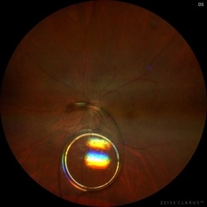
 Posterior Dislocated Intraocular Lens
Oct 23 2025 by Aditya S Kelkar, MS, FRCS, FASRS,FRCOphth
Fundus photograph of an 53-year-old man with a posteriorly dislocated intraocular lens near the posterior pole.
Photographer: Dr Tejal Rao, National Institute of Ophthalmology, Pune, India
Imaging device: Optos Daytona
Condition/keywords: dislocated intraocular lens (IOL), IOL drop
-
 Radiation Retinopathy
Oct 20 2025 by Meng-Hsin Chen
Fundus photograph of a 54-year-old woman with radiation retinopathy due to radiation exposure in-utero. Patient also presents with no light perception in the eye.
Photographer: Meng-Hsin Chen
Condition/keywords: No light perception, radiation retinopathy
-
 Retinal Hypotonia
Oct 15 2025 by Oftalmontt Clínica Láser
Color fundus photograph of a 57-year-old man with streaks on the retinal tapestry suggestive of retinal hypotonia.
Photographer: Ophthalmic Medical Technologist
Imaging device: Canon cx-1
Condition/keywords: retinal hypotonia, Retinography
-
 Folded Macula
Oct 13 2025 by Malvika Singh
Fundus photograph of a 25 year old male showing retinal detachment with macula off with retinal folds at the macula.
Photographer: Dr Malvika Singh, Retina Foundation, Ahmedabad, India
Imaging device: Mirante SLO/OCT
Condition/keywords: retinal detachment
-
 Retinal Arteriovenous Malformation
Oct 7 2025 by Korey Starkey
55 year-old patient presents with retinal arteriovenous malformation in the left eye and BRVO w/retinal neovascularization. Patient is asymptomatic. No edema or treatment necessary today, signs of old RVO with MAs along inferior arcade and dot heme.
Photographer: Korey Starkey
Imaging device: Topcon
Condition/keywords: branch retinal vein occlusion (BRVO), fundus photography, inferior arcade, microaneurysms, retinal arteriovenous malformations, retinal neovascularization, Topcon
-
 Chorioretinal Coloboma
Oct 6 2025 by Seif Allah Anwar
Fundus photograph of the patient left eye showing large, well-demarcated, excavated chorioretinal coloboma involving the inferior fundus, extending from the optic disc to the periphery. The lesion appears white due to bare sclera visibility, with absence of overlying choroid and retina. Retinal vessels course over the colobomatous area inferiorly.
Photographer: Dr. Seif Anwar, FRCSEd
Imaging device: Centervue Eidon
Condition/keywords: chorioretinal coloboma
-
 Retinal Detachment Associated with Proliferative Vitreoretinopathy
Oct 1 2025 by Kingston Rodolfo Ureña-Wong, MD, Opht, MSc
Fundus photograph of an 53-year-old woman with a Retinal detachment asociated with Proliferative Vitreoretinopathy after a silicon removal surgery 1 week prior.
Photographer: Ureña-Wong Kingston Rodolfo, APEC
Condition/keywords: Proliferative Vitreoretinopathy, Retinal Detachment
-
 Central Retinal Artery Occlusion
Sep 30 2025 by César Adrián Gomez Valdivia, MD
This fundus photograph demonstrates the classic retinal whitening due to inner retinal ischemia, with a cherry-red spot at the fovea. The fovea appears red because it is nourished by the intact choroidal circulation, while the surrounding ischemic retina turns pale.
Photographer: @eyemissu2
Imaging device: TOPCON TRX
Condition/keywords: central retinal artery occlusion (CRAO)
-
 Choroidal Rupture
Sep 30 2025 by César Adrián Gomez Valdivia, MD
This fundus photograph shows curvilinear streaks of choroidal rupture radiating from the fovea, associated with subretinal hemorrhage. The rupture lines appear as crescent-shaped, whitish streaks representing a break in Bruch’s membrane, choriocapillaris, and retinal pigment epithelium.
Photographer: @eyemissu2
Imaging device: TOPCON TRX
Condition/keywords: Choroidal, Rupture
-
 Strained Retina
Sep 27 2025 by Malvika Singh
Fundus photograph of a 44 year old male showing hemorrhages at different layers.
Photographer: Dr Malvika Singh, Retina Foundation, Ahmedabad, India
Imaging device: Mirante SLO/OCT
Condition/keywords: valsalva retinopathy
-
 Strained Retina
Sep 27 2025 by Malvika Singh
Fundus photograph of a 44 year old male showing hemorrhages at different layers.
Photographer: Dr Malvika Singh, Retina Foundation, Ahmedabad, India
Imaging device: Mirante SLO/OCT
Condition/keywords: valsalva retinopathy
-
 Giant Retinal Cyst
Sep 20 2025 by JORGE SOBERANES
Fundus photograph of a 45-year-old-man with a large cyst on the nasal superior side of the retina. The patient had a history of a pneumatic retinopexy two years ago and the cyst has been there since that.
Photographer: Dr. Jorge Soberanes, Asociación para Evitar la Ceguera en México (APEC), UNAM
Condition/keywords: abnormal retina, pneumatic retinopexy, retinal cyst
-
 Dislocated IOL
Sep 20 2025 by JORGE SOBERANES
Fundus photograph of a 65-year-old man with a history of cataract surgery one year ago and bad vision since that.
Photographer: Dr. Jorge Soberanes, APEC, Universidad Nacional Autónoma México
Condition/keywords: dislocated lens, intraocular lens dislocation
-
 Amelanotic Melanoma
Sep 19 2025 by Aditya S Kelkar, MS, FRCS, FASRS,FRCOphth
Widefield fundus photograph of a 37 year old showing a large, dome-shaped, intraocular mass involving the temporal retina. The lesion appears elevated and lacks surface pigmentation. Overlying retinal vessels are displaced and draped across the tumor surface, with surrounding retinal elevation noted. The appearance is suggestive of amelanotic variant of choroidal melanoma.
Photographer: Dr. Muskan Mangal
Imaging device: Optos Daytona
Condition/keywords: choroidal melanoma, intraocular tumor
-
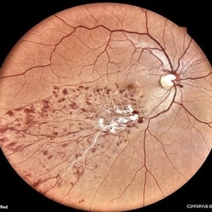 Lower Temporal Branch Retinal Vein Occlusion
Sep 16 2025 by Seif Allah Anwar
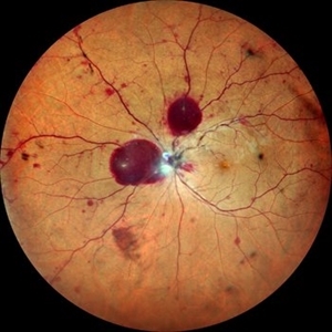
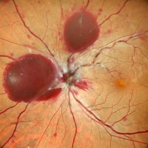
Fundus photograph of a 46-year old hypertensive male patient showing sheathed lower temporal retinal vein with whitish cotton wool spots and hemorrhages ( dots, blots and flame shaped) along the area drained by the obstructed vein with vein.
Photographer: Dr Seif Anwar. FRCSEd
Imaging device: CENTERVUE EIDON
Condition/keywords: Lower temporal branch retinal vein occlusion
-
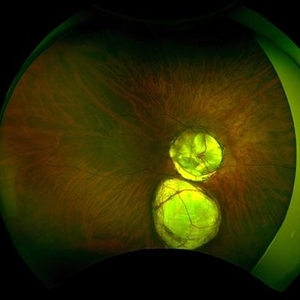 Double Bubble Coloboma Appearance
Sep 14 2025 by SHADAB HASAN
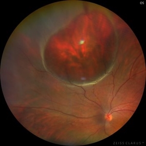
This ultra-widefield fundus photograph shows a large, well-demarcated, whitish excavated involving the inferior fundus with two distinct lobes, giving a “double-bubble” appearance. These features are characteristic of an inferior choroidal coloboma.
Photographer: SHADAB HASAN
Imaging device: OPTOS
Condition/keywords: Coloboma
 Loading…