Search results (60 results)
-
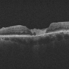
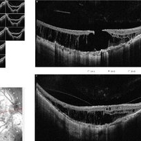
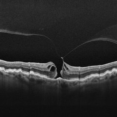 Stage 2 Macular Hole From VMT
Mar 21 2025 by Drew Mitchell
HD 1 line 100x OCT showcasing a full thickness macular hole caused by vitreomacular traction on fovea. Choroidal folds can also be seen on scan.
Photographer: Drew Mitchell OCT-C
Imaging device: Zeiss Cirrus 6000
Condition/keywords: Choroidal Folds, FTMH, macular hole, OCT, PVD
-
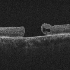
 Myopic Traction Maculopathy
Mar 17 2025 by Drew Mitchell
HD 1 line 100x 9 mm scan of a right eye with MTM at stage 3c. Macular Schisis Detachment.
Photographer: Drew Mitchell OCT-C
Imaging device: Zeiss Cirrus 5000
Condition/keywords: full thickness macular hole, Macular hole, myopic foveoschisis, myopic macular schisis, myopic traction maculopathy, PVD
-
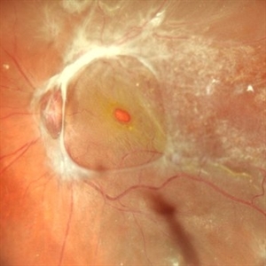
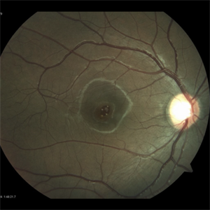
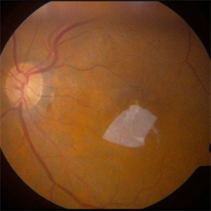
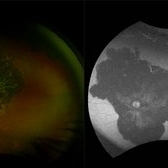
 Polyploidal Choroidal Vasculopathy
Dec 27 2024 by Tejaswita Verma
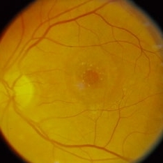
Fundus image of a 74 year old woman with CF1mt vision in right eye showing large PED in a case of PCV. There was associated full thickness macular hole in the same eye.
Photographer: DR. TEJASWITA VERMA
Imaging device: MIRANTE
Condition/keywords: PED, polypoidal choroidal vasculopathy (PCV)
-
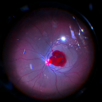
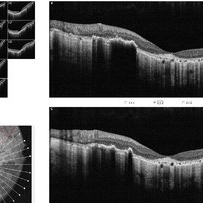
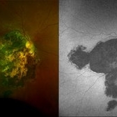
 Combined Retinal Detachment With Macular Hole
Sep 28 2024 by Tejaswita Verma
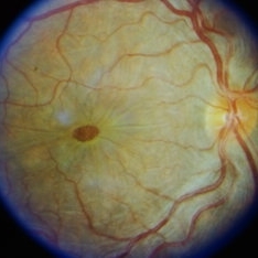
Fundus image of the LE of a 67 year old diabetic, hypertensive female with CF 3metres vision showing combined RD with FTMH, in a pseudophakic eye. She was lost to follow up status post 2 anti VEGF injections received 8 months back due to typhoid fever.
Photographer: DR. TEJASWITA VERMA
Imaging device: MIRANTE
Condition/keywords: full thickness macular hole, proliferative diabetic retinopathy (PDR), tractional retinal detachment
-
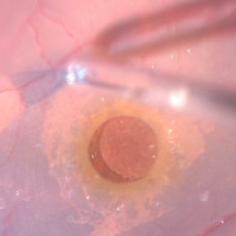 ILM Peeling in a Case of Large Macular Hole
Sep 28 2024 by Anjana Mirajkar, MS Ophthalmology
An intra operative still showing stained ILM peeling done with forceps in a case of large macular hole.
Photographer: Dr. Anjana Mirajkar -Retina Foundation, Ahmedabad
Condition/keywords: full thickness macular hole, internal limiting membrane (ILM) peeling
-
 Full Thickness Macular Hole
Sep 23 2024 by NOEMI JOSEFINA Dr CHACCA, Fellow
Right eye of a 63 year man patient came with blurring of vision of right eye since 5 years. Vision was 6/24, minimum diameter 336 µm
Photographer: Dra. Noemí Josefina Chacca Magaño,Instituto Mexicano de Oftalmología, Querétaro, México
Imaging device: Optopol REVO NX
Condition/keywords: OCTA
-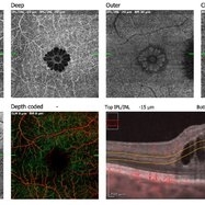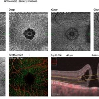
 Full Thickness Macular Hole
Sep 23 2024 by NOEMI JOSEFINA Dr CHACCA, Fellow
Right eye of a 63 year man patient came with blurring of vision of right eye since 5 years. Vision was 6/24, minimum diameter 336 µm
Photographer: Dra. Noemí Josefina Chacca Magaño,Instituto Mexicano de Oftalmología, Querétaro, México
Imaging device: OPTOPOL REVO NX
Condition/keywords: OCTA
-
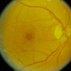
 Thickness Macular Hole
Apr 10 2024 by Tareq Alsulami
Fundus photography of an 14 years old man with Full Thickness Macular Hole after direct exposure of the blue laser to his eye
Photographer: Tareq alsulami ophthalmic technician-king Abdullah Medical City-Saudi Arabia
Imaging device: ZEISS
Condition/keywords: holes, macular
-
 Full Thickness Macular Hole OCT
Apr 10 2024 by Tareq Alsulami
OCT of an 14 years old man with Full Thickness Macular Hole after direct exposure of the blue laser to his eye
Photographer: Tareq alsulami ophthalmic technician-king Abdullah Medical City-Saudi Arabia
Imaging device: Heidelberg
Condition/keywords: Macular hole
-
 The Rose Of The Eye
Mar 14 2024 by SANDEEP KUMAR
Intraoperative capture of a failed full thickness macular hole being treated with human amniotic membrane graft and autologous blood under air.
Photographer: Sandeep Kumar , Shroff eye centre kailash colony New Delhi India
Imaging device: Callisto imaging system
Condition/keywords: autologous blood, full thickness macula hole, human amniotic graft
-
 Amniotic-Membrane Grafted Macular Hole
Oct 25 2023 by Jessica Hampton, BS
Optical-coherence tomography image of a 67-year old woman with a recurrent, chronic full-thickness macular hole in the left eye repaired with an amniotic membrane graft, seen at 2 years follow up.
Photographer: Dr. Diana Do, Stanford Medicine, Byers Eye Institute
Condition/keywords: amniotic membrane graft, full thickness macular hole
-
 Chronic Full Thickness Macular Hole
Oct 25 2023 by Jessica Hampton, BS
Optical-coherence tomography image of a 65-year old woman with a chronic full-thickness macular hole in the left eye, recurred following three attempts at repair with pars plana vitrectomy, membrane peel, and gas tamponade.
Photographer: Dr. Diana Do, Stanford Medicine, Byers Eye Institute
Condition/keywords: full thickness macular hole, optical coherence tomography (OCT)
-
 Full Thickness Macular Hole
Oct 25 2023 by Jessica Hampton, BS
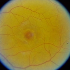
Fundus photograph of a 67-year-old woman with a history of recurrent, chronic full-thickness macular hole in the left eye repaired with an amniotic membrane graft, seen at 2 years follow-up.
Photographer: Dr. Diana Do, Stanford Medicine, Byers Eye Institute
Condition/keywords: amniotic membrane graft, full thickness macular hole, fundus photograph
-
 Full Thickness Macular Hole
Oct 25 2023 by Jessica Hampton, BS
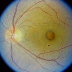
Fundus photograph of a 65-year-old woman with chronic, recurrent full-thickness macular hole repaired with an amniotic membrane graft.
Photographer: Diana Do M.D., Stanford Medicine, Byers Eye Institute
Condition/keywords: amniotic membrane graft, full thickness macular hole
-
 OCT IMAGE - Healed chorioretinitis
Sep 29 2023 by PUSHPANJALI BADOLE
36 yr old female with recent decline in right eye vision, with left eye having poor vision since childhood. Fundus examination revealed BE healed chororetinitis with RE OCT s/o maculoschisis with full thickness macular hole.
Photographer: NITIN DESLE, ISHA NETRALAYA, KALYAN
Imaging device: DAYTONA OPTOS
Condition/keywords: choroiditis
-
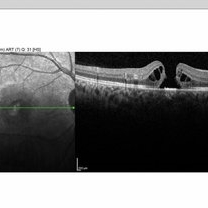
 MACULOSCHISIS WITH FULL THICKNESS MACULAR HOLE
Sep 29 2023 by PUSHPANJALI BADOLE
36 yr old female with recent decline in right eye vision, with left eye having poor vision since childhood. Fundus examination revealed BE healed chororetinitis with RE OCT s/o maculoschisis with full thickness macular hole.
Photographer: NITIN DESLE, ISHA NETRALAYA, KALYAN
Imaging device: DAYTONA OPTOS
Condition/keywords: choroiditis
-
 Choroiditis
Sep 29 2023 by PUSHPANJALI BADOLE
36 yr old female with recent decline in right eye vision, with left eye having poor vision since childhood. Fundus examination revealed BE healed chororetinitis with RE OCT s/o maculoschisis with full thickness macular hole.
Photographer: NITIN DESLE, ISHA NETRALAYA, KALYAN
Condition/keywords: chorioretinitis
-
 HEALED CHOROIDITIS
Sep 29 2023 by PUSHPANJALI BADOLE
36 yr old female with recent decline in right eye vision, with left eye having poor vision since childhood. Fundus examination revealed BE healed chororetinitis with RE OCT s/o maculoschisis with full thickness macular hole.
Photographer: NITIN DESLE, ISHA NETRALAYA, KALYAN
Condition/keywords: healed choroiditis
-
 Macular hole
Sep 26 2023 by Ben Serar
Fundus photograph of RE showing Full thickness Macular Hole (FTMH).
Condition/keywords: macular hole
-
 Macular hole
Sep 21 2023 by Ben Serar
Fundus Photograph of LE showing full thickness macular hole.
Condition/keywords: macular hole
-
 Macular hole
Sep 21 2023 by Ben Serar
Fundus Photograph of LE showing full thickness macular hole.
Condition/keywords: macular hole
-
 Macular hole
Sep 21 2023 by Ben Serar
Fundus Photograph of LE showing full thickness macular hole.
Condition/keywords: macular hole
-
 Macular hole
Sep 14 2023 by Ben Serar
Fundus photograph of the LE showing full thickness macular hole.
Condition/keywords: macular hole
-
 Macular Hole
Sep 12 2023 by Ben Serar
Fundus photograph of RE showing Full thickness Macular Hole (FTMH).
Condition/keywords: Full thickness Macular Hole (FTMH)
 Loading…