Search results (195 results)
-
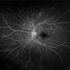
 BRVO-MCR-FFA
Jun 27 2025 by Gayathri M S
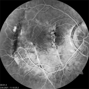
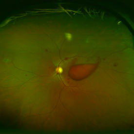
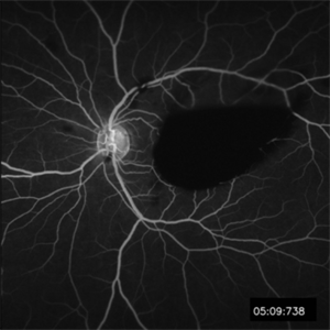
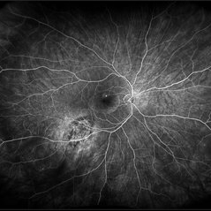
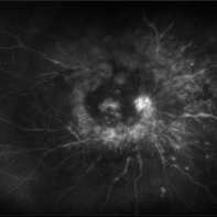
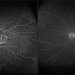
Case of impending Macular BRVO. 52 year old female on medication for Hypertension and Diabetes Mellitus since 2 years. BCVA 6/18,N6. IOP 16 mmHg. Multicolor Reflectance and Fundus Fluorescein Angiography picture shows mild dilated tortuous inferior vessels, small areas of capillary non perfusion and few microanurysms.
Photographer: Gayathri MS
Imaging device: Heidelberg spectralis
Condition/keywords: fluorescein angiogram (FA), macular branch retinal vein occlusion (BRVO), multicolor
-
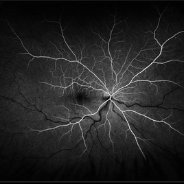
 Fluorescein Angiography in Choroidal Rupture
Jun 26 2025 by Hector Gabriel Moreno Solano, MD, MHA
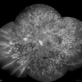
Fluorescein angiography of the right eye reveals three linear hypofluorescent lesions with progressive staining at the edges, consistent with choroidal ruptures. These lesions are temporally located in the posterior pole, with one of them situated near the fovea but without direct foveal involvement. The pattern is suggestive of previous blunt ocular trauma.
Photographer: Héctor Gabriel Moreno Solano, Instituto Mexicano de Oftalmología “IMO I.A.P”
Imaging device: CLARUS
Condition/keywords: Choroidal Rupture, fluorescein angiogram (FA)
-
 Post-traumatic Choroidal Rupture-Fluorescein Angiography
Jun 20 2025 by Alexander Babaev
Fluorescein angiography of an 46-year-man with a choroidal rupture after blunt trauma, complicated CNV. 00.31s, Dye leakage is visible along the edges of the rupture
Photographer: Babaev Alexander, Saint-Petersburg, medical clinic "Vision"
Imaging device: Carl Zeiss Visucam 500
Condition/keywords: fluorescein angiogram (FA)
-
 Ocular Ischemic Syndrome
Jun 18 2025 by Korey Starkey
58-year-old patient with OIS and Hollenhorst plaque.
Photographer: Korey Starkey
Imaging device: Optos
Condition/keywords: capillary nonperfusion, fluorescein angiogram (FA), hollenhorst plaque, NVD, ocular ischemic syndrome, Optos
-
 Ocular Ischemic Syndrome
Jun 18 2025 by Korey Starkey
58-year-old patient with OIS in both eyes. Patient has had PRP in the past, however, presence of NVD with peripheral nonperfusion remains despite PRP.
Photographer: Korey Starkey
Imaging device: Optos
Condition/keywords: DME, FA early phase, fluorescein angiogram (FA), NVD, ocular ischemic syndrome, ois, Optos, peripheral retinal nonperfusion
-
 VKH Syndrome
Jun 12 2025 by Virginia Gebhart
Fluorescein angiogram of 22 year old male with VKH syndrome. Significant cell in AC and vitreous, multiple punched-out CR scars in periphery, mild vascular leakage. Pt referred to rheumatology for immunomodulatory treatment.
Photographer: Virginia Gebhart, Retina Consultants of Carolina
Imaging device: Optos California
Condition/keywords: FA, fluorescein angiogram (FA), multifocal choroiditis, panuveitis, VKH, Vogt-Koyanagi-Harada
-
 Stargardt Disease (FA)
Jan 22 2025 by Virginia Gebhart
Fluorescein angiogram of 19 year old female with confirmed Stargardt Disease. Hyperfluorescence in the macula with staining defect and silent choroid.
Photographer: Virginia Gebhart, Retina Consultants of Carolina
Imaging device: Optos California
Condition/keywords: fluorescein angiogram (FA), Silent Choroid, Stargardt disease
-
 Choroidal Melanoma 3 Ways
Jan 16 2025 by Virginia Gebhart
RGB/FA/ICG of 76 year old female with a new choroidal melanoma. Pt scheduled for plaque radiation. BCVA 20/400
Photographer: Virginia Gebhart, Retina Consultants of Carolina
Imaging device: Optos California
Condition/keywords: fluorescein angiogram (FA), indocyanine green (ICG) angiography, OPTOS CALIFORNIA RGB
-
 MEWDS
MEWDS
Dec 11 2024 by Virginia Gebhart
28 year old female with new Multiple Evanescent White Dot Syndrome. Patient reports gray spot in vision, OCT shows RPE disruption centrally but no edema. FA shows early hyperfluorescent punctate spots throughout the posterior pole, but no leakage. Normal findings OD. Will observe for now
Photographer: Virginia Gebhart, Retina Consultants of Carolina
Imaging device: Optos California
Condition/keywords: FA, fluorescein angiogram (FA), multiple evanescent white dot syndrome (MEWDS)
-
 Hemiretinal Vein Occlusion
Nov 14 2024 by Brandon I Fram, MD, BS
40 year-old male with vision changes and observed hemiretinal vein occlusion.
Condition/keywords: branch retinal vein occlusion (BRVO), fluorescein angiogram (FA), Fluorescein angiography, hemi CRVO, hemicentral retinal vein occlusion
-
 Early FA/ICG at 1 Minute of Atypical ANCA Associated Retinal Vasculitis
Nov 13 2024 by Deepak Sambhara, MD
Fluorescein and Indocyanine Green Angiography of a 49-year-old male with high ANA titer, atypical ANCA positivity, who presented to clinic with 1 month of vision loss. Exam revealed anterior chamber cell, mild vitreous cell, sclerotic vessels along arterioles. Early FA/ICG at 1 minute demonstrates absent arteriole fill.
Photographer: Killian Roberts, Micaela Hertz; Eye Clinic of Wisconsin
Imaging device: Heidelberg Spectralis
Condition/keywords: A-ANCA, autoimmune vasculitis, fluorescein angiogram (FA), indocyanine green (ICG) angiography, retinal vasculitis
-
 Late FA/ICG at 4 Minutes of Atypical ANCA Associated Retinal Vasculitis
Nov 13 2024 by Deepak Sambhara, MD
Fluorescein and Indocyanine Green Angiography of a 49-year-old male with high ANA titer, atypical ANCA positivity, who presented to clinic with 1 month of vision loss. Exam revealed anterior chamber cell, mild vitreous cell, sclerotic vessels along arterioles. Late FA/ICG at 4 minutes demonstrates absent arteriole fill with venular periphlebitis.
Photographer: Killian Roberts, Micaela Hertz; Eye Clinic of Wisconsin
Imaging device: Heidelberg Spectralis
Condition/keywords: A-ANCA, autoimmune vasculitis, fluorescein angiogram (FA), indocyanine green (ICG) angiography, retinal vasculitis
-
 Best Disease
Nov 7 2024 by Virginia Gebhart
Fluorescein angiogram of 49 year female with Best Disease. Genetic testing done in 2000 confirms Best Disease and also possible Stargardts mutation. Characteristic bullseye maculopathy with surrounding yellowish flecks are present in both eyes.
Photographer: Virginia Gebhart, Retina Consultants of Carolina
Imaging device: Optos California
Condition/keywords: Best disease, fluorescein angiogram (FA)
-
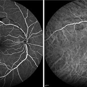
 Valsalva Retinopathy
Oct 28 2024 by Andrew Jin, MD
These photos depict the fundus photo and corresponding fluorescein angiogram for a 43 year old man with emesis after food poisoning. Note the blockage from the central preretinal hemorrhage and scattered peripheral intraretinal hemorrhages.
Condition/keywords: fluorescein angiogram (FA), fundus photograph, valsalva retinopathy
-
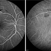
 Valsalva Retinopathy
Oct 28 2024 by Andrew Jin, MD
These photos depict the fundus photo and corresponding fluorescein angiogram for a 43 year old man with emesis after food poisoning. Note the blockage from the central preretinal hemorrhage and scattered peripheral intraretinal hemorrhages.
Condition/keywords: fluorescein angiogram (FA), fundus photograph, valsalva retinopathy
-
 Proliferative Diabetic Retinopathy
Oct 13 2024 by Brandon I Fram, MD, BS
28 year-old with florid neovascularization of the disc and extensive nonperfusion imaged with fluorescein angiography
Condition/keywords: florid type PDR, fluorescein angiogram (FA), neovascularization of the disc (NVD), PDR, proliferative diabetic retinopathy (PDR)
-
 Branch Retinal Vein Occlusion
Aug 22 2024 by Virginia Gebhart
Fluorescein angiogram of branch retinal vein occlusion in 75 year old female. Scattered microaneurysms with late CME and persistent SRF. Pt will consider laser treatment but is hesitant for injections at this time due to possible side effects.
Photographer: Virginia Gebhart
Imaging device: Optos California
Condition/keywords: branch retinal vein occlusion (BRVO), BRVO, cystoid macular edema (CME), FA, FA late phase, fluorescein angiogram (FA), macular edema, microaneurysms, retinal microaneurysms
-
 Suspicious Nevus / CSR
Aug 8 2024 by Virginia Gebhart
Fluorescein angiogram of 54 year old male with a suspicious appearing choroidal nevus and central serous retinopathy. Will monitor closely and follow up with serial exams.
Photographer: Virginia Gebhart
Imaging device: Optos California
Condition/keywords: central serous retinopathy (CSR), choroidal nevus, fluorescein angiogram (FA), FLUORESCEIN ANGIOGRAPHY, nevus
-
 Focal Chorioretinitis
Jul 11 2024 by Virginia Gebhart
67 year old female with punched-out CR scars. Hx of laser 3x for apparent peripapillary CNV. ESR, CRP, toxo, IgG/IgM all "normal." Bartonella, quant gold, and FTA-ABS ordered given possibility of neuroretinitis. Vision CF
Photographer: Virginia Gebhart
Imaging device: Optos California
Condition/keywords: FA, fluorescein angiogram (FA), FLUORESCEIN ANGIOGRAPHY, focal chorioretinitis, optic neuritis
-
 Posterior Uveitis with Macular Edema
Jul 9 2024 by Korey Starkey
Ultra-wide field angiography of a 70 year old female with cystoid macular edema secondary to posterior uveitis. Patient's vision was Dcc20/200 at time of visit.
Photographer: Korey Starkey
Imaging device: Optos
Condition/keywords: cystoid macular edema (CME), fluorescein angiogram (FA), FLUORESCEIN ANGIOGRAPHY, hyperfluorescence, posterior uveitis, ULTRA WIDE FIELD, ultra-widefield image, vitreous debris
-
 Posterior Uveitis
Jul 5 2024 by Zach Seim
Fluorescein Angiogram of a 73 year old Male with Posterior Uveitis OS. Patient presented with VA of DCC 20/80-1 OS at this visit.
Photographer: Zach Seim
Imaging device: Optos California
Condition/keywords: fa, fluorescein angiogram (FA), Optos, OPTOS CALIFORNIA, posterior uveitis
-
 Disseminated Retinitis and Retinochoroiditis, Pigment Epitheliopathy OS
Jul 5 2024 by Zach Seim
Optos Fluorescein Angiogram of a 94 year old female with Disseminated Retinitis and Retinochoroiditis, Pigment Epitheliopathy OS.
Photographer: Zach Seim
Imaging device: Optos California
Condition/keywords: fluorescein angiogram (FA), left eye, OPTOS CALIFORNIA, retinitis, retinochoroiditis
-
 Fluorescein Angiography Montage
Jun 21 2024 by BENITO VERGARA, MD
Montage of an angiography with fluorescein from the left eye of a 32 year-old male with diabetic retinopathy previously treated with panretinal photocoagulation, that shows leakage at optic nerve and upper nasal arcade.
Photographer: Benito Vergara, Asociación Para Evitar la Ceguera en México.
Imaging device: Zeiss Clarus 700
Condition/keywords: Angiography Montage, angiography with fluorescein, diabetic retinopathy, FA montage, fluorescein angiogram (FA), peripheral scars
-
 Retinal Arterial Anastamosis with Foveal Aneurysm - FA
Feb 22 2024 by Charles L. Tucker, MD, FACS, FASRS
Fluorescein angiogram of an anomalous arterial-arterial anastomosis with an aneurysm in the fovea.
Photographer: Isabel Anderson, Retina Institute of the Carolinas, Gastonia, NC
Condition/keywords: aneurysm, arterial anastamosis, fluorescein angiogram (FA), Perifoveal Exudative Vascular Anomalous Complex, PEVAC
-
 Venous Loop
Feb 20 2024 by Soobien Lee
A 77-year-old male with a history of bilateral optic neuropathy from bilateral optic nerve sheath meningiomas S/P radiation/proton-beam therapies. Presented with radiation retinopathy OS and a known venous loop OS.
Photographer: Gavin Bragdon, Elman Retina Group
Imaging device: Optos Ultra-Widefield Fluorescein Angiography
Condition/keywords: fluorescein angiogram (FA), Optos, radiation retinopathy, retinal vascular disease, venous loop
 Loading…