Search results (197 results)
-
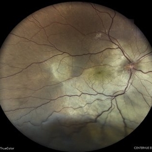
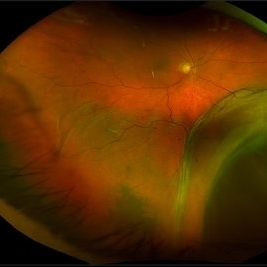
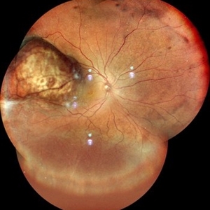
 Uveal Effusion Syndrome
Jun 13 2025 by Brandon I Fram, MD, BS
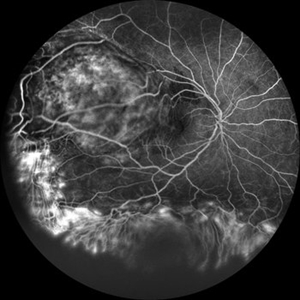
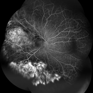
75 year-old with bilateral inferior serous detachments, right more than left. Scleral window with biopsy showed scleral thickening with stromal deposits of amorphous glycosaminoglycan-like material.
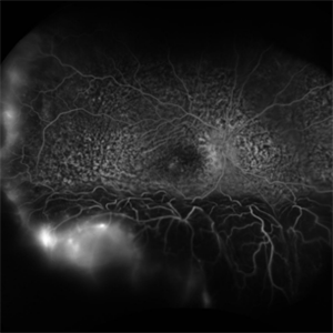
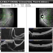
Imaging device: Fluorescein Angiography
Condition/keywords: exudative retinal detachment, idiopathic uveal effusion syndrome, leopard spots, uveal effusion, uveal effusion syndrome
-
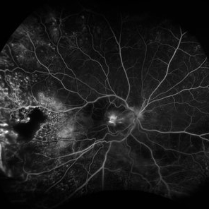
 Vogt Kayanagi Harada Disease
May 26 2025 by Malvika Singh
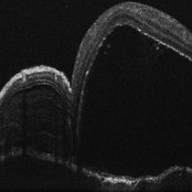
OCT image of the retina showing SRF in case of an exudative retinal detachment and bacillary layer detachment
Photographer: Dr Malvika Singh, Retina Foundation, Ahmedabad, India
Imaging device: Mirante SLO/OCT
Condition/keywords: OCT, Vogt-Koyanagi-Harada
-
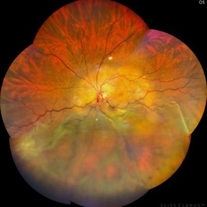
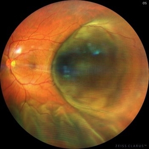
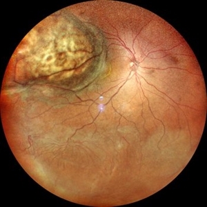
 Vortex-pattern Exudative Retinal Detachment
Feb 22 2025 by CUI YUELING
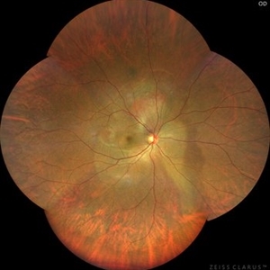
Patient: Male, 40 years old. Chief Complaint: Blurred vision and metamorphopsia in the left eye for more than 10 days. Past Medical History Hypertension for 4 years, with a highest recorded blood pressure of 160/80 mmHg. Currently controlled with oral "Nifedipine Sustained-Release Tablets, 2 tablets daily." Long-term history of heavy alcohol consumption and smoking. Ophthalmic Examination: Visual Acuity: Right eye (OD): 0.4 (uncorrected, no improvement with correction). Left eye (OS): 0.5 (-1.5DS = 1.0). Intraocular Pressure (IOP): OD: 15 mmHg. OS: 17 mmHg. Anterior Segment:Unremarkable. Fundus Examination: Right eye: Optic disc margins are clear, with a slightly reddish hue. Cup-to-disc ratio (C/D) = 0.2. A scalloped, orange-red elevated lesion is observed superior to the optic disc, with anterior displacement of the focal point. This is accompanied by a secondary, turbine-like exudative retinal detachment centered around the optic disc, involving the macula. The macular region shows scattered punctate yellow-white exudates. Diagnosis: Choroidal hemangioma with secondary exudative retinal detachment(OD).
Photographer: Cui yueling The First People's Hospital of Zunyi, Guizhou, Zunyi, China
Imaging device: Zeiss Clarus 500
Condition/keywords: choroidal hemangioma, exudative retinal detachment
-
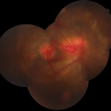
 Coat's Disease with Exudative RD
Feb 12 2025 by Tejaswita Verma
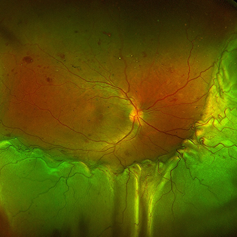
Fundus photo of a 7 year old boy with vision Counting fingers close to face in the right eye and intermittent outward deviation of the right eye observed by parents. Fundus examination shows subretinal exudates, telengiectatic vessels in superotemporal quadrant, intraretinal hemorrhages, macular scar, NVD.
Photographer: DR. TEJASWITA VERMA
Imaging device: MIRANTE
Condition/keywords: Coats' disease, exudative retinal detachment
-
 Coats` Disease
Dec 18 2024 by Thirumalesh Mochi Basavaraj, MD
Fundus photo graph of a 6 year old child with exudative retinal detachment with sub retinal lipid exudation and peripheral telengectasia.
Photographer: Puttaswamy N K
Condition/keywords: exudative detachment, peripheral telangiectasia
-
 Exudative Retinal Detachment
Oct 26 2024 by rahul saradge
41y/M, k/c/o TYPE 1 DM DOV SINCE 1 WEEK OU H/O TYPHOID 2 WEEKS BACK Here is the image showing Exudative RD in Right Eye We Planned PRP laser for this patient, advised him Carotid Doppler and 2D ECHO
Photographer: Anagha Wakode, Isha Netralaya Thane
Imaging device: optos
Condition/keywords: choroidals, exudative Retinal detachment
-
 Vogt-Koyanagi-Harada Disease
Sep 24 2024 by Gustavo Uriel Fonseca Aguirre
A 39-year-old female patient with no ophthalmologic history was diagnosed with Vogt-Koyanagi-Harada disease. Exudative retinal detachment was observed in the left eye.
Photographer: Gustavo U. Fonseca Aguirre, Fundación Hospital Nuestra Señora de la Luz, Ciudad de México
Condition/keywords: Vogt-Koyanagi-Harada
-
 Vogt-Koyanagi-Harada Disease
Sep 24 2024 by Gustavo Uriel Fonseca Aguirre
A 39-year-old female patient with no ophthalmologic history was diagnosed with Vogt-Koyanagi-Harada disease. Exudative retinal detachment was observed in the right eye.
Photographer: Gustavo U. Fonseca Aguirre, Fundación Hospital Nuestra Señora de la Luz, Ciudad de México
Condition/keywords: Vogt-Koyanagi-Harada
-
 Exudative Retinal Detachment
May 27 2024 by Akansha Sharma
Color fundus photograph of a 38 year old male with breast carcinoma leading to intraocular metastasis as represented by an exudative retinal detachment.
Photographer: Dr. Akansha Sharma, Bharati Eye Hospital
Condition/keywords: Disc Edema, exudative detachment, macular pucker, METASTATSIS
-
 Exudative Retinal Detachment, Secondary to Coat's Disease
May 10 2024 by Jeffrey Barker
15 year old Male with a exudative retinal detachment, secondary to Coat's Disease.
Photographer: Jeffrey P. Barker
Condition/keywords: Coat's disease, Retina detachment
-
 Exudative Retinal Detachment With Choroidal Metastasis
May 1 2024 by Vishal Agrawal, MD, FRCS,FACS,FASRS
Left eye fundus picture of a 65-year-old female with choroidal metastases and exudative retinal detachment. The patient is under treatment for breast carcinoma.
Photographer: Dr Ayushi
Imaging device: Clarus 700
Condition/keywords: choroidal metastasis, exudative detachment
-
 Von Hippel-Lindau
Feb 6 2024 by Thirumalesh Mochi Basavaraj, MD
8 Year old child with Multiple Capillary Haemangiomas with Exudative retinal detachment.
Photographer: Puttaswamy
Condition/keywords: exudative detachment, Von Hippel-Lindau
-
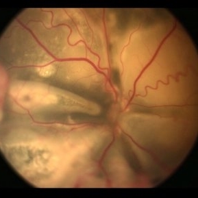
 Exudative RD
Feb 6 2024 by Thirumalesh Mochi Basavaraj, MD
26 yr old male with a exudative retinal detachment with the retina just behind the crystalline lens in a case of optic disc capillary hemangioma.
Photographer: Puttaswamy N K
Condition/keywords: exudative detachment
-
 New Choroidal Melanoma
Jan 4 2024 by Virginia Gebhart
77 year old male with a bilobed pigmented mass with exudative RD, and trace inflammation present in AV consistent with choroidal melanoma. Mass extends into ciliary body. Pt scheduled for MRI prior to plaque radiation to rule out metastasis.
Photographer: Virginia Gebhart
Imaging device: Optos California
Condition/keywords: ciliary body melanoma, exudative retinal detachment
-
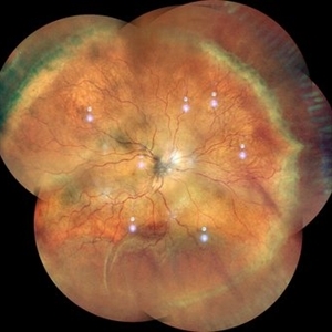 VKH
VKH
Sep 29 2023 by Anjana Mirajkar, MS Ophthalmology
Wide field color photo image of RE of a 41 year old female case of VKH showing exudative retinal detachment inferiorly with multiple fluid pockets in the posterior pole with ILM folds
Photographer: Dr. Anjana Mirajkar -Retina Foundation, Ahmedabad
Imaging device: Mirante-Nidek
Condition/keywords: Vogt-Koyanagi-Harada
-
 Choroidal Melanoma with exudative retinal detachment
Jul 19 2023 by Mariam Cernichiaro-Espinosa, MD
40-year old male with choroidal melanoma and secondary exudative retinal detachment. His visual acuity was 20/200. It was treated with enucleation.
Photographer: Mariam Cernichiaro-Espinosa, Asociación para Evitar la Ceguera en México, I.A.P. Mexico City, Mexico.
Imaging device: Zeiss Clarus
Condition/keywords: exudative retinal detachment
-
 Choroidal Melanoma with Exudative Retinal Detachment
Mar 2 2023 by Aditya S Kelkar, MS, FRCS, FASRS,FRCOphth
Color fundus photograph of the left eye of a 45 year old male showing choroidal melanoma with exudative retinal detachment.
Photographer: Dr. Pranali Surawase, National Institute of Ophthalmology, Pune, India.
Imaging device: Zeiss Clarus 500
Condition/keywords: choroidal mass, exudative retinal detachment, Retinal detachment
-
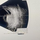 Choroidal Hemangioma
Jan 29 2023 by Anjana Mirajkar, MS Ophthalmology
USG BSCAN of RE of a 25 year old female with RE exudative retinal detachment with subretinal mass most likely a Choroidal Hemangioma.
Photographer: Dr. Anjana Mirajkar -Retina Foundation, Ahmedabad
Condition/keywords: unilateral exudative retinal detachment
-
 Choroidal Hemangioma
Jan 29 2023 by Anjana Mirajkar, MS Ophthalmology
OCT BE of RE of a 25 year old female with RE exudative retinal detachment with subretinal mass most likely a Choroidal Hemangioma.
Photographer: Dr. Anjana Mirajkar -Retina Foundation, Ahmedabad
Condition/keywords: choroidal hemangioma
-
 Choroidal Hemangioma
Jan 29 2023 by Anjana Mirajkar, MS Ophthalmology
Widefield fluorescein angiography image of RE of a 25 year old female with RE exudative retinal detachment with subretinal mass most likely a Choroidal Hemangioma.
Photographer: Dr. Anjana Mirajkar -Retina Foundation, Ahmedabad
Condition/keywords: choroidal hemangioma
-
 Choroidal Hemangioma
Jan 29 2023 by Anjana Mirajkar, MS Ophthalmology
Widefield fluorescein angiography image (montage) of RE of a 25 year old female with RE exudative retinal detachment with subretinal mass most likely a Choroidal Hemangioma.
Photographer: Dr. Anjana Mirajkar -Retina Foundation, Ahmedabad
Condition/keywords: choroidal hemangioma
-
 Choroidal Hemangioma
Jan 29 2023 by Anjana Mirajkar, MS Ophthalmology
Widefield color image of RE of a 25 year old female with RE exudative retinal detachment with subretinal mass most likely a Choroidal Hemangioma with fronds of vessels noted inferiorly.
Photographer: Dr. Anjana Mirajkar -Retina Foundation, Ahmedabad
Condition/keywords: choroidal hemangioma
-
 Choroidal Hemangioma
Jan 29 2023 by Anjana Mirajkar, MS Ophthalmology
Widefield color image (montage) of RE of a 25 year old female with RE exudative retinal detachment with subretinal mass most likely a Choroidal Hemangioma with fronds of vessels noted inferiorly.
Photographer: Dr. Anjana Mirajkar -Retina Foundation, Ahmedabad
Condition/keywords: choroidal hemangioma
-
 Exudative Retinal Detachment secondary to Leber's Miliary Aneurysm in a case of Retinitis Pigmentosa
Oct 13 2022 by Vaidehi Sathaye
Fundus Photograph of RE of a 23 year male patient , with an Exudative Retinal Detachment secondary to Leber's Miliary Aneurysm in a case of Retinitis Pigmentosa.
Photographer: Dr. Vaidehi Sathaye
Imaging device: Mirante
Condition/keywords: exudative detachment, Leber's miliary aneurysm, retinitis pigmentosa
-
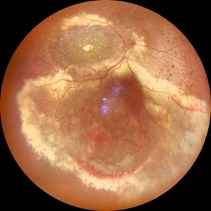
 Stage 3 Coats' Disease
Aug 7 2022 by Muhammad Amer Awan, MD, FRCSEd, FRCOphth, FRCS Glasgow, FACS, FASRS
Fundus photography of a 6 months old baby boy who presented with unilateral leucoria. There was right exudate retinal detachment with extensive hard exudates and tortuous retinal vessels. Diagnosis of Coats' disease was made that was externally drained and intravitreal rhanibizumab was given.
Photographer: Muhammad Amer Awan, Shifa Taamer e Millat University
Condition/keywords: Coats' disease, exudative retinal detachment, exudative retinopathy, unilateral exudative retinal detachment
 Loading…