Search results (28 results)
-
 West African Crystalline Maculopathy
Oct 22 2023 by Niloofar Piri, MD
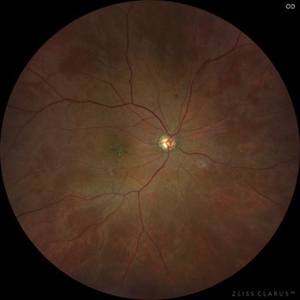
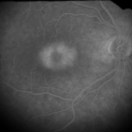
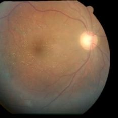
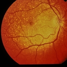
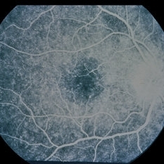
Wide field fundus photograph of the right eye of the same patient from Liberia demonstrating birefringent crystals in the fovea. Notice the partially fibrosed NVE inferiorly in the macula.
Photographer: Niloofar Piri, MD
Condition/keywords: crystalline maculopathy, crystalline retinopathy, West African Crystalline Maculopathy
-
 West African Crystalline Maculopathy
Oct 22 2023 by Niloofar Piri, MD
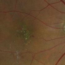
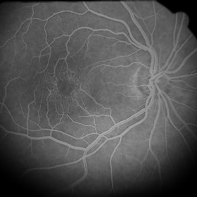
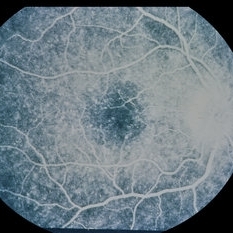
Fundus photograph of right eye of a patient from Liberia demonstrating multiple birefringent yellow green crystalline deposits in the fovea. Please notice the partially fibrosed NVE inferiorly. The disease has been shown to be associated with vascular disorders including diabetic retinopathy.
Photographer: Niloofar Piri, MD
Condition/keywords: crystalline maculopathy, crystalline retinopathy, West African Crystalline maculopathy
-
 Crystalline Retinopathy
Jun 27 2020 by Thirumalesh Mochi Basavaraj, MD
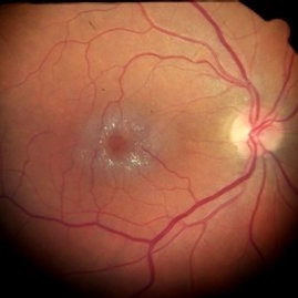
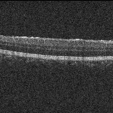
23-year-old asymptomatic female, came for routine examination. Inset shows OCT image with hyperreflective lesion scattered throughout the retina, choroidal sclerosis can also be noted.
Photographer: Ravikrishna, Puttaswamy
Imaging device: Heidelberg Spectralis
Condition/keywords: crystalline retinopathy
-
 Dry AMD with Central Geographic Atrophy, a Few Intraretinal Crystals, and Prominent Choroidal Vessels
Sep 18 2019 by John S. King, MD
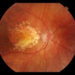
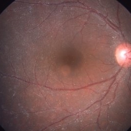
93-year-old white female routine AMD visit with VA of CF in OD due to central geographic atrophy.
Photographer: Gretchen Harper
Imaging device: Topcon 50
Condition/keywords: crystalline retinopathy, dry, geographic atrophy
-
 Retinal Crystals in Macular Telangiectasia
Jan 6 2019 by John S. King, MD
61-year-old white female, healthy, slow progressive central vision loss OD. 20/200 OD and 20/30 OS with mild-mod NSC OU. Retinal crystals around fovea in the posterior pole. FA shows 360 degrees of parafoveal telangiectatic vessels that leak without signs of CNVM. Minimal changes in fellow eye. Asymmetric MacTel 2. Monitoring and using AREDS 2.
Photographer: Kay Dalby
Imaging device: Topcon 50 (satellite office)
Condition/keywords: crystalline retinopathy, cystoid macular edema (CME), macular telangiectasia
-
 Unilateral Crystalline Retinopathy
Dec 20 2017 by Kaustubh B Harshey
SDOCT scan through the temporal retina passing through the crystals observed on fundus imaging. Crystals appear to be deposited on the ILM.
Photographer: Prem Kumar, Aravind Eye Hospital and PG Institute of Ophthalmology, Tirunelveli
Condition/keywords: crystalline retinopathy
-
 Unilateral Crystalline Retinopathy
Dec 20 2017 by Kaustubh B Harshey
Contralateral eye of the patient with crystalline retinopathy showing RPE alterations and pigment migration at the posterior pole.
Photographer: Prem Kumar, Aravind Eye Hospital and PG Institute of Ophthalmology, Tirunelveli
Condition/keywords: crystalline retinopathy
-
 Unilateral Crystalline Retinopathy
Dec 20 2017 by Kaustubh B Harshey
Fundus picture of the involved eye showing refractile crystals on the retinal surface.
Photographer: Prem Kumar, Aravind Eye Hospital and PG Institute of Ophthalmology, Tirunelveli
Condition/keywords: crystalline retinopathy
-
 Preretinal Triamcinolone Crystals Years after IVT
Jun 8 2016 by John S. King, MD
Preretinal crystals years after IVT.
Condition/keywords: crystalline retinopathy, crystals
-
 Ring 17 Chromosome
Nov 5 2014 by David Callanan, MD
3-year-old patient, crystalline retinopathy; 20/30 roughly; epilepsy.
Condition/keywords: crystalline retinopathy
-
 Ring 17 Chromosome
Nov 5 2014 by David Callanan, MD
3-year-old patient, crystalline retinopathy; 20/30 roughly; epilepsy.
Condition/keywords: crystalline retinopathy
-
 Ring 17 Chromosome
Nov 5 2014 by David Callanan, MD
3-year-old patient, crystalline retinopathy; 20/30 roughly; epilepsy.
Condition/keywords: crystalline retinopathy
-
 Ring 17 Chromosome
Nov 5 2014 by David Callanan, MD
3-year-old patient, crystalline retinopathy; 20/30 roughly; epilepsy.
Condition/keywords: crystalline retinopathy
-
 Ring 17 Chromosome
Nov 5 2014 by David Callanan, MD
3-year-old patient, crystalline retinopathy; 20/30 roughly; epilepsy.
Condition/keywords: crystalline retinopathy
-
 Ring 17 Chromosome
Nov 5 2014 by David Callanan, MD
3-year-old patient, crystalline retinopathy; 20/30 roughly; epilepsy.
Condition/keywords: crystalline retinopathy
-
 Ring 17 Chromosome
Nov 5 2014 by David Callanan, MD
3-year-old patient, crystalline retinopathy; 20/30 roughly; epilepsy.
Condition/keywords: crystalline retinopathy
-
 Ring 17 Chromosome
Nov 5 2014 by David Callanan, MD
3-year-old patient, crystalline retinopathy; 20/30 roughly; epilepsy.
Condition/keywords: crystalline retinopathy
-
 Ring 17 Chromosome
Nov 5 2014 by David Callanan, MD
3-year-old patient, crystalline retinopathy; 20/30 roughly; epilepsy.
Condition/keywords: crystalline retinopathy
-
---thumb.jpg/image-square;max$300,300.ImageHandler) Primary Hyperoxaluria and Oxalosis
Primary Hyperoxaluria and Oxalosis
Jul 24 2013 by Hamid Ahmadieh, MD
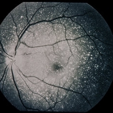
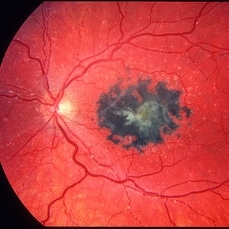
Color fundus photograph of the right eye of a 55-year-old man with primary hyperoxaluria and oxalosis. Characteristic crystalline retinopathy (flecked retina), black geographic maculopathy, and partial optic atrophy are visible. In addition, occluded branches of central retinal artery due to calcium oxalate deposition are visible.
Photographer: Hanieh Payab, Ophthalmic Research Center, Labbafinejad Medical Center, Tehran
Imaging device: Topcon Fundus Camera
Condition/keywords: oxalosis, primary hyperoxaluria
-
---thumb.jpg/image-square;max$300,300.ImageHandler) Primary Hyperoxaluria and Oxalosis
Primary Hyperoxaluria and Oxalosis
Jul 24 2013 by Hamid Ahmadieh, MD
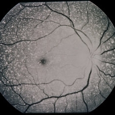
Color fundus photograph of the right eye of a 55-year-old man with primary hyperoxaluria and oxalosis. Characteristic crystalline retinopathy (flecked retina), black geographic maculopathy, and partial optic atrophy are visible. In addition, occluded branches of central retinal artery due to calcium oxalate deposition are visible.
Photographer: Hanieh Payab, Ophthalmic Research Center, Labbafinejad Medical Center, Tehran
Imaging device: Topcon Fundus Camera
Condition/keywords: oxalosis, primary hyperoxaluria
-
 Oxalosis
May 2 2013 by Henry J. Kaplan, MD
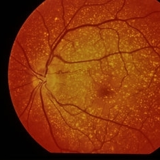
Crystalline retinopathy in oxalosis as a result of calcium oxalate deposits in the retina ; also deposition in RPE which causes fleck retina as pigmentary lesion in the center.
Condition/keywords: crystalline retinopathy, oxalosis
-
 Canthaxanthine maculopathy LE
Jan 11 2013 by Alex P. Hunyor, MD
Crystalline retinopathy due to canthaxanthine - left eye.
Condition/keywords: canthaxanthin maculopathy
 Loading…