Search results (90 results)
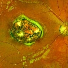
-
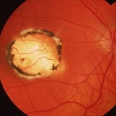
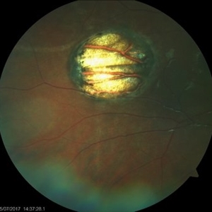
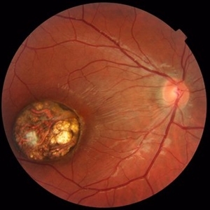
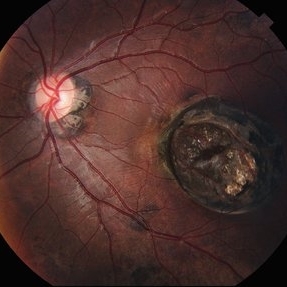 Wagon-Wheel Lesion
Jun 5 2025 by César Adrián Gómez Valdivia, MD
Wagon-wheel lesion found in a 12 YO male patient diagnosed with congenital toxoplasmosis. Findings were bilateral.
Photographer: @eyemissu2
Imaging device: TOPCON TRC-50DX
Condition/keywords: toxoplasmosis chorioretinitis, Wagon-wheel lesion
-
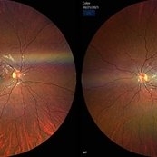
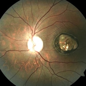
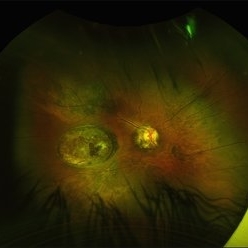 Wagon-Wheel Lesion
Jun 5 2025 by César Adrián Gómez Valdivia, MD
Wagon-wheel lesion found in a 12 year-old male patient diagnosed with congenital toxoplasmosis. Findings were bilateral.
Photographer: @eyemissu2
Imaging device: California ICG OPTOS
Condition/keywords: toxoplasmosis, Wagon-wheel lesion
-
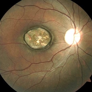
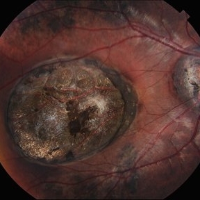 Wagon-Wheel Lesion
Jun 5 2025 by César Adrián Gómez Valdivia, MD
Wagon-wheel lesion found in a 12 year-old male patient diagnosed with congenital toxoplasmosis. Findings were bilateral.
Photographer: @eyemissu2
Imaging device: TOPCON TRC-50DX
Condition/keywords: toxoplasmosis, Wagon-wheel lesion
-
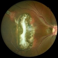 Toxoplasmosis
Nov 24 2021 by Catarina Monteiro
Fundus photograph of a 7-year-old child with a chorioretinal scar likely due to congenital toxoplasmosis.
Photographer: Catarina Monteiro
Condition/keywords: chorioretinal scar, congenital toxoplasmosis, ocular toxoplasmosis
-
 Congenital Toxoplasmosis Macular Scarring
Nov 6 2021 by Emmanouil Gavalas, MD
Fundus photographs of an 26-year-old female showing right eye macular scarring Incidental diagnosis Visual Acuity OD 6/12 OS 6/6
Photographer: Emmanouil Gavalas MD, Ophthalmos Reseach and Educational Institute,Nicosia,Cyprus
Imaging device: Zeiss Clarus 500
Condition/keywords: congenital toxoplasmosis, macular scar, ocular toxoplasmosis
-
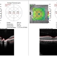 Congenital Toxoplasmosis Macular Scarring
Nov 6 2021 by Emmanouil Gavalas, MD
Right eye OCT image showing atrophy and loss of foveal neuroretinal tissue and RPE.
Photographer: Emmanouil Gavalas MD, Ophthalmos Reseach and Educational Institute,Nicosia,Cyprus
Imaging device: Heidelberg Spectralis OCT
Condition/keywords: congenital toxoplasmosis, macular scar, ocular toxoplasmosis
-
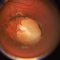
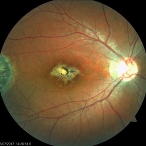
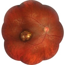
 Congenital Toxoplasmosis
Feb 2 2021 by Niloofar Piri, MD
43-year-old female with large oval chorioretinal scar in posterior pole with heavy RPE hyperplasia and history of hydrocephalus s/p VP shunt since birth. Findings are consistent with congenital toxoplasmosis.
Condition/keywords: congenital toxoplasmosis
-
 Congenital Toxoplasmosis
Dec 18 2019 by Yoshihiro Yonekawa, MD, FASRS
Widefield fundus image of a teenage girl's right eye with an inactive congenital toxoplasmosis macular lesion. Her vision is 20/400 in this eye.
Photographer: Netanya Lerner, COA, Wills Eye Hospital/Mid Atlantic Retina
Imaging device: Optos California
Condition/keywords: congenital toxoplasmosis, pediatric retina
-
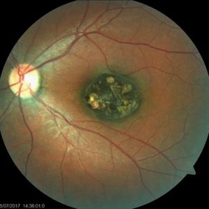
 Congenital Toxoplasmosis
Apr 8 2019 by Gary R. Cook, MD, FACS
Left eye of the same 38-year-old female with congenital toxoplasmosis lesion; V.A. = 20/40 due to temporal location of the Toxo scar.
Imaging device: Topcon VT-50
Condition/keywords: chorioretinal scar, congenital toxoplasmosis, inactive toxoplasmosis, macular scar, ocular toxoplasmosis
-
 Congenital Toxoplasmosis
Apr 8 2019 by Gary R. Cook, MD, FACS
Right eye of a 38-year-old female with bilateral congenital toxoplasmosis lesions; V.A. = 20/70 OD
Imaging device: Topcon VT-50
Condition/keywords: chorioretinal scar, congenital toxoplasmosis, inactive, inactive toxoplasmosis, macular scar, ocular toxoplasmosis
-
 Congenital Toxoplasmosis Scar
Apr 8 2019 by Gary R. Cook, MD, FACS
5-year-old white male with a typical, deep, pigmented chorioretinal scar secondary to congenital toxoplasmosis OS.
Condition/keywords: chorioretinal scar, congenital toxoplasmosis, inactive toxoplasmosis, macular scar, ocular toxoplasmosis
-
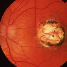
 Congenital Toxoplasmosis
Apr 8 2019 by Gary R. Cook, MD, FACS
23-year-old with congenital toxoplasmosis; view of optic disc and macular scar OS.
Condition/keywords: chorioretinal scar, congenital toxoplasmosis, inactive toxoplasmosis, macular scar, ocular toxoplasmosis
-
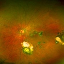
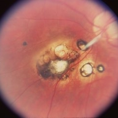 Congenital Toxoplasmosis
Apr 8 2019 by Gary R. Cook, MD, FACS
23-year-old with congenital toxoplasmosis; view of toxo scars inferonasal to optic disc OS.
Condition/keywords: chorioretinal scar, congenital toxoplasmosis, ocular toxoplasmosis
-
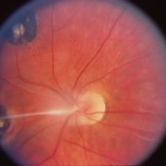 Congenital Toxoplasmosis
Apr 8 2019 by Gary R. Cook, MD, FACS
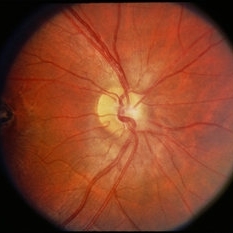
23-year-old male with congenital toxoplasmosis; view of optic disc and glial band OD.
Condition/keywords: congenital toxoplasmosis, inactive toxoplasmosis, ocular toxoplasmosis
-
 Congenital Toxoplasmosis
Apr 8 2019 by Gary R. Cook, MD, FACS
Right eye of a 23-year-old male with congenital toxoplasmosis OD; view of macular lesion.
Condition/keywords: congenital toxoplasmosis, inactive toxoplasmosis, macular scar, ocular toxoplasmosis
-
 Slide 4-9
Feb 20 2019 by Lancaster Course in Ophthalmology
Congenital toxoplasmosis, a congenital defect of environmental origin. The chorioretinal scar is not specific but is identical to that produced by acquired infections.
Condition/keywords: chorioretinal scar, congenital defect, congenital toxoplasmosis
-
 Congenital Toxoplasmosis
Jul 22 2017 by Akif Erol
Color fundus photograph of the right eye of an 18-year-old girl with decreased vision due to a large chorioretinal scar involving the macula. The lesion is typical for a congenital ocular toxoplasmosis
Photographer: Mehmet Akif Erol, Afyon Kocatepe University Ophthalmology Clinic
Condition/keywords: color fundus photograph, congenital toxoplasmosis
-
 Congenital Toxoplasmosis
Jul 22 2017 by Akif Erol
Color fundus photograph of the left eye of an 18-year-old girl with decreased vision due to a large chorioretinal scar involving the macula. The lesion is typical for a congenital ocular toxoplasmosis
Photographer: Mehmet Akif Erol, Afyon Kocatepe University Ophthalmology Clinic
Condition/keywords: color fundus photograph, congenital toxoplasmosis
-
 Toxoplasmosis
Jun 3 2017 by Gabriel Costa Andrade, PhD
Fundus photograph of an 14-year-old boy with multiple chorioretinal scars secondary to toxoplasmosis.
Photographer: Gabriel Costa de Andrade
Imaging device: Optos® California
Condition/keywords: congenital toxoplasmosis, ocular toxoplasmosis, toxoplasmosis chorioretinitis, toxoplasmosis uveitis
-
 Congenital Toxoplasmosis
Oct 10 2015 by Hamid Ahmadieh, MD
Color fundus photograph of the right eye of a 15 -year-old boy with decreased vision due to a large chorioretinal scar involving the macula . The lesion is typical for a congenital ocular toxoplasmosis .
Photographer: Solmaz Shahmohammad, Negah Eye Center, Tehran, Iran
Condition/keywords: color fundus photograph, congenital toxoplasmosis
-
 Congenital Toxoplasmosis
Oct 10 2015 by Hamid Ahmadieh, MD
Wide-field color fundus photograph of the right eye of a 15 -year-old boy with decreased vision due to a large chorioretinal scar involving the macula . The lesion is typical for a congenital ocular toxoplasmosis .
Photographer: Solmaz Shahmohammad, Negah Eye Center, Tehran, Iran
Condition/keywords: color fundus photograph, congenital toxoplasmosis
-
 Congenital Toxoplasmosis LE
Apr 29 2015 by Neha Goel, MS DNB FRCS (Glasg)
Fundus photograph of the left eye of a 25-year-old male with decreased vision since early childhood.
Photographer: Neha Goel
Imaging device: Zeiss visucam
Condition/keywords: congenital toxoplasmosis, inactive toxoplasmosis, toxoplasmosis
-
 Congenital Toxoplasmosis RE
Apr 29 2015 by Neha Goel, MS DNB FRCS (Glasg)
Fundus photograph of the right eye of a 25-year-old male with decreased vision since early childhood.
Photographer: Neha Goel
Imaging device: Zeiss visucam
Condition/keywords: congenital toxoplasmosis, inactive toxoplasmosis, toxoplasmosis
-
 Congenital Toxoplasmosis / New Lesion
Mar 14 2014 by David Callanan, MD
15-year-old Hispanic male with congenital Toxoplasmosis / new lesion.
Condition/keywords: congenital toxoplasmosis
 Loading…