Search results (33 results)
-
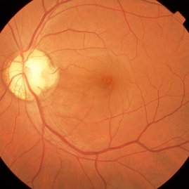
 Optic Nerve Pit
Feb 21 2024 by Virginia Gebhart
65 year old female with optic nerve pit. Asymptomatic, continued observation.
Photographer: Virginia Gebhart
Imaging device: Topcon TRC 50DX
Condition/keywords: congenital optic nerve pit, Optic nerve pit
-
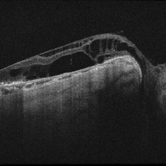
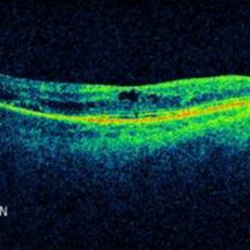
 Optic Disc Pit OCT
Aug 1 2023 by Aditya S Kelkar, MS, FRCS, FASRS,FRCOphth
Optical Coherence Tomography of an 21 year old male with a Optic Disc Pit.
Photographer: Dr. Ajinkya Rawale. National institute of Ophthalmology, Pune, India.
Imaging device: Zeiss Plex
Condition/keywords: congenital optic nerve pit
-
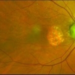
 Optic Disc Pit Associated with Optic Disc Coloboma and Retinochoroidal Coloboma
Jul 22 2020 by Deepak Bhojwani, MS
Fundus photograph of a 32-year-old male showing large optic disc pit in a colobomatous optic nerve head along with isolated inferior retino-choroidal coloboma. (A rare / coincidental occurrence of multiple congenital anomalies of optic disc and retina)
Photographer: DEEPAK BHOJWANI
Condition/keywords: coloboma of choroid, coloboma of optic disc, congenital optic nerve pit
-
 Colobomatous Optic Disc Maculopathy
Feb 13 2020 by Yoshihiro Yonekawa, MD, FASRS
EDI-OCT of a teenage girl with submacular fluid from a colobomatous optic disc. Note the subtle tracking of the subretinal fluid into the disc.
Photographer: Netanya Lerner, COA, Wills Eye Hospital/Mid Atlantic Retina
Imaging device: Topcon
Condition/keywords: chorioretinal coloboma, coloboma of optic disc, congenital optic nerve pit, subretinal fluid
-
 Colobomatous Optic Disc Maculopathy
Feb 13 2020 by Yoshihiro Yonekawa, MD, FASRS
Fluorescein angiography, late frame, of a teenage girl with submacular fluid from a colobomatous optic disc. The camera is focused is on the elevated macula, and the disc is subtly defocused.
Photographer: Netanya Lerner, COA, Wills Eye Hospital/Mid Atlantic Retina
Imaging device: Topcon
Condition/keywords: chorioretinal coloboma, coloboma of optic disc, congenital optic nerve pit, subretinal fluid
-
 Colobomatous Optic Disc Maculopathy
Feb 13 2020 by Yoshihiro Yonekawa, MD, FASRS
Beautifully focused fundus photograph of a teenage girl with submacular fluid from a colobomatous optic disc.
Photographer: Netanya Lerner, COA, Wills Eye Hospital/Mid Atlantic Retina
Imaging device: Topcon
Condition/keywords: chorioretinal coloboma, coloboma of optic disc, congenital optic nerve pit, subretinal fluid
-
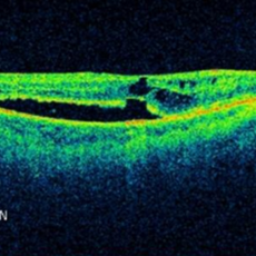
 Retinoschisis and Subretinal Fluid in Optic Disc Pit Related Maculopathy
Aug 20 2018 by DIEGO A BUESO PONCE, MD
OCT B-scan of a 19-year-old female with congenital optic disc pit maculopathy.
Photographer: Diego Bueso Ponce, Clinica Unidad Laser, Barranquilla Colombia
Imaging device: Topcon DRI OCT Triton, Swept source OCT
Condition/keywords: B scan ultrasound, congenital optic nerve pit, retinoschisis
-
 Optic Disc Pit Maculopathy
Aug 20 2018 by DIEGO A BUESO PONCE, MD
Fundus photograph of an 19-year-old female with congenital optic disc pit and associated maculopathy with subretinal fluid and retinoschisis.
Photographer: Diego Bueso Ponce, Clinica Unidad Laser, Barranquilla Colombia
Imaging device: Topcon DRI OCT Triton, Swept source OCT
Condition/keywords: congenital optic nerve pit, maculopathy, optic disc pit, retinoschisis
-
 Optic Nerve Pit OD - OCT
Aug 6 2018 by Hosam Attia, MD
65-year-old white male, presented for a second opinion for possible cataract extraction OD. BCVA: OD: 20/70 OS: 20/60 WRx: OD: -3.75 +1.50 x 5 OS: -1.75 +1.50 x 178 SLE: +2 NS OD>OS DFE: OD: Nasal macular GA, connected by milder track of RPE changes to an optic nerve pit OD (no fluid seen clinically) OS: enlarged C/D w/ no pits, macular RPE change w/ No heme, CME/ SRF OCT: OD: Peri-papillary cystoid changes & outer retinal atrophy (corresponding to the area of GA on the pseudocolor photo) w/ No SRF (mimicking PP CNVM), connected to the optic disc pit by shallow sinus/ tract. OS: Drusenoid RPE changes, No cystoid changes/ SRF
Imaging device: Zeiss Cirrus -5000
Condition/keywords: congenital optic nerve pit
-
 Optic Nerve Pit OD - Pseudocolor Photo
Aug 6 2018 by Hosam Attia, MD
65-year-old white male, presented for a second opinion for possible cataract extraction OD. BCVA: OD: 20/70 OS: 20/60 WRx: OD: -3.75 +1.50 x 5 OS: -1.75 +1.50 x 178 SLE: +2 NS OD>OS DFE: OD: Nasal macular GA, connected by milder track of RPE changes to an optic nerve pit OD (no fluid seen clinically) OS: enlarged C/D w/ no pits, macular RPE change w/ No heme, CME/ SRF OCT: OD: Peri-papillary cystoid changes & outer retinal atrophy (corresponding to the area of GA on the pseudocolor photo) w/ No SRF (mimicking PP CNVM), connected to the optic disc pit by shallow sinus/ tract. OS: Drusenoid RPE changes, No cystoid changes/ SRF
Imaging device: Optos California
Condition/keywords: congenital optic nerve pit
-
 Optic Nerve Pit With Sub-Retinal Fluid
Sep 17 2015 by Jason S. Calhoun
Young female with blurred vision in the left eye. Fundus photograph shows optic nerve pit adjacent to the macula where there is sub retinal fluid visible.
Photographer: Jason Calhoun, Mayo Clinic, Department of Ophthalmology
Imaging device: TOPCON-TRC50EX
Condition/keywords: congenital optic nerve pit
-
 Optic Nerve Pit with Sub-Retinal Fluid
Sep 17 2015 by Jason S. Calhoun
Young female with blurred vision in the left eye. Fundus photograph shows optic nerve pit adjacent to the macula where there is sub retinal fluid visible.
Photographer: Jason Calhoun, Mayo Clinic, Department of Ophthalmology
Imaging device: TOPCON-TRC50EX
Condition/keywords: congenital optic nerve pit
-
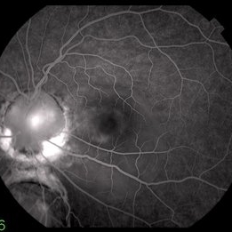
 Optic Nerve Pit Related Serous Retinal Detachment
Sep 15 2014 by Thomas A. Ciulla, MD, MBA, FASRS
Pre-op late phase angiogram showing optic nerve pit and prior laser treatment at temporal aspect of optic nerve
Photographer: Thomas Steele
Condition/keywords: congenital optic nerve pit, pre-op, serous retinal detachment, vitrectomy, vitreomacular surgery
-
 Optic Nerve Pit Related Serous Retinal Detachment
Sep 15 2014 by Thomas A. Ciulla, MD, MBA, FASRS
Pre-op early phase angiogram showing optic nerve pit and prior laser treatment at temporal aspect of optic nerve
Photographer: Thomas Steele
Condition/keywords: congenital optic nerve pit, pre-op, serous retinal detachment, vitrectomy, vitreomacular surgery
-
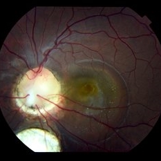
 Optic Nerve Pit Related Serous Retinal Detachment
Sep 15 2014 by Thomas A. Ciulla, MD, MBA, FASRS
Pre-op color fundus photograph showing optic nerve pit and prior laser treatment at temporal aspect of optic nerve.
Photographer: Thomas Steele
Condition/keywords: congenital optic nerve pit, pre-op, serous retinal detachment, vitrectomy, vitreomacular surgery
-
 Optic Nerve Pit Related Serous Retinal Detachment
Sep 15 2014 by Thomas A. Ciulla, MD, MBA, FASRS
Pre-op red-free photo showing optic nerve pit and prior laser treatment at temporal aspect of optic nerve. Note the subtle outline of the serous retinal detachment.
Photographer: Thomas Steele
Condition/keywords: congenital optic nerve pit, pre-op, red-free, serous retinal detachment, vitrectomy, vitreomacular surgery
-
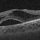
 Optic Nerve Pit Related Serous Retinal Detachment
Sep 10 2014 by Thomas A. Ciulla, MD, MBA, FASRS
Pre-op OCT. The visual acuity measured 20/200. Note the severe subretinal fluid with chronic features, including the cystoid spaces.
Condition/keywords: congenital optic nerve pit
-
 Optic Nerve Pit Related Serous Retinal Detachment
Sep 8 2014 by Thomas A. Ciulla, MD, MBA, FASRS
Post-op OCT at 1 year. Visual acuity measured 20/40.
Condition/keywords: congenital optic nerve pit, post-op, serous retinal detachment, vitrectomy, vitreomacular surgery
-
 Optic Nerve Pit Related Serous Retinal Detachment
Sep 8 2014 by Thomas A. Ciulla, MD, MBA, FASRS
Post-op OCT at 1 month. Visual acuity improved to 20/80.
Condition/keywords: congenital optic nerve pit
-
 Optic Nerve Pit Related Serous Retinal Detachment
Sep 8 2014 by Thomas A. Ciulla, MD, MBA, FASRS
Post-op OCT at 3 months. Visual acuity measured 20/80.
Condition/keywords: congenital optic nerve pit
-
 Optic Nerve Pit Related Serous Retinal Detachment
Sep 8 2014 by Thomas A. Ciulla, MD, MBA, FASRS
Post-op OCT at 9 months. Visual acuity improved to 20/40.
Condition/keywords: congenital optic nerve pit
-
 Disc Pit With Maculopathy
Jun 3 2014 by Neha Goel, MS DNB FRCS (Glasg)
Fundus photograph of the right eye of a 28-year-old male.
Photographer: Neha Goel
Imaging device: Zeiss Visucam
Condition/keywords: congenital optic nerve pit, neurosensory detachment of retina
-
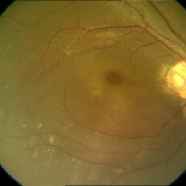
 Pit Macular Syndrome
Mar 21 2013 by Yusuke Oshima, MD, PhD
Fundus photograph of a 38-year-old man with macular detachment associated with an optic disc pit.
Photographer: Yusuke Takada, Osaka University Graduate School of Medicine
Condition/keywords: congenital optic nerve pit
-
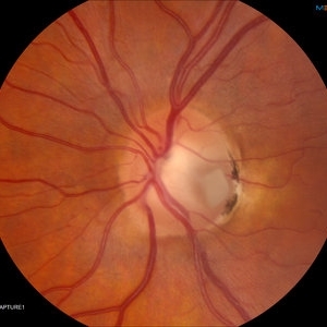
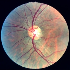 Optic disc pit 3
Jan 11 2013 by Alex P. Hunyor, MD
Optic disc pit left eye.
Condition/keywords: congenital optic nerve pit, optic disc pit
 Loading…