Search results (17 results)
-
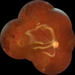
 Combined Retinal Detachment With a Butterfly Shaped Configuration
Mar 13 2025 by S. Natarajan, MD, FASRS, FRCS (GLASGOW) , FICO, D.Sc, FELA
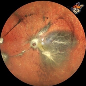
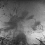
A 46 year old female presented to us with diminished vision in both the eyes. Her blood glucose levels were deranged. She had bilateral proliferative diabetic retinopathy and pan retinal photocoagulation was done elsewhere. Left eye showed a combined retinal detachment with fibrovascular proliferation on the disc and along inferior arcade with a convex configuration of retinal detachment. Patient was planned for surgical intervention. The image shows a butterfly like configuration of combined retinal detachment with the subretinal fluid pocket appearing like the wings of the butterfly.
Photographer: ASHWINI SUTAR ADITYA JYOT EYE HOSPITAL
Imaging device: Mirante ( PLEASE SELECT COVER PAGE )
Condition/keywords: retinal detachment with a butterfly shaped
-
 Combined Traction Rhegmatogenous Detachment
Oct 17 2024 by Hemanth Murthy, MBBS, MD, FASRS
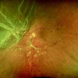
A 68 year old male presented with a shadow in the left eye since 3 days. He was a known diabetic and hypertensive for 20 years. Vision was 20/40 in right eye and 20/60 in left eye. Fundus examination showed Proliferative diabetic retinopathy in right eye and Proliferative diabetic retinopathy with combined traction rhegmatogenous detachment in left eye.
Photographer: Mr Veda Vyas
Condition/keywords: combined retinal detachment, proliferative diabetic retinopathy (PDR)
-
 Combined Retinal Detachment With Macular Hole
Sep 28 2024 by Tejaswita Verma
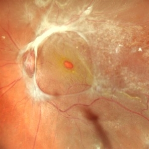
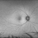
Fundus image of the LE of a 67 year old diabetic, hypertensive female with CF 3metres vision showing combined RD with FTMH, in a pseudophakic eye. She was lost to follow up status post 2 anti VEGF injections received 8 months back due to typhoid fever.
Photographer: DR. TEJASWITA VERMA
Imaging device: MIRANTE
Condition/keywords: full thickness macular hole, proliferative diabetic retinopathy (PDR), tractional retinal detachment
-
 Cutter Segmentation in a case of Diabetic Combined Retinal Detachment | Intra-Operative Still
Apr 25 2023 by Veer Singh, MS, FVRS, FMRF, FICO (Retina)
Cutter Segmentation in a case of Diabetic Combined Retinal Detachment | Intra-Operative Still Patient underwent Vitrectomy with Silicone Oil
Photographer: Dr. Veer Singh
Condition/keywords: combined retinal detachment, cutter, diabetic retinopathy, intraoperative, pars plana vitrectomy (PPV)
-
 360 retinotomy for combined closed funnel tractional and rhematogenous retinal detachment
Jan 1 2023 by Malek Yassine, MD
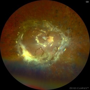
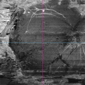
This is Fundus Autofluorecence, showing the residual hypoautofluorescent spots on the exposed choroid, relating to the previous panretinal photocoagulation, as well as the limits of the retinotomy with continuous laser which appeasr hypoautofluorecent with hyperautofluorecent margins.
Photographer: Malek Yassine, HMOED, Agadir, Morocco.
Imaging device: Zeiss Clarus
Condition/keywords: combined retinal detachment, rhegmatogenous retinal detachment, tractional retinal detachment
-
 360 Retinotomy in a closed Funnel combined Tractional and rhegmatogenous retinal detachment
Jan 1 2023 by Malek Yassine, MD
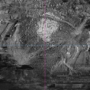
This is the results at 6 months of a Bimanual 23 G-PPV with a very extensive and posterior 360 retinotomy for the management of a combined longstanding closed funnel RD, with submacular membranes, intraretinal PVR. Preop VA was a doubtful light perception. Borders of the retinotomy are stable at 6 months under 1300 Cs Silicon oil with some pigmented PVR developping the edges. Macula appears spared. Silicon oil emulsification droplets are well visualized beneath the superior temporal arcade.
Imaging device: Zeiss Clarus 700
Condition/keywords: combined retinal detachment, retinotomy, silicone oil
-
 OCT Angiography of a 360 retinotomy for closed funnel combined retinal detachment
Jan 1 2023 by Malek Yassine, MD
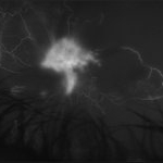
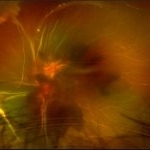
this is an OCTA image of 12X12 MM, showing all the 3 vascular plexi of the residual posterior retinal, with a good perfusion in the superior and central area, a ratification in the intermediate plexus in the inferior area, a non perfused temporal area, and some macular cysts. There's almost none macular translocation
Imaging device: Topcon Triton DRI-OCT
Condition/keywords: combined retinal detachment, OCT Angiography, retinotomy
-
 OCT en face of a 360 retinotomy for closed funnel combined retinal detachment
Jan 1 2023 by Malek Yassine, MD
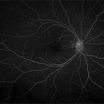
Swept Source OCT en face at deep capillary plexus, shows foveal and parafoveal intraretinal cysts corresponding to macular edema under silicon oil
Imaging device: Topcon Triton DRI-OCT
Condition/keywords: combined retinal detachment, OCT EN FACE
-
 OCT en face of a 360 retinotomy for closed funnel combined retinal detachment
Jan 1 2023 by Malek Yassine, MD
Swept source OCT en face at the silicon oil - Retina Interface shows droplets of SO emulsification around the fovea and at the superior arcade, with some inferior striae corresponding to ERM formation
Imaging device: Topcon Triton DRI-OCT
Condition/keywords: oct en face
-
 Combined Traction-Rhegmatogenous Retinal Detachment
Apr 8 2019 by Gary R. Cook, MD, FACS
White female with a combined diabetic-related traction-rhegmatogenous retinal detachment; s/p full laser PRP in the past; V.A. = 20/400
Imaging device: Topcon VT-50
Condition/keywords: combined retinal detachment, pan-retinal photocoagulation (PRP), proliferative diabetic retinopathy (PDR)
-
 Tractional vs Combined Tractional/Rhegmatogenous Retinal Detachment with Active Neovascularization OS
Jun 1 2018 by Hosam Attia, MD
47-year-old African American, with history of diabetes mellitus of unknown duration and control, was referred for initial evaluation for conjunctival laceration in his left eye, following accidental finger nail injury, 6 days prior to presentation. - On exam, his vision was 20/50 OD and Bare HM/ LP OS. - Fundus color photos OD: No significant pathology, aside from attenuated vasculature OS: Chronic, Mac-Off, almost closed funnel tractional vs combined tractional/rhegmatogenous retinal detachment with large neovascularization (NVE) superiorly, detached ghost vessels, mild fresh vitreous hemorrhage, sub-retinal bands and inferior white vitreous debris from old hemorrhage (not shown) - FA OD: No significant pathology aside from possible mild capillary non-perfusion in the extreme periphery, attenuated vasculature and possible tiny microaneurysms, nasally. OS: Extensive, wide spread capillary non- perfusion (correlate w/ detached Ghost vessels on color photos), and leakage from the NVE. - B/L Carotid Duplex was recommended due to the striking asymmetry in pathology with unknown medical history, diabetes duration and control, etc (even in absence of any signs suggestive of possible ocular ischemic syndrome OD)
Imaging device: Optos California
Condition/keywords: combined retinal detachment, tractional retinal detachment
-
 Tractional vs Combined Tractional/Rhegmatogenous Retinal Detachment with Active Neovascularization OS
Jun 1 2018 by Hosam Attia, MD
47-year-old African American, with history of diabetes mellitus of unknown duration and control, was referred for initial evaluation for conjunctival laceration in his left eye, following accidental finger nail injury, 6 days prior to presentation. - On exam, his vision was 20/50 OD and Bare HM/ LP OS. - Fundus color photos OD: No significant pathology, aside from attenuated vasculature OS: Chronic, Mac-Off, almost closed funnel Tractional vs Combined Tractional/Rhegmatogenous Retinal Detachment with large neovascularization (NVE) superiorly, detached ghost vessels, mild fresh vitreous hemorrhage, sub-retinal bands and inferior white vitreous debris from old hemorrhage (Not shown) - FA OD: No significant pathology aside from possible mild capillary non-perfusion in the extreme periphery, attenuated vasculature and possible tiny microaneurysms, nasally. OS: Extensive, wide spread capillary non- perfusion (correlate w/ detached Ghost vessels on color photos), and leakage from the NVE. - B/L Carotid Duplex was recommended due to the striking asymmetry in pathology with unknown medical history, diabetes duration and control, etc (even in absence of any signs suggestive of possible ocular ischaemic syndrome OD)
Imaging device: Optos California
Condition/keywords: combined retinal detachment, tractional retinal detachment
-
 Tractional vs Combined Tractional/Rhegmatogenous Retinal Detachment with Active Neovascularization OS
Jun 1 2018 by Hosam Attia, MD
47-year-old African American, with history of diabetes mellitus of unknown duration and control, was referred for initial evaluation for conjunctival laceration in his left eye, following accidental finger nail injury, 6 days prior to presentation. - On exam, his vision was 20/50 OD and Bare HM/ LP OS. - Fundus color photos OD: No significant pathology, aside from attenuated vasculature OS: Chronic, Mac-Off, almost closed funnel Tractional vs Combined Tractional/Rhegmatogenous Retinal Detachment with large Neovascularization (NVE) superiorly, detached ghost vessels, mild fresh vitreous hemorrhage, sub-retinal bands and inferior white vitreous debris from old hemorrhage (Not shown) - FA OD: No significant pathology aside from possible mild capillary non-perfusion in the extreme periphery, attenuated vasculature and possible tiny microaneurysms, nasally. OS: Extensive, wide spread capillary non- perfusion (correlate w/ detached Ghost vessels on color photos), and leakage from the NVE. - B/L Carotid Duplex was recommended due to the striking asymmetry in pathology with unknown medical history, diabetes duration and control, etc (even in absence of any signs suggestive of possible ocular ischaemic syndrome OD)
Imaging device: Optos California
Condition/keywords: combined retinal detachment, tractional retinal detachment
-
 Tractional vs Combined Tractional/Rhegmatogenous Retinal Detachment with Active Neovascularization OS
Jun 1 2018 by Hosam Attia, MD
47-year-old African American, with history of diabetes mellitus of unknown duration and control, was referred for initial evaluation for conjunctival laceration in his left eye, following accidental finger nail injury, 6 days prior to presentation. - On exam, his vision was 20/50 OD and Bare HM/ LP OS. - Fundus color photos OD: No significant pathology, aside from attenuated vasculature OS: Chronic, Mac-Off, almost closed funnel tractional vs combined tractional/rhegmatogenous retinal detachment with large neovascularization (NVE) superiorly, detached ghost vessels, mild fresh vitreous hemorrhage, sub-retinal bands and inferior white vitreous debris from old hemorrhage (Not shown) - FA OD: No significant pathology aside from possible mild capillary non-perfusion in the extreme periphery, attenuated vasculature and possible tiny microaneurysms, nasally. OS: Extensive, wide spread capillary non- perfusion (correlate w/ detached ghost vessels on color photos), and leakage from the NVE. - B/L Carotid Duplex was recommended due to the striking asymmetry in pathology with unknown medical history, diabetes duration and control, etc (even in absence of any signs suggestive of possible ocular ischemic syndrome OD)
Imaging device: Optos California
Condition/keywords: combined retinal detachment, tractional retinal detachment
-
 Tractional vs Combined Tractional/Rhegmatogenous Retinal Detachment with Active Neovascularization OS
Jun 1 2018 by Hosam Attia, MD
47-year-old African American, with history of diabetes mellitus of unknown duration and control, was referred for initial evaluation for conjunctival laceration in his left eye, following accidental finger nail injury, 6 days prior to presentation. - On exam, his vision was 20/50 OD and Bare HM/ LP OS. - Fundus color photos OD: No significant pathology, aside from attenuated vasculature OS: Chronic, Mac-Off, almost closed funnel tractional vs combined tractional/rhegmatogenous retinal detachment with large neovascularization (NVE) superiorly, detached ghost vessels, mild fresh vitreous hemorrhage, sub-retinal bands and inferior white vitreous debris from old hemorrhage (Not shown) - FA OD: No significant pathology aside from possible mild capillary non-perfusion in the extreme periphery, attenuated vasculature and possible tiny microaneurysms, nasally. OS: Extensive, wide spread capillary non- perfusion (correlate w/ detached Ghost vessels on color photos), and leakage from the NVE. - B/L Carotid Duplex was recommended due to the striking asymmetry in pathology with unknown medical history, diabetes duration and control etc (even in absence of any signs suggestive of possible ocular ischaemic syndrome OD)
Imaging device: Optos California
Condition/keywords: combined retinal detachment, tractional retinal detachment
-
 Tractional vs Combined Tractional/Rhegmatogenous Retinal Detachment with Active Neovascularization OS
Jun 1 2018 by Hosam Attia, MD
47-year-old African American, with history of diabetes mellitus of unknown duration and control, was referred for initial evaluation for conjunctival laceration in his left eye, following accidental finger nail injury, 6 days prior to presentation. - On exam, his vision was 20/50 OD and Bare HM/ LP OS. - Fundus color photos OD: No significant pathology, aside from attenuated vasculature OS: Chronic, Mac-Off, almost closed funnel tractional vs combined tractional/rhegmatogenous retinal detachment with large neovascularization (NVE) superiorly, detached ghost vessels, mild fresh vitreous hemorrhage, sub-retinal bands and inferior white vitreous debris from old hemorrhage (Not shown) - FA OD: No significant pathology aside from possible mild capillary non-perfusion in the extreme periphery, attenuated vasculature and possible tiny microaneurysms, nasally. OS: Extensive, wide spread capillary non- perfusion (correlate w/ detached Ghost vessels on color photos), and leakage from the NVE. - B/L Carotid Duplex was recommended due to the striking asymmetry in pathology with unknown medical history, diabetes duration and control, etc (even in absence of any signs suggestive of possible ocular ischemic syndrome OD)
Imaging device: Optos California
Condition/keywords: combined retinal detachment, neovascularization elsewhere (NVE), tractional retinal detachment
-
 Relaxing Retinectomy
Jul 7 2015 by Hamid Ahmadieh, MD
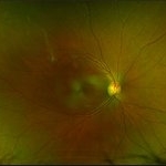
Color fundus photograph of the right eye of a 25-year-old woman who underwent pars plana vitrectomy , relaxing retinectomy , endolaser photocoagulation and silicone oil injection for management of a very advanced vasoproliferative vitreoretinopathy with combined traction / rhegmatogenous RD secondary to a very severe occlusive retinal vasculitis. Retinal reattachment was achieved after surgery.
Photographer: Soulmaz Shahmohammad, Negah Eye Center, Tehran, Iran
Condition/keywords: color fundus photograph, combined retinal detachment, occlusive retinal vasculitis, relaxing retinectomy, vasoproliferative vitreoretinopathy
 Loading…