Search results (466 results)
-
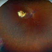
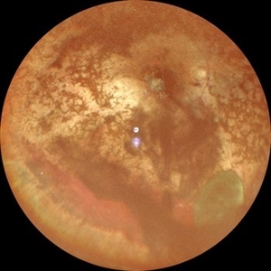
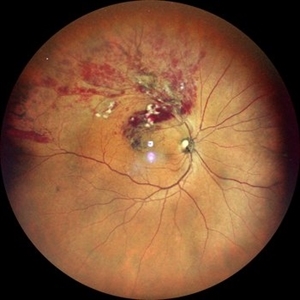
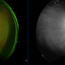 Retinal Macroaneurysm (Left Eye)
Apr 29 2025 by Daniela Bogenschutz
72 year-old female has visual complaints of central vision changes ongoing for 4 days. Patient was acutely symptomatic with an intraretinal hemorrhage due to the retinal macroaneurysm. We had a fun little laugh as this retinal macroaneurysm form a shape of a tick in her left eye. This photo is a side-by-side of the color photos and the autofluorescence done. She is being treated by her general doctor for elevated blood pressure.
Photographer: Daniela Bogenschutz, OSC; Retina Consultants of Carolina, P.A.
Imaging device: Optos
Condition/keywords: retinal macroaneurysm
-
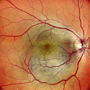
 Extensive Chorioretinal Scarring with Partial Macular Sparring
Apr 22 2025 by Maxwell J Wingelaar, MD
A multicolor photo showing chorioretinal scarring with partial macular sparing in the left eye.
Photographer: Killian Roberts
Imaging device: Heidelberg Spectralis Multicolor Photo
Condition/keywords: chorioretinal atrophy, chorioretinal inflammations
-
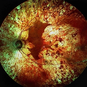
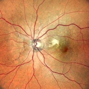
 Extensive Chorioretinal Scarring in the Right Eye
Apr 22 2025 by Maxwell J Wingelaar, MD
A multicolor photo showing chorioretinal scarring with macular involvement in the right eye
Photographer: Killian Roberts
Imaging device: Heidelberg Spectralis Multicolor Photo
Condition/keywords: chorioretinal atrophy, chorioretinal inflammations
-
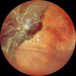
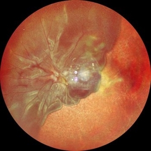
 A Large Break at the Posterior Pole With RD With PVR (S/p Old Blunt Trauma)
Jan 16 2025 by Anand Temkar
Right eye widefield fundus color photo of a 10 year old kid who noticed diminution of vision in right eye since a month. We can see the large break at the posterior pole with rolled up margins associated with retinal detachment and PVR changes.
Photographer: Dr.Anand Temkar- Retina Foundation, Ahmedabad
Imaging device: Mirante
Condition/keywords: posterior pole break, proliferative vitreoretinopathy (PVR), Retinal Detachment
-
 A Large Break at the Posterior Pole With RD With PVR (S/p Old Blunt Trauma)
Jan 16 2025 by Anand Temkar
Right eye central fundus color photo of a 10 year old kid who noticed diminution of vision in right eye since a month. We can see the large break at the posterior pole with rolled up margins associated with retinal detachment and PVR changes.
Photographer: Dr.Anand Temkar- Retina Foundation, Ahmedabad
Imaging device: Mirante
Condition/keywords: Posterior pole break, proliferative vitreoretinopathy (PVR), Retinal Detachment
-
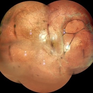
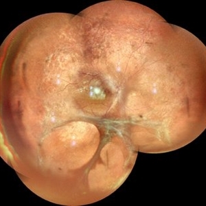
 Coat's Disease
Jan 14 2025 by Kimberly Wakester
Fundus photographs of an 7-year-old boy with Coat's Disease in the right eye. There is subfoveal lipid end scarring in the macula and "light bulb" type telangiectasias temporally noted on exam and shown in Optos color photos. FA findings show anastomoses, capillary dropout, and "light bulb" type telangiectasias temporally with mild late leakage. Patient will be monitored at this time and have repeat imaging in 4 months.
Photographer: Kimberly Wakester, COA
Imaging device: Optos California
Condition/keywords: Coat's disease
-
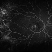
 Retinal Detachment with Multiple OCT Overlays
Jan 7 2025 by Drew Mitchell
Optos 360* Color photo montage with multiple Zeiss Cirrus OCT scan overlays. Retinal Detachment with multiple breaks and a Epiretinal Membrane.
Photographer: Drew Mitchel, OCT-C
Imaging device: Optos California
Condition/keywords: ERM, macular pucker, montage, Optos, OPTOS CALIFORNIA, RD, Retinal Detachment
-
 Uveal Effusion Syndrome
Jan 7 2025 by Drew Mitchell
Optos Color Montage of Uveal Effusion Syndrome
Photographer: Drew Mitchell, OCT-C
Imaging device: Optos California
Condition/keywords: color photo, montage, OPTOS, uveal effusion
-
 Right Eye Color Photo in Case of Choroidal Hemangioma
Nov 29 2024 by Anand Temkar
Right eye color photo of a 42 year old male in case of choroidal hemangioma. We can see the reddish-orange, round to oval choroidal tumor located completely in the posterior half of the fundus.
Photographer: Dr.Anand Temkar- Retina Foundation, Ahmedabad
Imaging device: Mirante
Condition/keywords: Choroidal Hemangioma
-
 Left Eye Color Photo With Extrafoveal CNVM (Stable) in Case of Angioid Streaks
Nov 29 2024 by Anand Temkar
A 45 year old male came with chief complaint of blurring vision in right eyes since past 4 days. His vision is 6/12 in right eye and 6/9 in left eye. His vision was 14 mmHg in right eye and 16 mmHg in left eye. He was diagnosed with Angioid Streaks in both eyes about a year ago, then he developed choroidal neovascularization in his left eye 8 months ago, for which he received AntiVEGF injections x 3. Left eye is a stable eye now. Patient presented with right eye choroidal neovascularization in a case of Angioid Streaks on recent follow up. We have advised him right eye AntiVEGF injections x 3. In this image, the left eye color photo shows angioid streaks with extrafoveal CNVM ( stable ) ( status post antiVEGF x 3 )
Photographer: Dr.Anand Temkar- Retina Foundation, Ahmedabad
Imaging device: Mirante
Condition/keywords: Angioid Streaks, choroidal neovascular membrane (CNVM)
-
 Right Eye Color Photo With Hemorrhages in Case of CNVM With Angioid Streaks
Nov 29 2024 by Anand Temkar
A 45 year old male came with chief complaint of blurring vision in right eyes since past 4 days. His vision is 6/12 in right eye and 6/9 in left eye. His vision was 14 mmHg in right eye and 16 mmHg in left eye. He was diagnosed with Angioid Streaks in both eyes about a year ago, then he developed choroidal neovascularization in his left eye 8 months ago, for which he received AntiVEGF injections x 3. Left eye is a stable eye now. Patient presented with right eye choroidal neovascularization in a case of Angioid Streaks on recent follow up. We have advised him right eye AntiVEGF injections x 3. In this image, the right eye color photo shows bleed from CNVM in case of angioid streaks.
Photographer: Dr.Anand Temkar- Retina Foundation, Ahmedabad
Imaging device: Mirante
Condition/keywords: Angioid Streaks, choroidal neovascular membrane (CNVM)
-
 Vitreous Cyst With Vitreous Hemorrhage in a Case of Retinitis Pigmentosa
Oct 5 2024 by Anand Temkar
This picture shows RE color photo of a 30 year old female with vitreous cyst with spontaneous vitreous hemorrhage in a case of Retinitis Pigmentosa.
Photographer: Dr.Anand Temkar- Retina Foundation, Ahmedabad
Condition/keywords: vitreous cyst, vitreous hemorrhage
-
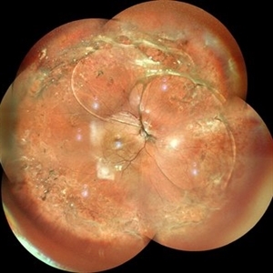
 X-Linked Juvenile Retinoschisis
Oct 5 2024 by Anand Temkar
This is a color photo montage of LE of a 15 year old child with X-linked juvenile retinoschisis.
Photographer: Dr.Anand Temkar- Retina Foundation, Ahmedabad
Imaging device: Mirante
Condition/keywords: x-linked retinoschisis (XLRS)
-
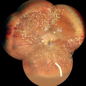
 X-Linked Juvenile Retinoschisis
Oct 5 2024 by Anand Temkar
This is a color photo montage of RE of a 15 year old child with X-linked juvenile retinoschisis.
Photographer: Dr.Anand Temkar- Retina Foundation, Ahmedabad
Imaging device: Mirante
Condition/keywords: x-linked retinoschisis (XLRS)
-
 Re-Retinal Detachment
Sep 20 2024 by Anand Temkar
A 52 year old male came with chief complaints of DOV in LE since past 6 months. He gave history of LE BMV ( no documents ) about a year ago. His vision was 6/9 in RE and PL +ve and PR faulty in LE. In this widefield color photo of LE we can see the detached retina folded on itself revealing bare choroid.
Photographer: Dr.Anand Temkar- Retina Foundation, Ahmedabad
Imaging device: Mirante
-
 Re-Retinal detachment
Sep 20 2024 by Anand Temkar
A 52 year old male came with chief complaints of DOV in LE since past 6 months. He gave history of LE BMV ( no documents ) about a year ago. His vision was 6/9 in RE and PL +ve and PR faulty in LE. In this widefield color photo of LE we can see the detached retina folded on itself revealing bare choroid.
Photographer: Dr.Anand Temkar- Retina Foundation, Ahmedabad
Imaging device: Mirante
-
 OCT in Case of Macular Coloboma (LE)
Sep 18 2024 by Anand Temkar
A 24 year old male came with chief complaint of diminution of vision in both eyes since childhood. Vision in both eyes was 6/24. IOP in RE was 12 and LE was 14 mm of Hg. On fundus examination periphery was within normal limits and central fundus revealed this picture. The serology testing such as serum IgM, IgG for toxoplasma and cytomegalovirus was negative. I have also uploaded LE color photo and BE OCT of this patient.
Photographer: Dr.Anand Temkar- Retina Foundation, Ahmedabad
Imaging device: Mirante
Condition/keywords: Coloboma
-
 OCT in Case of Macular Coloboma (RE)
Sep 18 2024 by Anand Temkar
A 24 year old male came with chief complaint of diminution of vision in both eyes since childhood. Vision in both eyes was 6/24. IOP in RE was 12 and LE was 14 mm of Hg. On fundus examination periphery was within normal limits and central fundus revealed this picture. The serology testing such as serum IgM, IgG for toxoplasma and cytomegalovirus was negative. I have also uploaded LE color photo and BE OCT of this patient.
Photographer: Dr.Anand Temkar- Retina Foundation, Ahmedabad
Imaging device: Mirante
Condition/keywords: coloboma
-
 Macular Coloboma (LE)
Sep 18 2024 by Anand Temkar
A 24 year old male came with chief complaint of diminution of vision in both eyes since childhood. Vision in both eyes was 6/24. IOP in RE was 12 and LE was 14 mm of Hg. On fundus examination periphery was within normal limits and central fundus revealed this picture. The serology testing such as serum IgM, IgG for toxoplasma and cytomegalovirus was negative. I have also uploaded LE color photo and BE OCT of this patient.
Photographer: Dr.Anand Temkar- Retina Foundation, Ahmedabad
Imaging device: Mirante
Condition/keywords: macular coloboma
-
 Macular Coloboma (RE)
Sep 18 2024 by Anand Temkar
A 24 year old male came with chief complaint of diminution of vision in both eyes since childhood. Vision in both eyes was 6/24. IOP in RE was 12 and LE was 14 mm of Hg. On fundus examination periphery was within normal limits and central fundus revealed this picture. The serology testing such as serum IgM, IgG for toxoplasma and cytomegalovirus was negative. I have also uploaded LE color photo and BE OCT of this patient.
Photographer: Dr.Anand Temkar- Retina Foundation, Ahmedabad
Imaging device: Mirante
Condition/keywords: coloboma of macula
-
 Membranes Formed Under Silicon Oil and Retina
Jul 18 2024 by Anjana Mirajkar, MS Ophthalmology
A montage color photo of RE of a 7 year old male with membranes formed between silicon and the retina injected for retinal detachment.
Photographer: Dr. Anjana Mirajkar -Retina Foundation, Ahmedabad
Imaging device: Mirante-Nidek
Condition/keywords: sub-silicon membranes
-
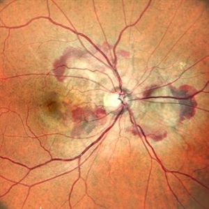
 Idiopathic Retinal Vasculitis
Jun 9 2024 by Anjana Mirajkar, MS Ophthalmology
A color photo montage of an 32 year old male of LE showing laser marks in inferior and superior half with an floating ozurdex implant (inferiorly) in a case of idiopathic retinal vasculitis.
Photographer: Dr. Anjana Mirajkar -Retina Foundation, Ahmedabad
Imaging device: Mirante-Nidek
Condition/keywords: idiopathic retinal vasculitis, laser photocoagulation, Ozurdex implant
-
 Branch Retinal Vein Occlusion
Apr 28 2024 by Anjana Mirajkar, MS Ophthalmology
A widefield color photo of a 55 year old male case of supero-temporal BRVO showing venous tortuosity, cotton wool spots, flame shaped hemorrhages and macular edema.
Photographer: Dr. Anjana Mirajkar -Retina Foundation, Ahmedabad
Imaging device: Mirante-Nidek
Condition/keywords: branch retinal vein occlusion (BRVO), ST BRVO
 Loading…