Search results (404 results)
-
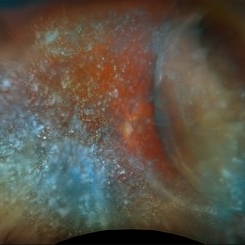
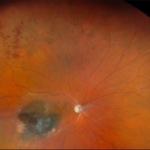
 Ciliochoroidal Melanoma
Jan 15 2026 by Virginia Gebhart
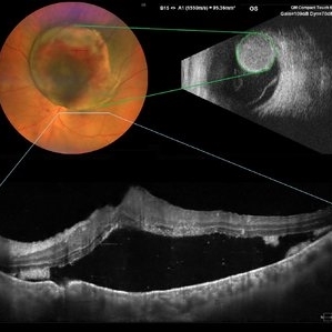
54 year old female referred for choroidal lesion with possible retinal detachment. Pt states she did not notice any vision changes until she failed exam at the DMV. On exam there is a large pigmented ciliary body mass with a height measuring 13.2 mm. Ultrasound shows large mass with low internal reflectivity and vascularity. Due to tumor size enucleation was recommended. Pt will be scheduled for surgery pending CT scan results. Also ordered blood test HLA-A0201 to check eligibility for Kimmtrak if cancer spreads.
Photographer: Virginia Gebhart, Retina Consultants of Carolina
Imaging device: Optos California
Condition/keywords: ciliary body mass, ciliary body melanoma, ciliochoroidal melanoma
-
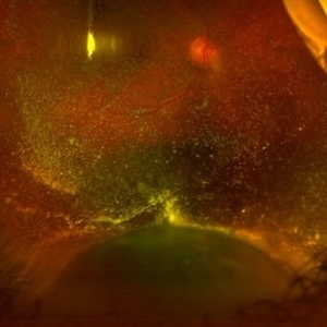
 New Collar Button Melanoma
Jan 7 2026 by Virginia Gebhart
67 year old female referred for possible choroidal melanoma. Clinical exam, photos and ultrasound findings consistent with melanoma. Large pigmented tumor with collar button configuration, subretinal fluid inferior and vitreous hemorrhage. Left eye has scleral pigmentation 360 degrees, pt states she has had "brown spots" on her eye since childhood. Iris and fundus are also noticeably darker in the left eye consistent with oculodermal melanocytosis. Pt states she has had regular eye exams and has never been diagnosed. Pt will be scheduled for plaque brachytherapy pending CT scan results.
Photographer: Virginia Gebhart, Retina Consultants of Carolina
Imaging device: Optos California
Condition/keywords: choroidal melanoma, collar button, hemorrhage, melanoma, Oculodermal Melanocytosis
-
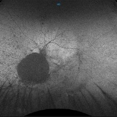
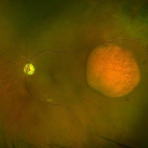
 Nevus to Melanoma in 1 Year
Dec 12 2025 by Virginia Gebhart
83 year old male presents with partially amelanotic choroidal nevus, which has been followed for the last 10 years. On exam there is a new area of orange pigment, and change in diameter compared to photos from 2024. Ultrasound shows small increase in height, medium to low internal reflectivity, and vascularity. Pt is asymptomatic. Due to personal matters pt wishes to reassess in few months and schedule treatment.
Photographer: Virginia Gebhart, Retina Consultants of Carolina
Imaging device: Optos California
Condition/keywords: choroidal melanoma, choroidal nevus
-
 Retinal Plumage
Nov 18 2025 by DR APOORVA JADHAV, MBBS , DNB
A 35 year-old male came in with diminished vision in right eye. On multicolor we can appreciate large pigmented choroidal mass lesion with surrounding srf which is reaching fovea. Diagnosed to be Choroidal Melanoma.
Photographer: Dr Apoorva Jadhav
Imaging device: Heidelberg Spectralis
Condition/keywords: choroidal melanoma, srf
-
 Choroidal and Near Total RD, Severe Asteroid Hyalosis, Treated Melanoma
Oct 22 2025 by Virginia Gebhart
78 year old male with sudden decrease in vision. Poor view due significant asteroid hyalosis. Bscan showed large nasal choroidal and near total retinal detachments, attached temporally. No obvious break found. Regressed tumor inferiorly s/p brachytherapy in April 2023. BCVA 20/320, IOP of 03. Pt schedule for primary PPV and possible SB placement vs. GFE
Photographer: Virginia Gebhart, Retina Consultants of Carolina
Imaging device: Optos California
Condition/keywords: asteroid hyalosis, brachytherapy, choroidal detachment, choroidal melanoma, melanoma, RD, retinal detachment, sub-total retinal detachment
-
Venous Pulsations in Large Choroidal Melanoma
Oct 9 2025 by Virginia Gebhart
81 year old female diagnosed with large, pigmented collar button tumor. Given size of lesion enucleation was recommended. CT scans show no evidence of metastatic disease. Pt is doing well s/p enucleation.
Condition/keywords: bscan ultrasound, choroidal melanoma
-
 Echoes of Malignancy: Mushroom-Pattern Uveal Melanoma
Oct 3 2025 by Claudio Brancato, MD
B-scan ultrasound image depicting a choroidal melanoma with the classic “mushroom-shaped” configuration, resulting from Bruch’s membrane rupture and dome-shaped tumor growth. The lesion shows internal echogenicity and well-defined borders, consistent with the typical echographic appearance of malignant uveal tumors.
Photographer: Gregorio Lo Giudice, MD - University of Palermo, ARNAS Civico Hospital
Condition/keywords: Choroidal, ecography, melanoma
-
 Choroidal Melanoma
Oct 3 2025 by Virginia Gebhart
63 year old male with new choroidal melanoma. Pt states vision has been poor for 2 years, worsening in the last year. Bscan ultrasound shows lesion extends into the macula up to the optic nerve. Recommended enucleation due to size of lesion (7.7 x 15.6 x 15.2) and poor prognosis of visual recovery. Surgery will be scheduled pending CT scan results.
Photographer: Virginia Gebhart, Retina Consultants of Carolina
Imaging device: Optos California
Condition/keywords: choroidal melanoma, exudative detachment
-
 Choroidal Melanoma
Oct 1 2025 by Virginia Gebhart
60 year old male referred by optometrist for retinal detachment. Pt had been having symptoms of flashing lights and shadow in vision for approximately 1 month. Exam and diagnostics consistent with choroidal melanoma with exudative detachment inferior. Due to size of lesion (10.8 x 14.8 x 13.2) enucleation was recommended. Pt will be scheduled for surgery pending CT scan results.
Photographer: Virginia Gebhart, Retina Consultants of Carolina
Imaging device: Optos California
Condition/keywords: choroidal melanoma, exudative detachment, melanoma
-
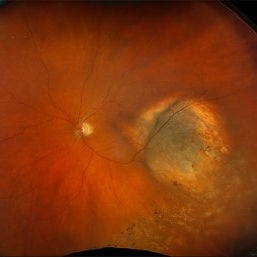
 Amelanotic Melanoma
Sep 19 2025 by Aditya S Kelkar, MS, FRCS, FASRS,FRCOphth
Widefield fundus photograph of a 37 year old showing a large, dome-shaped, intraocular mass involving the temporal retina. The lesion appears elevated and lacks surface pigmentation. Overlying retinal vessels are displaced and draped across the tumor surface, with surrounding retinal elevation noted. The appearance is suggestive of amelanotic variant of choroidal melanoma.
Photographer: Dr. Muskan Mangal
Imaging device: Optos Daytona
Condition/keywords: choroidal melanoma, intraocular tumor
-
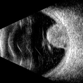
 B-scan Ultrasound of Choroidal Melanoma with Serous Retinal Detachment
Sep 5 2025 by Kristen Wagner
B-scan ultrasound of a choriodal melanoma with serous retinal detachment.
Photographer: Kristen Wagner, COT Tennessee Retina
Condition/keywords: B scan ultrasound, Choroidal melanoma, serous retinal detachment
-
 Jupiter and the Sun
Aug 20 2025 by Gustavo Uriel Fonseca Aguirre
This ultra-widefield fundus photograph demonstrates a peripheral inferior choroidal melanoma with overlying asteroid hyalosis. The lesion shows characteristic pigmentation and irregular borders, while the asteroid bodies appear as numerous refractile opacities distributed throughout the vitreous cavity.
Photographer: Gustavo U. Fonseca Aguirre, Hospital Conde de Valenciana, Ciudad de México
Condition/keywords: asteroid hyalosis, choroidal melanoma
-
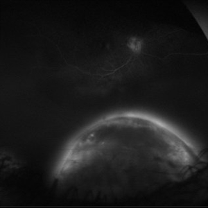 Full Moon
Aug 20 2025 by Gustavo Uriel Fonseca Aguirre
This ultra-widefield fluorescein angiography reveals a hyperfluorescent peripheral inferior choroidal melanoma. The lesion demonstrates early heterogeneous hyperfluorescence with progressive late staining and diffuse leakage.
Photographer: Gustavo U. Fonseca Aguirre, Hospital Conde de Valenciana, Ciudad de México
Condition/keywords: choroidal melanoma, FLUORESCEIN ANGIOGRAPHY
-
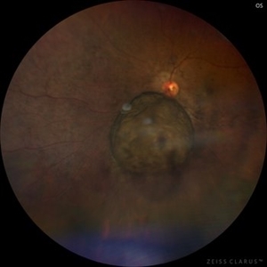
 Choroidal Melanoma
Aug 19 2025 by JEFFERSON R SOUSA, Tecg.º (Biomedical Systems Technology)
A 54-year-old woman with progressive visual acuity loss in her left eye was admitted to the institution with a significant elevated lesion in the upper arch with macular involvement, confirmed by wide-angle fundus photography, ultrasound, and optical coherence tomography.
Photographer: JEFFERSON ROCHA DE SOUSA - Retinal Department at Lens Oftalmologia, Sao Paulo-Brazil
Imaging device: Clarus 700 - Zeiss, composite of four 135 degree images.
Condition/keywords: melanoma
-
 Amelanotic Melanoma
Aug 12 2025 by César Adrián Gómez Valdivia, MD
This FAF image reveals a hypoautofluorescent mass with areas of dense hyperautofluorescent stippling—a classic pattern suggestive of an amelanotic choroidal melanoma. Amelanotic melanoma is a rare variant of uveal melanoma, accounting for only a minority of cases. Unlike pigmented melanomas, these lesions lack melanin, making them more challenging to detect on conventional color fundus imaging. FAF Characteristics: • Central hypoautofluorescence: due to loss or compression of the RPE • Peripheral hyperautofluorescent speckling: consistent with lipofuscin accumulation or RPE disruption • Often associated with subretinal fluid or orange pigment seen clinically Location: Juxtapapillary, with potential optic nerve involvement—a factor that complicates both diagnosis and
Photographer: @eyemissu2
Imaging device: California ICG OPTOS
Condition/keywords: amelanotic melanoma
-
 Amelanotic Melanoma
Aug 12 2025 by César Adrián Gómez Valdivia, MD
This case highlights an amelanotic melanoma, an atypical presentation of a choroidal melanoma lacking the characteristic pigmentation. These lesions can easily be mistaken for choroidal hemangiomas, metastases, or inflammatory masses. Clinically, the lesion appears as a dome-shaped, yellowish subretinal mass, often associated with subretinal fluid, lipofuscin deposition, or retinal detachment. The absence of pigment can delay diagnosis, making multimodal imaging essential. Diagnostic tools: • B-scan ultrasound: low to medium internal reflectivity • OCT: overlying subretinal fluid and RPE elevation • FAF: orange pigment and RPE disruption • ICG/FA: variable, often hypofluorescent core Important: Prompt referral to ocular oncology is critical for management and prognosis.
Photographer: @eyemissu2
Imaging device: TOPCON TRC-50DX
Condition/keywords: amelanotic melanoma
-
 New Choroidal Melanoma
Jul 16 2025 by Virginia Gebhart
78 year old male with a partially amelanotic dome-shaped lesion with RPE changes, hard exudates, overlying intraretinal fluid and minimal SRF temporally. Exam and ultrasound findings consistent with choroidal melanoma. Pt will be scheduled for brachytherapy pending CT scan results.
Photographer: Virginia Gebhart, Retina Consultants of Carolina
Imaging device: Optos California
Condition/keywords: amelanotic melanoma, choroidal melanoma
-
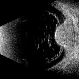
 Choroidal Melanoma (USG)
Jul 5 2025 by Gustavo Uriel Fonseca Aguirre
This B-mode transverse ultrasound scan reveals a mushroom-shaped choroidal tumor in the inferior nasal quadrant adjacent to the optic nerve head. The lesion appears solid with homogeneous internal reflectivity and is associated with minimal surrounding subretinal fluid and scleral excavation. It measures 6.54 mm in height × 7.52 mm in base diameter (transverse view) and extends 9.52 mm longitudinally. The vitreous contains abundant punctate opacities consistent with pigment dispersion. The retina and choroid remain attached elsewhere.
Photographer: Gustavo U. Fonseca Aguirre, Hospital Conde de Valenciana, Ciudad de México
Condition/keywords: choroidal melanoma
-
 Choroidal Hemangioma (AF)
Jul 5 2025 by Gustavo Uriel Fonseca Aguirre
This wide-field fundus autofluorescence image demonstrates a mushroom-shaped choroidal melanoma adjacent to the optic nerve head, exhibiting hypo-autofluorescence (melanin). Vitreous pigment dispersion (tobacco dust sign) is evident, indicating tumor activity.
Photographer: Gustavo U. Fonseca Aguirre, Hospital Conde de Valenciana, Ciudad de México
Condition/keywords: choroidal melanoma
-
 Choroidal Melanoma
Jul 5 2025 by Gustavo Uriel Fonseca Aguirre
This 50° central fundus photograph reveals a mushroom-shaped choroidal melanoma adjacent to the optic nerve head. The lesion demonstrates characteristic pigmentation with overlying vitreous pigment dispersion (tobacco dust sign).
Photographer: Gustavo U. Fonseca Aguirre, Hospital Conde de Valenciana, Ciudad de México
Condition/keywords: choroidal melanoma
-
 Choroidal Melanoma
Jul 3 2025 by Gustavo Uriel Fonseca Aguirre
This B-mode transverse ultrasound scan shows asteroid hyalosis with partial posterior vitreous detachment. A dome-shaped choroidal melanoma is observed in the inferior quadrant (preequatorial to equatorial region), appearing as a solid, regularly bordered lesion with heterogeneous internal structure and mild acoustic attenuation. Standardized A-mode reveals medium-to-low internal reflectivity. The tumor measures 11.62 mm in base diameter and 6.60 mm in height. The retina and choroid remain attached, with minimal suprachoroidal fluid in the inferior quadrant.
Photographer: Gustavo U. Fonseca Aguirre, Hospital Conde de Valenciana, Ciudad de México
Condition/keywords: choroidal melanoma
-
 RPE Rip s/p Brachytherapy for Malignant Melanoma
Jun 20 2025 by Virginia Gebhart
77 year old female with regressing tumor 4 months s/p brachytherapy. RPE rip at inferior edge of lesion.
Photographer: Virginia Gebhart, Retina Consultants of Carolina
Imaging device: Optos California
Condition/keywords: brachytherapy, choroidal melanoma, RPE Rip
-
 Massive Choroidal Melanoma
Jun 18 2025 by Corey R Lacher, MD
A 57-year-old patient presented with no light perception vision in her right eye. B-scan ultrasonography revealed evidence of a large choroidal melanoma. External photography demonstrated detached retina visible just posterior to the lens. The patient subsequently underwent enucleation, and histopathologic examination confirmed the diagnosis of choroidal melanoma. The tumor measured 24 mm anteroposteriorly, 24 mm horizontally, and 25 mm vertically.
Photographer: Beth Malpica
Condition/keywords: choroidal melanoma
-
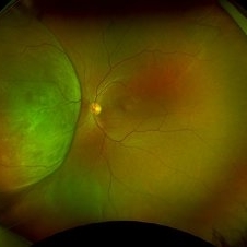
 Amelanotic Choroidal Melanoma with Optic Atrophy
Jun 11 2025 by Aditya S Kelkar, MS, FRCS, FASRS,FRCOphth
Fundus photograph of a 64-year-old woman with optic atrophy and amelanotic choroidal melanoma temporal to the macula.
Photographer: Dr Harsh Jain, National Institute of Ophthalmology
Imaging device: Optos Daytona
Condition/keywords: amelanotic melanoma, optic atrophy
-
 Radiation Retinopathy with BRVO
May 28 2025 by Virginia Gebhart
46 year old male with regressing choroidal melanoma. Stable pigment dispersion over biopsy site, BRVO secondary to radiation retinopathy. BCVA CF, will continue to observe.
Photographer: Virginia Gebhart, Retina Consultants of Carolina
Imaging device: Optos California
Condition/keywords: brachytherapy, branch retinal vein occlusion (BRVO), BRVO, Choroidal melanoma, melanoma, radiation retinopathy
 Loading…