Search results (27 results)
-
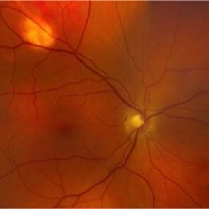
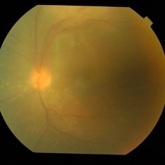
 Congenital hypertrophy of the retinal pigment epithelium (CHRPE)
Jun 22 2022 by Dawson Winter
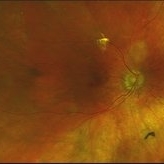
Ultrawide field fundus autofluorescence optos image of the left eye of a 28 year old female. Patient admits to floaters that come and go, but has no other ocular symptoms at this time. At the time of the appointment the patient was seeing 20/30+1 OS. Patient underwent MRI testing of the ocular orbit and results were found to be normal.
Photographer: Dawson Winters
Imaging device: Optos California
Condition/keywords: autofluorescence imaging, choroidal lesions, congenital hypertrophy of the retinal pigment epithelium (CHRPE), fundus autofluorescence (FAF), left eye, lesion, Optos, temporal retina, ultra-wide field imaging
-
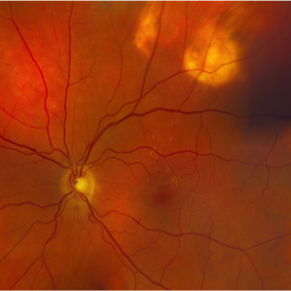
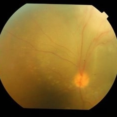
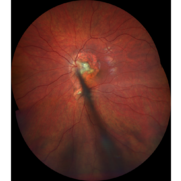 Nerve Stalk
Jun 18 2021 by Kristen Wagner
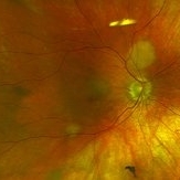
Fundus photograph of a hyperplastic primary vitreous, nerve stalk, and choroidal lesion of a 7-year-old male.
Photographer: Kristen Wagner, COT Tennessee Retina Nashville TN
Imaging device: Clarus
Condition/keywords: choroidal lesions, nerve, vitreous
-
 Rare Bilateral Choroidal Metastasis from Occult Primary Lung Cancer
May 5 2021 by Deependra Vikram Singh, MD FASRS
Fundus photographs and OCT scans of a 73-year-old non-smoker Indian male who presented to our retina clinic in 2013 with blurred vision in left eye for past 2 weeks. BCVA was 20/20 in right eye and 20/40 in left eye. Slit lamp exam was unremarkable for both eyes with no cells in aqueous or anterior vitreous. Fundus examination revealed creamy yellow choroidal lesions in both eyes. Lesion in right eye was one disc diameter (DD) in size and was located close to fovea (Fig-1a). Lesion in the left eye was bigger with a size of 2 DD located superior to fovea (Fig-1b). OCT scan for left eye revealed neurosensory detachment involving fovea (Fig-1c). Fundus fluorescein angiography was inconclusive for right eye and showed late hyper fluorescence the choroidal lesion in left eye. Patient underwent detailed systemic work up for malignancy that revealed primary lung non-small cell carcinoma. He had widespread metastasis affecting liver and brain. Palliative chemotherapy and radiotherapy were initiated 4 weeks after he presented to us. The choroidal lesions show progression on fundus picture and OCT scans done at 4 weeks follow up after initial presentation (Fig – 1d, e, f). The lesions in both eyes show regression at 4 weeks and 12 weeks follow up after initiation of therapy. Unfortunately, patient succumbed at 13 weeks follow up due to disease progression. The case demonstrates rare bilateral choroidal metastasis from primary lung cancer and also highlights that lesions can be asymptomatic till they develop neurosensory detachment as evident from asymptomatic lesion in right eye despite proximity to fovea and symptomatic lesion in left eye with NSD.
Photographer: Deependra Vikram Singh, Eye-Q Superspecialty Eye Hospitals, Gurugram
Imaging device: Topcon
Condition/keywords: choroidal mass, choroidal metastasis
-
 Pneumocystis Carinii Choroiditis
Dec 12 2019 by McGill University Health Centre
38-year-old HIV positive patient with AIDS. Fundoscopy showing large and small subretinal (choroidal) nodular lesions throughout the retina in particular between the arcades
Photographer: Miguel N. Burnier, McGill University Health Center-McGill University Ocular Pathology & Translational Research Laboratory
Imaging device: Fundoscopy
Condition/keywords: AIDS, choroidal lesions, HIV, pneumocystis carinii choroiditis
-
 Pneumocystis Carinii Choroiditis
Dec 12 2019 by McGill University Health Centre
38-year-old HIV positive patient with AIDS. Fundoscopy shows large and small subretinal (choroidal) nodular lesions through out the retina in particular between the arcades.
Photographer: Miguel N. Burnier, McGill University Health Center-McGill University Ocular Pathology & Translational Research Laboratory
Imaging device: Fundoscopy
Condition/keywords: AIDS, choroidal lesions, HIV
-
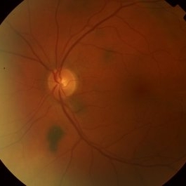
 Choroidal Metastasis from Breast Cancer
Oct 1 2019 by John S. King, MD
60-year-old white female with four year history of breast cancer associated with metastases to many organs including the CNS, was sent her to r/o melanoma, found on routine exam. Visual acuity was HM; there was NSC/PSC; there was a unilateral, large choroidal lesion in the posterior pole that was yellow, well circumscribed, with plateau configuration associated with SRF adn heme.
Photographer: Kay Dalby
Imaging device: Optos CA
Condition/keywords: breast cancer, choroidal lesions, choroidal metastasis
-
 Choroidal Metastasis from Breast Cancer
Oct 1 2019 by John S. King, MD
60-year-old white female with four year history of breast cancer associated with metastasis to many organs including the CNS, was sent her to r/o melanoma, found on routine exam. Visual acuity was HM; there was NSC/PSC; there was a unilateral, large choroidal lesion in the posterior pole that was yellow, well circumscribed, with plateau configuration associated with SRF adn heme.
Photographer: Kay Dalby
Imaging device: Optos CA
Condition/keywords: breast cancer, choroidal lesions, choroidal metastasis
-
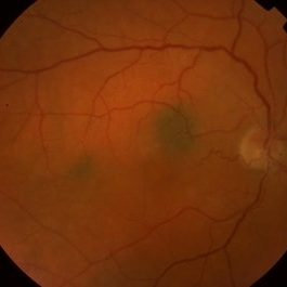
 Metastatic NSCLCA to the Choroid: lesions regressing while undergoing chemotherapy
May 27 2019 by John S. King, MD
Two small, yellow, choroidal lesions can be seen above the nerve and IT arcade can be seen that have regressed compared to the initial photos. Vision 20/20.
Photographer: Shelly Blair
Imaging device: Optos CA
Condition/keywords: choroidal metastasis, lung cancer metastasis
-
 Metastatic NSCLCA to the Choroid: Initial Appearance
May 27 2019 by John S. King, MD
60-year-old white male non-smoker presented to Dr. Zocchi with acute transient decreased vision in the right eye. Background history includes metastatic NSCLC (adenocarcinoma). Acuity OD 20/60, and posterior segment had two small, yellow, choroidal lesions, above the nerve and IT arcade (these had a fairly smooth and dome shaped appearance on the OCT, and top lesion had mild SRF) (see photo)
Photographer: Shelly Blair
Imaging device: Optos CA
Condition/keywords: choroidal metastasis, lung cancer metastasis
-
 BDUMP
BDUMP
Dec 11 2018 by John S. King, MD
67-year-old white female with normal vision four months ago, consulted for dry AMD. She reported that vision in the left eye had worsened over the last two months and had progressively gotten worse. Denied history of cancer, or her primary eye doctor ever mentioning choroidal nevi. Va cc was 20/30 OD and 20/100 OS. No RAPD. IOP 9-10 OU. Anterior segment had some stellate like pigmented dusting of the endothlium, a/c was quiet, 2+NSC OU. Vitreous quiet; multiple, flat, pigmented choroidal lesions varying in size was seen the in fundus. Area in the temporal macula extending up to the superior arcade in the left eye that was suspicious for a mass; it did have a "giraffe like" pattern on one of the early FA pics; the OCT in this area showed thickening of the choroid without a definite mass lesion, and overlying thickening of the RPE, or exudative like scar, with SRF directly above. Consulted with Dr. Matt Wilson, who confirmed diagnosis, and had patient evaluated by oncology, who diagnosed non-small cell lung cancer.
Photographer: Stacey Coleman
Imaging device: Topcon
Condition/keywords: bilateral diffuse uveal melanocytic proliferation (BDUMP)
-
 BDUMP
BDUMP
Dec 11 2018 by John S. King, MD
67-year-old white female with normal vision four months ago, consulted for dry AMD. She reported that vision in the left eye had worsened over the last two months and had progressively gotten worse. Denied history of cancer, or her primary eye doctor ever mentioning choroidal nevi. Va cc was 20/30 OD and 20/100 OS. No RAPD. IOP 9-10 OU. Anterior segment had some stellate like pigmented dusting of the endothlium, a/c was quiet, 2+NSC OU. Vitreous quiet; multiple, flat, pigmented choroidal lesions varying in size was seen the in fundus. Area in the temporal macula extending up to the superior arcade in the left eye that was suspicious for a mass; it did have a "giraffe like" pattern on one of the early FA pics; the OCT in this area showed thickening of the choroid without a definite mass lesion, and overlying thickening of the RPE, or exudative like scar, with SRF directly above. Sent patient to Dr. Matt Wilson, who confirmed BDUMP, and had patient sent to oncology to find a possible primary lesion. Mass seen on CT chest; biopsy revealed non-small cell lung cancer, and is getting chemo/radio treatment. Ocular findings have not progressed over the last few months.
Photographer: Stacey Coleman
Imaging device: Topcon 50
Condition/keywords: bilateral diffuse uveal melanocytic proliferation (BDUMP)
-
 Sclerochoroidal Calcification OD
Nov 9 2016 by Courtney Crawford, MD, FACS
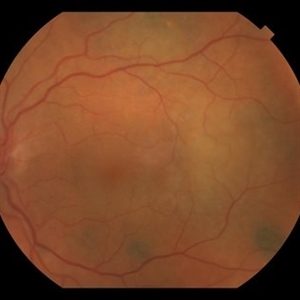
55-year-old male with stable choroidal lesions in the superotemporal quadrant of both eyes.
Condition/keywords: idiopathic sclerochoroidal calcification
-
 Sclerochoroidal Calcification OS
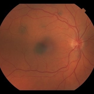
Nov 9 2016 by Courtney Crawford, MD, FACS
55-year-old male with stable choroidal lesions in the superotemporal quadrant of both eyes
Condition/keywords: idiopathic sclerochoroidal calcification
-
 Renal Cell Carcinoma
Dec 29 2015 by David W. Faber, MD
Wide angle image of a 65-year-old male with a history of metastatic renal cell carcinoma, presented with decreased vision and multiple choroidal lesions consistent with metastatic lesions.
Imaging device: Optos California
Condition/keywords: renal cell, tumor
-
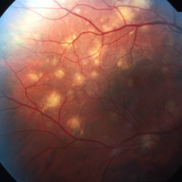
 Pneumocystis Carinii Choroiditis
Jan 5 2015 by H. Michael Lambert, MD
Multiple, cream colored, flat choroidal lesions.
Condition/keywords: pneumocystis carinii choroiditis
-
 Small Choroidal Nevi
Jul 9 2014 by Susanna S. Park, MD, PhD
Asymptomatic 58-year-old woman with subtle small flat pigmented choroidal lesions around the disc and in the periphery consistent with choroidal nevi.
Photographer: Ellen Redenbo
Condition/keywords: choroidal nevus
-
 Multiple Choroidal Nevi
Jul 9 2014 by Susanna S. Park, MD, PhD
Asymptomatic 45-year-old woman with multiple small pigmented choroidal lesions in the posterior pole consistent with nevi.
Photographer: Ellen Redenbo
Condition/keywords: choroidal nevus
-
 Macular Nevi
Jul 9 2014 by Susanna S. Park, MD, PhD
Fundus photograph of an asymptomatic 40-year-old woman noted with subtle pigmented choroidal lesions in the macula consistent with small nevi.
Photographer: Ellen Redenbo
Condition/keywords: choroidal nevus
-
 Tubercular Granuloma
May 15 2014 by Mallika Goyal, MD
Left Eye Fundus of a 35-year-old HIV sero-positive male with visual symptoms for 3 months shows multiple punctate choroidal lesions in the mid-periphery, likely disseminated tuberculosis lesions. Not on ART, CD4 34, starting antitubercular drugs for pulmonary and disseminated tuberculosis.
Photographer: Mallika Goyal, MD, Apollo Health City, Jubilee Hills, Hyderabad, India
Condition/keywords: tubercular choroidal granuloma
-
 Tubercular Granuloma
May 15 2014 by Mallika Goyal, MD
Left eye fundus of a 35-year-old HIV sero-positive male with visual symptoms for 3 months shows multiple punctate choroidal lesions in the mid-periphery, likely disseminated tuberculosis lesions. Not on ART, CD4 34, starting antitubercular drugs for pulmonary and disseminated tuberculosis.
Photographer: Mallika Goyal, MD, Apollo Health City, Jubilee Hills, Hyderabad, India
Condition/keywords: tubercular choroidal granuloma
-
 Tubercular Granuloma
May 15 2014 by Mallika Goyal, MD
Left Eye Fundus of a 35-year-old HIV sero-positive male with visual symptoms for 3 months shows multiple punctate choroidal lesions in the mid-periphery, likely disseminated tuberculosis lesions. Not on ART, CD4 34, starting antitubercular drugs for pulmonary and disseminated tuberculosis.
Photographer: Mallika Goyal, MD, Apollo Health City, Jubilee Hills, Hyderabad, India
Condition/keywords: tubercular choroidal granuloma
-
---thumb.jpg/image-square;max$300,300.ImageHandler) Birdshot Retinochoroidopathy
Birdshot Retinochoroidopathy
Feb 26 2013 by Henry J. Kaplan, MD
Birdshot retinochoroidopathy. Multiple ovoid yellow choroidal lesions spread out radially most prominent on nasal side.
Condition/keywords: birdshot, birdshot retinochoroidopathy
-
---thumb.jpg/image-square;max$300,300.ImageHandler) Multifocal Choroiditis and Panuveitis Syndrome
Multifocal Choroiditis and Panuveitis Syndrome
Feb 26 2013 by Henry J. Kaplan, MD
Multifocal choroiditis and panuveitis: left eye. Acute stage: haziness of the media due to vitritis and multiple active yellow and also inactive choroidal lesions are present.
Condition/keywords: multifocal choroiditis
-
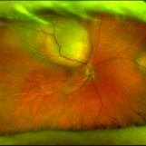
---thumb.jpg/image-square;max$300,300.ImageHandler) late-phase FA showing arteriolar attenuation and late staining of choroidal lesions
late-phase FA showing arteriolar attenuation and late staining of choroidal lesions
Feb 14 2013 by From the Collections of Thomas M. Aaberg, MD and Thomas M. Aaberg Jr., MD
late-phase FA showing arteriolar attenuation and late staining of choroidal lesions
Condition/keywords: multifocal choroiditis, posterior segment inflammation, white dot syndrome
 Loading…