Search results (83 results)
-
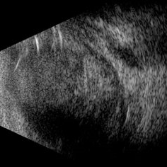 Scleral Rupture
May 9 2025 by Gustavo Uriel Fonseca Aguirre
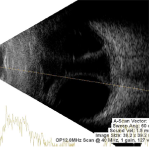
This B-mode longitudinal ultrasound scan reveals dense vitreous hemorrhage, subretinal fluid, annular choroidal detachment, and scleral wall discontinuity with adjacent scleral folds. These findings indicate severe ocular trauma with probable scleral rupture and multi-compartment involvement.
Photographer: Gustavo U. Fonseca Aguirre, Hospital Conde de Valenciana, Ciudad de México
Condition/keywords: ocular trauma, scleral rupture
-
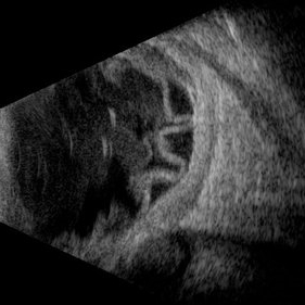 Traumatic Retinal Detachment
May 5 2025 by Gustavo Uriel Fonseca Aguirre
This B-mode longitudinal ultrasound scan over the macular area reveals vitreous hemorrhage, retinal detachment with folding, peripheral annular choroidal detachment, and sub-Tenon's fluid in the setting of blunt ocular trauma. The findings indicate severe posterior segment disruption with multi-compartment involvement.
Photographer: Gustavo U. Fonseca Aguirre, Hospital Conde de Valenciana, Ciudad de México
Condition/keywords: blunt trauma, Retinal Detachment
-
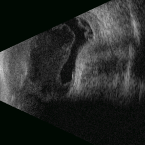 Posterior Nodular Scleritis
Apr 23 2025 by Gustavo Uriel Fonseca Aguirre
This B-mode ultrasound scan demonstrates a posterior scleral nodule accompanied by vitritis, serous retinal detachment, and annular choroidal detachment. The nodule appears as a localized hypoechoic scleral thickening, while the serous retinal detachment shows a smooth convex configuration. The choroidal detachment presents with the characteristic ring-shaped elevation, suggesting significant intraocular inflammation or hypotony.
Photographer: Gustavo U. Fonseca Aguirre, Hospital Conde de Valenciana, Ciudad de México
Condition/keywords: posterior nodular scleritis, posterior scleritis
-
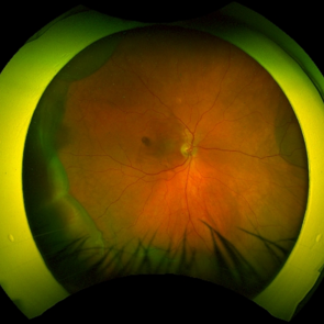 Serous Choroidal Detachment
Apr 18 2025 by Tracy Vu
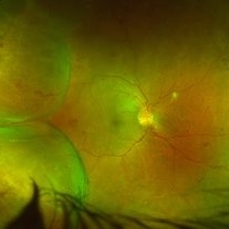
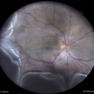
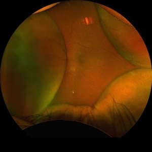
Fundus photograph of a 77-year-old male with near 360 degree choroidal effusions accompanied by a mild serous choroidal detachment in the inferotemporal quadrant.
Condition/keywords: choroidal effusion, serous choroidal detachment, serous retinal detachment
-
Hemorrhagic Choroidal Detachment
Apr 14 2025 by Gustavo Uriel Fonseca Aguirre
This B-mode transverse ultrasound scan demonstrates a hemorrhagic choroidal detachment with a characteristic wreath-like configuration, accompanied by concurrent retinal detachment. The choroidal lesion shows dome-shaped elevation with heterogeneous internal reflectivity, while the detached retina appears as a hyperechoic, undulating membrane.
Photographer: Gustavo U. Fonseca Aguirre, Hospital Conde de Valenciana, Ciudad de México
Condition/keywords: hemorrhagic choroidal detachment
-
360 Choroidal Detachment in Uveal Effusion Syndrome
Apr 11 2025 by Siri Uppuluri
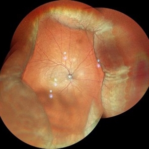
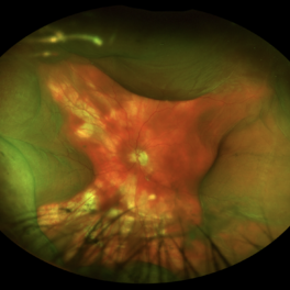
Fundus photograph of a phakic left eye in an 82-year-old man demonstrating 360 choroidal detachment secondary to uveal effusion syndrome. He underwent sclerectomy and drainage of the choroidal effusions with resolution after surgical intervention.
Photographer: Siri Uppuluri, MD; Rutgers New Jersey Medical School
Condition/keywords: choroidal detachment
-
 Uveal Effusion Syndrome in a Nanophthalmic Eye
Apr 3 2025 by Gustavo Uriel Fonseca Aguirre
B-mode ultrasonography of a nanophthalmic eye reveals diffuse choroidal and scleral thickening, annular ciliochoroidal detachment, and sub-Tenon fluid accumulation.
Photographer: Gustavo U. Fonseca Aguirre, Hospital Conde de Valenciana, Ciudad de México
Condition/keywords: nanophthalmos, uveal effusion syndrome
-
 Three Kisses
Mar 18 2025 by Gustavo Uriel Fonseca Aguirre
Cross-section of a B-mode ultrasound showing a kiss-shaped choroidal detachment; three lobes, giving the appearance of three kisses.
Photographer: Gustavo U. Fonseca Aguirre, Hospital Conde de Valenciana, Ciudad de México
Condition/keywords: Kissing Choroidal Detachment
-
 Hemorrhagic Choroidals
Jan 22 2025 by Danish Shabbir, Ophthalmic Technologist
78 year old female complains of suddenly vision decrease 2 days ago.
Photographer: Danish Shabbir,Retina-EyeCare Centre
Imaging device: Optos California
Condition/keywords: choroidal detachment, Retinal Detachment, retinal detachment with choroidal
-
 Combined Pathology
Oct 26 2024 by rahul saradge
53 year old male patient was presented with a complaints of diminished vision in LE since 1 month. The BCVA in RE was 6/36p and LE was CF 1/2m. Ocular dilated examination showed RE temporal CD with ?CRVO,OIS and OS showed TRD and old Hemi CRVO. Patient was injected with PST tricot followed by PRP laser at an interval of 1 week. Patient improved to BCVA 6/9.
Photographer: Aishwarya Bangar Isha Netralaya Thane
Imaging device: optos
Condition/keywords: choroidal detachment, crvo, ois, optos, pan retinal photocoagulation, tractional retinal detachment
-
 RE-OCT in Choroidal Detachment
Sep 22 2024 by Anand Temkar
A 52 year old male came with chief complaints of diminution of vision in RE since past 15 days. He gave history of ( RE ) cataract surgery + IOL about 2 months ago. His vision was 6/9 in RE and PL + ve, PR inaccurate in LE. His IOP was 10 mm of Hg in RE and 20 mm of Hg in LE.
Photographer: Dr.Anand Temkar- Retina Foundation, Ahmedabad
Imaging device: Mirante
Condition/keywords: choroidal detachment, choroidals, serous choroidal detachment
-
 360 Degrees Choroidal Detachment
Sep 22 2024 by Anand Temkar
A 52 year old male came with chief complaints of diminution of vision in RE since past 15 days. He gave history of ( RE ) cataract surgery + IOL about 2 months ago. His vision was 6/9 in RE and PL +ve, PR inaccurate in LE. His IOP was 10 mm of Hg in RE and 20 mm of Hg in LE.
Photographer: Dr.Anand Temkar- Retina Foundation, Ahmedabad
Imaging device: Mirante
Condition/keywords: choroidal detachment, choroidals, serous choroidal detachment
-
 Choroidal Detachment
Aug 14 2024 by STEFANY DAVILA
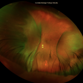
Montage of fundus photography of an elderly male with choroidal detachment 360 degrees after trabeculectomy surgery.
Photographer: Stefany Dávila Avila, Instituto Mexicano de Oftalmología, Querétaro
Imaging device: Zeiss Clarus 700
Condition/keywords: Choroidal, detachment
-
 Retinal Detachment With Choroidal Detachment
Aug 6 2024 by Akansha Sharma
Color fundus photograph of a 49 year old male with retinal detachment and choroidal detachment.
Photographer: Dr. Akansha Sharma, Bharati Eye Hospital
Condition/keywords: CD, choroidal detachment, RD, Retinal Detachment
-
 Choroidal Detachment OS
Jul 5 2024 by Zach Seim
Optos Fundus Photograph of a Choroidal Detachment OS in a 75 year old male. VA at presentation was DCC HM.
Photographer: Zach Seim
Imaging device: Optos California
Condition/keywords: choroidal detachment, choroidal mass, left eye, optos, OPTOS CALIFORNIA
-
 Giant Retinal Tear with Choroidal Detachment
Jun 12 2024 by Anand Temkar
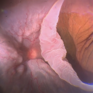
Intra operative still of a 34 year old male showing Giant Retinal Tear with Choroidal Detachment.
Photographer: Dr.Anand Temkar- Retina Foundation, Ahmedabad
Condition/keywords: choroidal detachment, giant retinal tear
-
 Proliferative Vitreoretinopathy
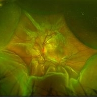
Jun 9 2024 by Marcelo Zas, MD PhD
We present a case of a 20-year-old patient who underwent surgery for congenital cataract when he was born and 20 years after he developed a retinal detachment with proliferative vitreoretinopathy. Proliferative vitreoretinopathy (PVR), a major complication of rhegmatogenous retinal detachment (RRD), is an abnormal process whereby proliferative, contractile cellular membranes form in the vitreous and on both sides of the retina, resulting in tractional retinal detachment with fixed retinal folds. PVR arises in an estimated 5-10% of RRD cases, and therefore represents a major complication of retinal detachment. The best treatment of PVR is its prevention. Clinical factors associated with increased risk of PVR include: • Chronic RRD • 2 o more horseshoe retinal tears and RRD exposing three-disc diameters or more of RPE • RD associated with giant retinal • RD associated with choroidal detachment • Ocular Trauma • RRD associated with vitreous hemorrhage • Aphakia and RRD • Failure of previous surgery or multiple retinal surgeries • Aggressive retinitis, etc.
Photographer: Luciano Scorsetti MD
Condition/keywords: proliferative vitreoretinopathy (PVR)
-
 Choroidal and Retinal Detachment Secondary to Full-thickness Macular Hole
May 24 2024 by Tony Y Chen, MD
Optos photograph of a 61-year-old woman with choroidal and retinal detachment secondary to full-thickness macular hole.
Condition/keywords: choroidal detachment, Retinal detachment with macular hole
-
 Choroidal Detachment
Feb 6 2024 by Thirumalesh Mochi Basavaraj, MD
60 year old gentleman post trab, presenting with serous choroidal detachment.
Photographer: Puttaswamy
Condition/keywords: serous choroidal detachment
-
 Choroidal Detachment
Jan 12 2024 by ANKIT JAIN
Membranous echoes with moderate to high spikes with restricted after movements suggestive of choroidal detachment.
Photographer: Dr Ankit Jain
Condition/keywords: choroidal detachment
-
 Choroidal Detachment
Nov 23 2023 by Anand Temkar
LE color photo montage showing choroidal detachment of a 63 years old male who gives history of LE filtration surgery with mitomycin c and anterior vitrectomy elsewhere a month ago.
Photographer: Dr.Anand Temkar- Retina Foundation, Ahmedabad
Imaging device: Mirante
Condition/keywords: choroidal detachment
-
 CD
CD
Oct 27 2023 by Anand Temkar
Membranous echoes with moderate to high spikes with restricted after movements suggestive of choroidal detachment.
Photographer: Dr.Anand Temkar- Retina Foundation, Ahmedabad
Condition/keywords: A-scan ultrasound, B scan ultrasound, choroidal detachment
-
 Uveal Effusion Syndrome
Oct 23 2023 by Gustavo Aguirre-Suarez
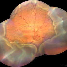
Fundus photograph of a 58-year-old man with Type 1 Uveal Effusion Syndrome, showing 360º bullous choroidal detachment.
Photographer: Dr. Gustavo Aguirre-Suarez
Imaging device: Zeiss Clarus 700
Condition/keywords: choroidal effusion, idiopathic uveal effusion syndrome
-
 Choroidal Detachment
Aug 14 2023 by Omar Toncel Churio
Fundus photograph of a woman patient with a choroidal detachment.
Photographer: Omar Toncel Churio, Hospital Militar de Especialidades Oftalmológicas, Ciudad de México
Imaging device: Optos California Retinal Camera
Condition/keywords: choroid, detachment, retina
-
 Hypotony with Hemorrhagic Choroidal Detachments
May 21 2023 by Ethan K Sobol, MD
Hypotony with hemorrhagic choroidal detachments
Condition/keywords: hemorrhagic choroidal detachment, hypotony
 Loading…