Search results (4 results)
-
 Cilioretinal Artery
Sep 20 2020 by Barton L Blackorby, MD
Early FA just after the choroidal flush. Notice the prominent filling of this cilioretinal artery well in advance of the retinal circulation. This image highlights that the source of blood flow to the cilioretinal artery is from the choroidal circulation as the retinal circulation is just beginning to fill with dye in this image.
Imaging device: Zeiss Clarus
Condition/keywords: cilioretinal artery, fluorescein angiogram (FA)
-
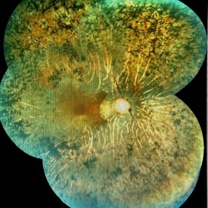 Retinitis Pigmentosa
Feb 26 2020 by Manuel Ángel Alcántara Delgado, MD
Merged color fundus photograph of a 68-year-old woman with advanced retinitis pigmentosa. It is appreciated bone spicule-shaped pigment deposits, optic disc pallor, retinal atrophy, attenuated retinal vessels and surface wrinkling retinopathy.
Photographer: Manuel Ángel Alcántara Delgado
Condition/keywords: chorioretinal atrophy, choroidal circulation, optic disc pallor, pericentral retinitis pigmentosa, retina, retinitis pigmentosa, retinitis pigmentosa (RP) dystrophy, sector retinitis pigmentosa
-
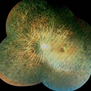 Retinitis pigmentosa
Feb 26 2020 by Manuel Ángel Alcántara Delgado, MD
Merged color fundus photograph of a 68-year-old woman with advanced retinitis pigmentosa. It is appreciated bone spicule-shaped pigment deposits, optic disc pallor, retinal atrophy and attenuated retinal vessels.
Photographer: Manuel Ángel Alcántara Delgado
Condition/keywords: choroidal circulation, optic disc pallor, pericentral retinitis pigmentosa, retina, retinitis pigmentosa, retinitis pigmentosa (RP) dystrophy, sector retinitis pigmentosa
-
 Vortex Vein In A Patient With A Blond Fundus
Oct 2 2013 by Jerald A. Bovino, MD
The vortex vein and vortex vein ampulla are visible in this patient with a blonde (lightly pigmented) fundus.
Condition/keywords: choroidal circulation, fundus photograph, vortex vein
 Loading…