Search results (632 results)
-
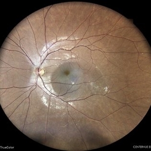
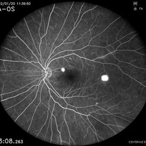
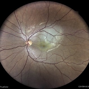
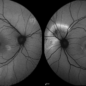
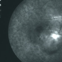
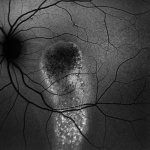 Serous River of Light
Sep 30 2025 by Malvika Singh
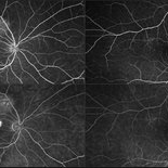
Fundus autofluorescence of a 55 year old man with history of chronic central serous chorioretinopathy showing an old gravitational track.
Photographer: Dr Malvika Singh, Retina Foundation, Ahmedabad, India
Imaging device: Mirante SLO/OCT
Condition/keywords: central serous retinopathy (CSR)
-
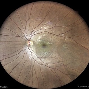
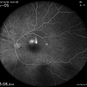
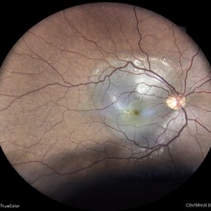
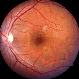
 CSR with multiple PEDs
Dec 5 2024 by Tejaswita Verma
Fundus picture of the right eye of a 40 year old female with 6/12 vision showing exudative subretinal fluid and multiple PEDs in a case of CSR.
Photographer: DR. TEJASWITA VERMA
Imaging device: MIRANTE
Condition/keywords: central serous retinopathy (CSR), pigment epithelial detachment (PED)
-
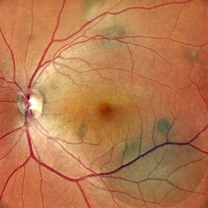
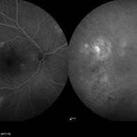
 Suspicious Nevus / CSR
Aug 8 2024 by Virginia Gebhart
Fluorescein angiogram of 54 year old male with a suspicious appearing choroidal nevus and central serous retinopathy. Will monitor closely and follow up with serial exams.
Photographer: Virginia Gebhart
Imaging device: Optos California
Condition/keywords: central serous retinopathy (CSR), choroidal nevus, fluorescein angiogram (FA), FLUORESCEIN ANGIOGRAPHY, nevus
-
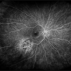
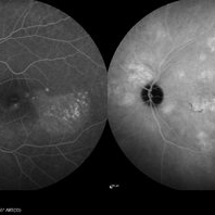
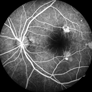
 Central Serous Retinopathy
Apr 9 2024 by Akansha Sharma
Fundus fluorescein angiography of a 39 year old male patient with smoke stack pattern of central serous retinopathy.
Photographer: Dr. Akansha Sharma, Bharati Eye Hospital
Condition/keywords: Central Serous Chorioretinopathy (CSR), central serous retinopathy (CSR)
-
 Central Serous Retinopathy
Apr 9 2024 by Akansha Sharma
Color fundus photograph of a 39 year old male patient with smoke stack pattern of central serous retinopathy.
Photographer: Dr. Akansha Sharma, Bharati Eye Hospital
Condition/keywords: Central Serous Chorioretinopathy (CSR), central serous retinopathy (CSR)
-
 Central Serous Retinopathy
Apr 9 2024 by Akansha Sharma
Fundus fluorescein angiography of a 35 year old male with central serous retinopathy demonstrating leaks.
Photographer: Dr. Akansha Sharma, Bharati Eye Hospital
Condition/keywords: Central Serous Chorioretinopathy (CSR), central serous retinopathy (CSR)
-
 Central Serous Retinopathy
Apr 9 2024 by Akansha Sharma
Color fundus photograph of a 35 year old male with central serous retinopathy.
Photographer: Dr. Akansha Sharma, Bharati Eye Hospital
Condition/keywords: Central Serous Chorioretinopathy (CSR), central serous retinopathy (CSR)
-
 Central Serous Retinopathy
Mar 19 2024 by Corey Grant
Ultra Wide-Field Fundus Autofluorescence Imaging of a 37 year old female with Central Serous Retinopathy affecting her right eye. Patient Visual Acuity was 20/20 in both eyes. Patient reported black spots in her vision onset three years ago, with associating flashes of light. Patient reports history of cortisone back injections a few years ago and denies Flonase use. The physician stated that there is hyperautofluorescence in the area of gutter of Sub-Retinal Fluid which likely happened from CSR.
Photographer: Corey Grant, OSC
Imaging device: OPTOS CALIFORNIA RGB
Condition/keywords: Central Serous Chorioretinopathy (CSR), central serous retinopathy (CSR), fundus autofluorescence (FAF), Guttering, hyperautofluorescence, inferior retina, OPTOS, Retina, Right Eye, subretinal fluid, ULTRA WIDE FIELD
-
 Central Serous Retinopathy
Mar 6 2024 by Akansha Sharma
Fluorescein angiography of a 34 year old male with smoke stack leak in the late phase in a case of central serous retinopathy.
Photographer: Dr. Akansha Sharma, Bharati Eye Hospital
Condition/keywords: Central Serous Chorioretinopathy (CSR), central serous retinopathy (CSR)
-
 Central Serous Retinopathy
Mar 6 2024 by Akansha Sharma
Color fundus photograph of a 34 year old male with central serous retinopathy.
Photographer: Dr. Akansha Sharma, Bharati Eye Hospital
Condition/keywords: Central Serous Chorioretinopathy (CSR), central serous retinopathy (CSR)
-
 CENTRAL SEROUS RETINOPATHY
May 31 2023 by Akansha Sharma
COLOUR FUNDUS PHOTOGRAPH OF A 33 YEAR OLD MALE WITH CENTRAL SEROUS RETINOPATHY
Photographer: Dr. Denish Patel, Dr. Akansha Sharma, Dr. Urmil Shah, Bharati Eye Hospital
Condition/keywords: central serous retinopathy (CSR)
-
 Central Serous Chorioretinopathy
Jan 25 2022 by Olivia Rainey
Late phase widefield fluorescein angiography of a 60-year-old male with Central Serous Chorioretinopathy. Chronic history of CSR followed with observation without treatment prior to presenting at our office. The physician noted significant findings on exam and imaging with multifocal areas of inactive and active changes OD. FA shows superotemporal macular leakage, subtle inferonasal macular leakage and staining as well as multifocal hypercyanescence on ICG. Fortunately foveal sparing and thus observation is recommended at this time OD.
Photographer: Olivia Rainey, OCT-C, COA
Imaging device: Heidelberg Spectralis
Condition/keywords: 55-degrees, central serous chorioretinopathy (CSCR), central serous retinopathy (CSR), chronic central serous chorioretinopathy (CSCR), fluorescein angiogram (FA), fluorescein leakage, heidelberg spectralis, indocyanine green (ICG) angiography, late phase
-
 Central Serous Chorioretinopathy
Jan 25 2022 by Olivia Rainey
Late phase widefield fluorescein angiography of a 60-year-old male with Central Serous Chorioretinopathy. Chronic history of CSR followed with observation without treatment prior to presenting at our office. The physician noted subfoveal subretinal fluid with recent visual decline. FA shows multifocal leakage and ICG shows hypercyanescence. OCTA, ICG, and FA consistent with CSR, and without concern for CNVM thus will observe without anti-VEGF at this time. PDT therapy recommended.
Photographer: Olivia Rainey, OCT-C, COA
Imaging device: Heidelberg Spectralis
Condition/keywords: 55-degrees, central serous chorioretinopathy (CSCR), central serous retinopathy (CSR), chronic central serous chorioretinopathy (CSCR), fluorescein angiogram (FA), heidelberg spectralis, indocyanine green (ICG) angiography, left eye
-
 Central Serous Chorioretinopathy
Jan 25 2022 by Olivia Rainey
Widefield fundus autofluorescence of a 60-year-old male with Central Serous Chorioretinopathy affecting both eyes. Chronic history of CSR followed with observation without treatment prior to presenting at our office. The physician noted significant findings on exam and imaging with multifocal areas of inactive and active changes in the right eye and subfoveal subretinal fluid with recent visual decline in the left eye. There are hyper and hypoautofluorescent changes, consistent with CSR.
Photographer: Olivia Rainey, OCT-C, COA
Imaging device: Heidelberg Spectralis
Condition/keywords: 55-degrees, central serous chorioretinopathy (CSCR), central serous retinopathy (CSR), chronic central serous chorioretinopathy (CSCR), fundus autofluorescence (FAF), heidelberg spectralis, left eye
-
 Chronic CSR
May 15 2021 by Deepak Bhojwani, MS
Fundus image of a 38-year-old gentlemen with features of chronic CSR and massive subretinal fluid.
Photographer: DEEPAK BHOJWANI; VISHAL PATEL FOR OCCURA EYE CARE
Condition/keywords: central serous retinopathy (CSR)
-
 CSR Post Laser
May 15 2021 by Deepak Bhojwani, MS
Post focal laser fundus image of 38-year-old gentlemen with chronic CSR showing regression of entire subretinal fluid.
Photographer: Deepak Bhojwani
Condition/keywords: central serous retinopathy (CSR)
-
 CSR FA
May 15 2021 by Deepak Bhojwani, MS
Fundus fluoroscein angiography image of a 38-year-old gentlemen with features of chronic CSR with multiple old areas of RPE mottling and staining. Also note there is a single active leakage site along inferotemporal arcade.
Photographer: Deepak Bhojwani
Condition/keywords: central serous retinopathy (CSR)
-
 Central Serous Chorioretinopathy
Mar 3 2020 by Sham Talati, DOMS
A patient who had CSR in RE.
Photographer: Dr. Ashok Talati, Dr.Talati's Eye Hospital, Ahmedabad
Condition/keywords: central serous chorioretinopathy (CSCR), central serous retinopathy (CSR)
-
 Central Serous Retinopathy with Smokestack
Nov 5 2019 by Nichole Lewis
40-year-old male with central serous retinopathy with smokestack.
Photographer: Nichole Lewis
Imaging device: Optos
Condition/keywords: central serous retinopathy (CSR), smokestack
-
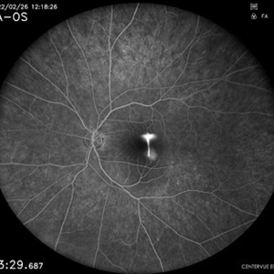
 Central Serous Retinopathy
Sep 3 2019 by Manish Nagpal, MD, FRCS (UK), FASRS
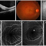
Patient having CSR with a classic inkblot leak and sensory elevation seen on the OCT.
Photographer: Akshar Soni
Condition/keywords: central serous retinopathy (CSR)
-
 Central Serous Retinopathy
Sep 3 2019 by Manish Nagpal, MD, FRCS (UK), FASRS
Patient having CSR with a classic smokestack leak and sensory elevation seen on the OCT along with a small PED and OCTA images.
Photographer: Akshar Soni
Condition/keywords: central serous retinopathy (CSR)
-
 Chronic Central Serous Chorioretinopathy
Mar 29 2019 by Nichole Lewis
54-year-old male with chronic central serous retinopathy with focal sub-retinal fluid and widespread retinal pigment epithelium changes. History of micropulse laser. Patient is HLA-B27 positive with quiescent iritis. VA 20/20.
Photographer: Nichole Lewis
Imaging device: Optos
Condition/keywords: central serous retinopathy (CSR), retinal pigment epithelium (RPE) changes, subretinal fluid
-
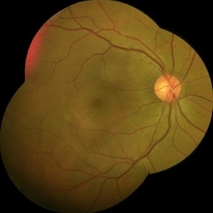
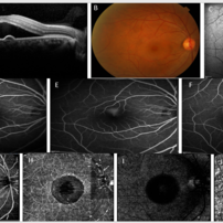
 CSR with large RPED
Mar 26 2019 by Gary R. Cook, MD, FACS
Late-phase FA frame showing mild pooling of dye beneath a large RPED inferonasal to the optic disc, blocked fluorescence from the pigment figures (black lines), and late dye leakage from the RPED.
Imaging device: Topcon VT-50
Condition/keywords: central serous retinopathy (CSR), FA late phase, fluorescein angiogram (FA), retinal pigment epithelium (RPE) detachment
-
 CSR with large RPED
Mar 26 2019 by Gary R. Cook, MD, FACS
Mid-phase FA showing large RPED inferonasal to optic disc with overlying cruciate pigment figures (black lines) and neurosensory macular detachment OD.
Imaging device: Topcon VT-50
Condition/keywords: central serous retinopathy (CSR), neurosensory detachment of retina, retinal pigment epithelium (RPE) detachment
-
 CSR with RPED
Mar 26 2019 by Gary R. Cook, MD, FACS
White male with acute CSR with a RPED beneath the NSRD OD.
Imaging device: Topcon VT-50
Condition/keywords: central serous retinopathy (CSR), retinal pigment epithelium (RPE) detachment
 Loading…