Search results (104 results)
-
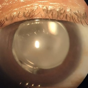
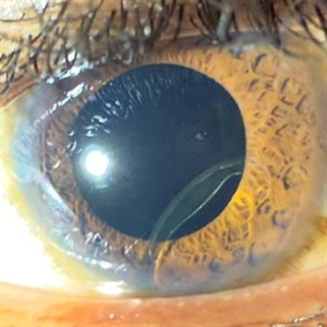 IOL Dislocation
Sep 29 2025 by Carmen R. Negrin-Martin, MD
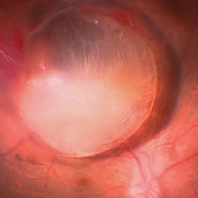
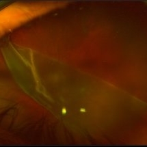
Male patient with a history of ocular trauma at 12 years of age. Underwent cataract surgery as an adult and now reports decreased vision of a few days’ duration. Referred to the retina service due to IOL dislocation. Visual acuity with +9.00: 20/40. Biomicroscopy: clear cornea, formed anterior chamber. The IOL haptic is observed hooked to the iris, with the optic in the posterior segment behind the iris. Posterior segment without alterations.
Photographer: Dr. Vizcaino MD
Imaging device: iPhone 16 pro Max
Condition/keywords: IOL dislocation
-
 Dislocated IOL
Sep 20 2025 by JORGE SOBERANES
Fundus photograph of a 65-year-old man with a history of cataract surgery one year ago and bad vision since that.
Photographer: Dr. Jorge Soberanes, APEC, Universidad Nacional Autónoma México
Condition/keywords: dislocated lens, intraocular lens dislocation
-
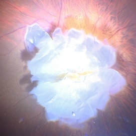
 Vitreous Cavity Migrated IOL
Sep 20 2025 by Thiago Mazzeo
Intraoperative image of a pars plana vitrectomy for the removal of migrated IOL during complicated cataract surgery.
Photographer: Thiago Mazzeo, Centro de Oftalmologia Especializade de Macaé (COEM)
Condition/keywords: scleral fixation, vitrectomy
-
 Suprachoroidal Hemorrhage
Aug 4 2025 by Anjana Mirajkar, MS Ophthalmology
A fundus photograph of a 56 year old female with a 360 degree suprachoroidal hemorrhage with a 360 degree crumpled retina during cataract surgery.
Photographer: Dr. Anjana Mirajkar- HV Desai eye hospital ,Pune
Imaging device: optos
Condition/keywords: giant retinal tear, suprachoroidal hemorrhage
-
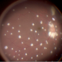
 PCIOL Opacification
Mar 31 2025 by DR Rohit Gupta
A pseudophakic patient visiting after 6 months of cataract surgery. On slit lamp examination a complete hazy white PCIOL was seen, which is a rare complication after cataract surgery.
Photographer: Dr Rohit gupta
Imaging device: Samsung S21
Condition/keywords: posterior chamber intraocular lens (PCIOL)
-
 Seedlings of Fungal Endophthalmitis
Mar 14 2025 by SHILPI H NARNAWARE, ICO ( Retina) , FAICO ( Vitreo-Retina)
57 year diabetic female , was treated as a case of recurrent vitreous post cataract surgery. Patient was posted for vitrectomy 3 months post cataract surgery. Intra-operatively, multiple yellowish colonies were seen all over the posterior pole were seen, which were later found to be Aspergillus colonies.
Photographer: Shilpi Narnaware, Sarakshi Netralaya , Nagpur, Maharashtra , India
Imaging device: Ngenuity
Condition/keywords: endophthalmitis, fungal
-
 Lacteocrumenasia
Mar 11 2025 by Gustavo Uriel Fonseca Aguirre
A 75-year-old female with a history of cataract surgery with intraocular lens implantation 20 years ago presented with progressive visual loss. On slit lamp examination, opaque material was found in the capsular bag behind the intraocular lens. Ultrasound biomicroscopy revealed hyperechoic material contained in the temporal-posterior sector of the capsular bag corresponding to lacteocrumenasia.
Photographer: Gustavo U. Fonseca Aguirre, Hospital Conde de Valenciana, Ciudad de México
Condition/keywords: Lacteocrumenasia, ultrasound biomicroscopy
-
 Multimodal Imaging in CHRPE
Mar 6 2025 by Gerardo - Montante Montelongo, MD
Fundus photograph of an 83-year-old male with a history of Diabetes, smoking, cataract surgery on the right eye in 2022, and open-angle glaucoma. Asymptomatic. Indirect ophthalmoscopy revealed 80% excavation, peripapillary atrophy, and a hyperpigmented perifoveal lesion with 35% atrophy, 10% drusen, and 5.1 mm diameter, corresponding to a CHRPE. At multimodal imaging, FFA shows hypoautofluorescence of the lesion, OCT shows preservation of internal retinal layers, atrophy of external retinal layer, with an RPE disruption, and posterior shadowing. USG shows a flat hyperechoic lesion 5.1 mm in diameter and 1.32 mm in thickness, solid and with high internal reflectance.
Photographer: Gerardo Montante-Montelongo, MD, Mexican Institute of Ophthalmology
Imaging device: Clarus 700
Condition/keywords: congenital hypertrophy of the retinal pigment epithelium (CHRPE), multimodal imaging
-
 Bullous Keratopathy
Jan 4 2025 by Mosab Salah
Corneal Slit photograph of an 84-year-old man underwent uneventful cataract surgery 1 year ago elsewhere, with a multiple fluid filled Bullae, not responding on conservative management and planned for KP.
Photographer: Abu-Ismail, Luai MD, The Islamic Hospital, Amman, Jordan
Imaging device: smartphone photography through SLB
Condition/keywords: bullous keratopathy, corneal edema
-
 Macular Degeneration
Dec 3 2024 by Sarah D Kang
Fundus photograph of an 85-year-old female patient with macular degeneration observed for retinal clearance before cataract surgery.
Condition/keywords: floaters, macular degeneration
-
 Macular Degeneration
Dec 3 2024 by Sarah D Kang
Fundus photograph of an 85-year-old female patient with macular degeneration observed for retinal clearance before cataract surgery.
Condition/keywords: floaters, macular degeneration
-
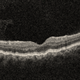 RE-OCT in Choroidal Detachment
Sep 22 2024 by Anand Temkar
A 52 year old male came with chief complaints of diminution of vision in RE since past 15 days. He gave history of ( RE ) cataract surgery + IOL about 2 months ago. His vision was 6/9 in RE and PL + ve, PR inaccurate in LE. His IOP was 10 mm of Hg in RE and 20 mm of Hg in LE.
Photographer: Dr.Anand Temkar- Retina Foundation, Ahmedabad
Imaging device: Mirante
Condition/keywords: choroidal detachment, choroidals, serous choroidal detachment
-
 360 Degrees Choroidal Detachment
Sep 22 2024 by Anand Temkar
A 52 year old male came with chief complaints of diminution of vision in RE since past 15 days. He gave history of ( RE ) cataract surgery + IOL about 2 months ago. His vision was 6/9 in RE and PL +ve, PR inaccurate in LE. His IOP was 10 mm of Hg in RE and 20 mm of Hg in LE.
Photographer: Dr.Anand Temkar- Retina Foundation, Ahmedabad
Imaging device: Mirante
Condition/keywords: choroidal detachment, choroidals, serous choroidal detachment
-
 IOL on Retinal Surface
Sep 8 2024 by Cesar Augusto Rocha Rojas, MD
Dislocated lens into the vitreous cavity during cataract surgery.
Photographer: Cesar Augusto Rocha Rojas, Hospital General de Zona #20, Instituto Mexicano del Seguro Social (IMSS)
Imaging device: Surgical microscope and smartphone
Condition/keywords: IOL drop
-
 Dislocated Cataractous Lens
Jun 16 2024 by Anjana Mirajkar, MS Ophthalmology
An intra operative still image of a 65 year old male showing an dislocated cataractous lens during cataract surgery which was removed during vitrectomy and and secondary IOL was placed.
Photographer: Dr. Anjana Mirajkar -Retina Foundation, Ahmedabad
Condition/keywords: dislocated crystalline lens
-
 Nucleus Drop
Jun 14 2024 by Tejaswita Verma
Intraoperative still of lens drop in a 63 year old female after an eventful cataract surgery.
Photographer: DR. TEJASWITA VERMA
Condition/keywords: intraoperative, lens matter, nucleus drop
-
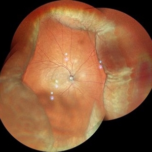
 Congenital Retinal Macro Vessel
May 15 2024 by KANWALJEET HARJOT MADAN, M.S. (Ophthalmology); FAICO (Vitreous - Retina)
This is the Fundus Picture of a 56 years Female, who had come for Cataract Surgery of her left eye. Her best corrected vision in left eye was 6/12. She was diagnosed to have Congenital Retinal Macro vessel in her left eye on fundus exam. She underwent cataract surgery and vision improved to 6/9. She is kept under regular follow up.
Photographer: Dr Kanwaljeet Harjot Madan
Condition/keywords: congenital, RETINAL MACROVESSEL
-
 Congenital Retinal Macro Vessel
May 15 2024 by KANWALJEET HARJOT MADAN, M.S. (Ophthalmology); FAICO (Vitreous - Retina)
This is the Fundus Picture of a 56 years Female, who had come for Cataract Surgery of her left eye. Her best corrected vision in left eye was 6/12. She was diagnosed to have Congenital Retinal Macro vessel in her left eye on fundus exam. She underwent cataract surgery and vision improved to 6/9. She is kept under regular follow up.
Photographer: Dr. Kanwaljeet Harjot Madan
Condition/keywords: congenital, RETINAL MACROVESSEL
-
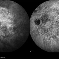
 Retinal Detachment with Giant Retinal Tear
Mar 26 2024 by Xitlali Caterina
Ultra-widefield fundus photograph of a 43-year-old male with a Retinal Detachment with Giant Retinal Tear affecting his left eye. Patient presented to the office with count fingers vision at 2 feet. He stated that about 8-9 days ago, he developed a clear curtain/veil and his vision started to get blurry. He also noted that he had floaters and flashes for about 8-9 days as well. The patient had cataract surgery a month prior to his visit. He stated that since his surgery, his vision had been better, but he had an area where he was not able to see well. The physician recommended a complex retinal detachment repair.
Photographer: Xitlali Caterina
Imaging device: OPTOS California RGB
Condition/keywords: fundus photograph, giant retinal tear, left eye, Optos, OPTOS CALIFORNIA, retinal detachment of the macula, retinal detachment with tear, ultra-wide field imaging, ultra-widefield image
-
 Chronic Regmatogenous Retinal Detachment
Mar 21 2024 by Mauricio Bayram-Suverza, MD
A 65-year-old man came to our department with a complaint of chronic visual loss in his right eye. He had undergone cataract surgery in the same eye at another facility 8 years ago. During the examination, it was observed that the patient had no light perception in the affected eye. Further, slit lamp examination revealed a chronic anteriorized retinal detachment.
Photographer: Mauricio Bayram-Suverza, Fundación Hospital Nuestra Señora de la Luz
Imaging device: iphone
Condition/keywords: proliferative vitreoretinopathy (PVR), vitreoretinal surgery
-
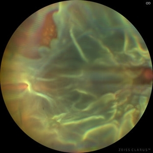 Chronic Open Funnel Retinal Detachment With Horse Shoe Tear
Feb 7 2024 by Harsh Vardhan Singh, MS
67 year old male with history of cataract surgery 1 year presented with old chronic retinal detachment with open funnel configuration with multiple breaks.
Photographer: Harsh Vardhan Singh
Imaging device: Clarus 700
Condition/keywords: chronic retinal detachment, Retinal Detachment, Retinal Detachment with multiple breaks
-
 Post Cataract surgery macular atrophy with unknown cause
Dec 8 2023 by Vanessa Evans-Jones
Fundus image of 78 year old woman who had cataract surgery followed by vitrectomy . macular hole and macular atrophy identified 2 weeks post vitrectomy and no signs of any issues pre operation
Condition/keywords: cataract, macular atrophy, unknown
-
 Monochrome-frame
Nov 21 2023 by Nassim Alejandro Abreu Arbaje, MD
Frame grab from a 68 year male, who underwent cataract surgery and vitrectomy with ilm peel because of PDR. In the picture we can see the enhanced contrast we can get while doing an ILM peel with a monochromatic digital filter
Photographer: Nassim Abreu, Hospital Dr. Elías Santana
Imaging device: NGenuity 3D system
Condition/keywords: enhaced contrast, Macular surgery, monochrome
-
 Retinal Detachment OD
Aug 23 2023 by Angela Rico
59 year-old male presents with Flashes and floaters OD for 2-3 weeks after Cataract surgery
Photographer: Angela Rico M.D.
-
 Lens Drop
Aug 21 2023 by Harsh Vardhan Singh, MS
Intra-operative image of Posterior lens dislocation as a complication of cataract surgery
Photographer: Harsh Vardhan Singh
Condition/keywords: lens dislocation, Lens Drop
 Loading…