Search results (187 results)
-
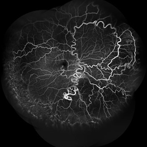
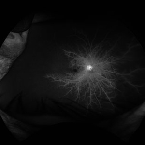
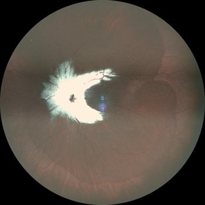
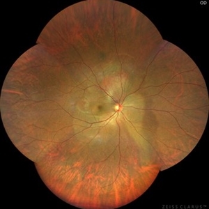 Vortex-pattern Exudative Retinal Detachment
Feb 22 2025 by CUI YUELING
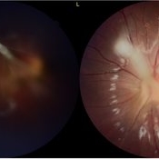
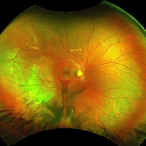
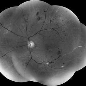
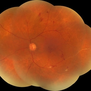
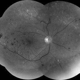
Patient: Male, 40 years old. Chief Complaint: Blurred vision and metamorphopsia in the left eye for more than 10 days. Past Medical History Hypertension for 4 years, with a highest recorded blood pressure of 160/80 mmHg. Currently controlled with oral "Nifedipine Sustained-Release Tablets, 2 tablets daily." Long-term history of heavy alcohol consumption and smoking. Ophthalmic Examination: Visual Acuity: Right eye (OD): 0.4 (uncorrected, no improvement with correction). Left eye (OS): 0.5 (-1.5DS = 1.0). Intraocular Pressure (IOP): OD: 15 mmHg. OS: 17 mmHg. Anterior Segment:Unremarkable. Fundus Examination: Right eye: Optic disc margins are clear, with a slightly reddish hue. Cup-to-disc ratio (C/D) = 0.2. A scalloped, orange-red elevated lesion is observed superior to the optic disc, with anterior displacement of the focal point. This is accompanied by a secondary, turbine-like exudative retinal detachment centered around the optic disc, involving the macula. The macular region shows scattered punctate yellow-white exudates. Diagnosis: Choroidal hemangioma with secondary exudative retinal detachment(OD).
Photographer: Cui yueling The First People's Hospital of Zunyi, Guizhou, Zunyi, China
Imaging device: Zeiss Clarus 500
Condition/keywords: choroidal hemangioma, exudative retinal detachment
-
 Photic Retinopathy
Jan 30 2025 by Juan Alberto Olivera Cueva
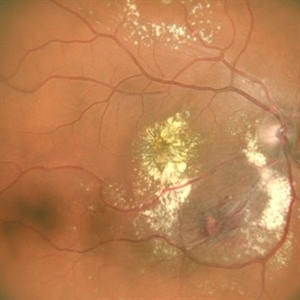
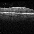
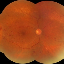
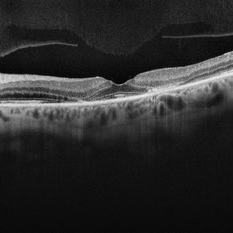
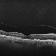
A 23-year-old male with a history of direct exposure to sunlight on several occasions, presenting blurred vision, comes for evaluation due to metamorphopsias of 3 months' evolution. The fundus photograph shows the presence of an epiretinal membrane. The OCT shows a hyperreflective line at the vitreomacular interface that causes traction to the layers of the inner retina, as well as distortion in the architecture of the macular region, with the presence of subfoveal detachment of the RPE.
Photographer: Dr. Juan Alberto Olivera Cueva, Escuela Militar de Medicina, Hospital Militar de Especialidades Oftalmológicas
Condition/keywords: epiretinal membrane (ERM)
-
 Photic Retinopathy
Jan 30 2025 by Juan Alberto Olivera Cueva
A 23 year-old male with a history of direct exposure to sunlight on several occasions, presenting blurred vision, comes for evaluation due to metamorphopsias of 3 months' evolution. The fundus photograph shows the presence of an epiretinal membrane. The OCT shows a hyperreflective line at the vitreomacular interface that causes traction to the layers of the inner retina, as well as distortion in the architecture of the macular region, with the presence of subfoveal detachment of the RPE.
Photographer: Dr. Juan Alberto Olivera Cueva, Escuela Militar de Medicina, Hospital Militar de Especialidades Oftalmológicas
Condition/keywords: MER, photic retinopathy
-
 Photic Retinopathy
Jan 29 2025 by Juan Alberto Olivera Cueva
A 23 year-old male with a history of direct exposure to sunlight on several occasions, presenting blurred vision, comes for evaluation due to metamorphopsias of 3 months' evolution. The fundus photograph shows the presence of an epiretinal membrane. The OCT shows a hyperreflective line at the vitreomacular interface that causes traction to the layers of the inner retina, as well as distortion in the architecture of the macular region, with the presence of subfoveal detachment of the RPE.
Photographer: Dr. Juan Alberto Olivera Cueva, Escuela Militar de Graduados de Sanidad, Hospital Militar de Especialidades Oftalmológico
Condition/keywords: Membrana Epirretiniana, MER, Photic retinopathy
-
 Photic Retinopathy
Jan 29 2025 by Juan Alberto Olivera Cueva
A 23-year-old male with a history of direct exposure to sunlight on several occasions, presenting blurred vision, comes for evaluation due to metamorphopsias of 3 months' evolution. The fundus photograph shows the presence of an epiretinal membrane. The OCT shows a hyperreflective line at the vitreomacular interface that causes traction to the layers of the inner retina, as well as distortion in the architecture of the macular region, with the presence of subfoveal detachment of the RPE.
Photographer: Dr. Juan Alberto Olivera Cueva, Escuela Militar de Graduados de Sanidad, Hospital Militar de Especialidades Oftalmológico
Condition/keywords: Membrana epirretiniana, MER, Photic Retinopathy
-
 Racemose Angioma
Jan 23 2025 by SHILPI H NARNAWARE, ICO ( Retina) , FAICO ( Vitreo-Retina)
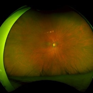
42 year old male, presented with blurred vision . Examination revealed Racemose angioma. FFA was done which revealed tortuosity of blood vessels.
Photographer: Shilpi Narnaware, Sarakshi Netralaya , Nagpur, Maharashtra , India
Imaging device: Mirante ( by Nidek)
Condition/keywords: FFA in a case of Racemose angioma
-
 Posterior Scleritis
Jan 4 2025 by Tejaswita Verma
Left eye fundus photo of a 49 year old female presenting with 5 days history of blurred vision, Vision was finger counting 2 mts. and nodular posterior scleritis with T sign on USG was present. OCT revealed altered foveal contour with septations and SRF pockets and bacillary layer detachment.
Photographer: DR. TEJASWITA VERMA
Imaging device: MIRANTE
Condition/keywords: posterior scleritis
-
 Treatment of Ocular Ischemic Syndrome with Hyperbaric Oxygen Therapy
Dec 2 2024 by Catherine S Kang
A 66-year-old female with past medical history significant for hypertension and ocular ischemic syndrome. She presented in emergency department (ED) reporting eye pain and blurred vision in both eyes since earlier that morning. On examination, best corrected visual acuity in each eye was counting fingers (20cm). Further investigation was performed and fluorescein angiography revealed a delay in choroidal filling. The patient has been followed for ocular ischemic syndrome since the onset of the condition and hyperbaric oxygen therapy was promptly initiated. Final best corrected visual acuity was 20/150 and macula developed atrophy.
Photographer: Catherine Kang
Condition/keywords: hyperbaric oxygen therapy, ocular ischemic syndrome
-
 Myelinated Nerve Fibres With Combined Hamartoma of Retina and RPE
Jul 31 2024 by Tejaswita Verma
Fundus image of a 20 year old female who presented with metamorphopsia ,slightly blurred vision. BCVA was 6/9, epiretinal membrane present on central fundus examination with myelinated nerve fibres.
Photographer: DR. TEJASWITA VERMA
Imaging device: MIRANTE
Condition/keywords: combined hamartoma of retina and RPE, myelinated nerve fibers
-
 Posterior-PFV
Jul 27 2024 by Gokcen Deniz Gulpinar Ikiz
7 Year old girl presented with blurred vision on the left eye, with intermittent esotopia. She had been followed conservatively for intermittent esotropia on the left eye, recently advised for patching of the right eye. The vision is 1.0 on the right eye and 0.4 (Snellen) on the left eye. Anterior segment is natural bilaterally, except 20 PD esotropia on the left eye, with alternation and fixation. Refraction was +0.25 +0.25 x180 and +1.00-1.50 x60 on the right and left eyes respectively. Dilated fundus examination was natural on the right eye. However, there was a fibrotic stalk originating from the optic nerve head extending to the vitreous, terminating in the middle of the vitreous cavity, in a spider web configuration. Which also causes nasal dragging of the macula, leading to partial shallow detachment of the fovea nasally. Vitrectomy is advised for the left eye, with lens preserving approach, to preserve the current functional potential and the anatomy of the globe in long term.
Photographer: Gokcen Deniz Gulpinar Ikiz, Special Eye Clinic
Condition/keywords: amblyopia, posterior PFV, vitrectomy
-
 Retinal Artery Macroaneurysm
Jul 13 2024 by Tejaswita Verma
A 53 year old female presented with blurred vision in RE since a month ,with borderline DM and HTN not on medications .H/o highest BP recording was 160/90 mm Hg.Vision 6/60 .FFA revealed leakages. She was advised RE focal laser with intravitreal anti-VEGF injections
Photographer: DR. TEJASWITA VERMA
Imaging device: MIRANTE
Condition/keywords: RETINAL ARTERY MACROANEURYSM
-
 Cilioretinal Artery Occlusion
May 14 2024 by Eloy Mata-Cortes, MD
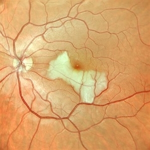
Color image capturing the left eye of a 32-year-old female. Despite a negative ophthalmological and medical history, she reported three days of blurred vision and a paracentral scotoma in her left eye, while maintaining central vision. The image reveals retinal whitening, extends from the parafoveal region to the inferotemporal arcade indicative of cilioretinal artery occlusion. Following this observation, the patient was referred for systemic assessment to explore the underlying etiology of the occlusion.
Photographer: Eloy Mata-Cortes, MD, Instituto Mexicano de Oftalmología, Querétaro, México
Imaging device: Nidek Mirante
Condition/keywords: cilioretinal artery occlusion, oclussion, retinal whitening
-
 Choroidal Metastasis
Apr 11 2024 by Corey Grant
Ultra-Widefield fundus photography and fundus autofluorescence images of a 61 year old female with Choroidal Metastasis affecting both eyes. Patient presented with blurred vision and flashes for a few weeks. Patient visual acuity was cc20/100 PH20/60 in the right eye and cc20/200 in the left eye. Patient admits to history of smoking for many years bit no known history of cancer prior to the visit. Physician recommended going to the ER for full body PET CT and stated that the first line of treatment is usually systemic chemo therapy. Patient will be reassessed in one month.
Photographer: Corey Grant
Imaging device: OPTOS CALIFORNIA RGB
Condition/keywords: cancer, choroidal metastasis, fundus autofluorescence (FAF), fundus photography, hyperautofluorescence, hypoautofluorescence, Optos, OPTOS CALIFORNIA RGB, Retina, ULTRA WIDE FIELD
-
 Ocular Syphilis
Feb 21 2024 by Nikhil K Bommakanti, MD
A monocular man in his sixties presented with blurred vision in the right eye for two months. Optical coherence tomography demonstrated vitreous cells and characteristic inflammatory deposits of the outer retina and retinal pigment epithelium, and laboratory testing confirmed the diagnosis of syphilis. He was admitted for intravenous penicillin and consultation with a specialist in infectious diseases.
Condition/keywords: syphilis
-
 Blistered Retina
Jan 27 2024 by prathibha hande, MS DNB
Fundus photo of a 32 year old male presenting with blurred vision. Undiagnosed renal hypertension. Blood pressure at the time of presentation 210/120 mmhg.
Photographer: Mr Prathap K
Imaging device: Mirante SLO fundus camera
Condition/keywords: hypertensive choroidopathy
-
 Congenital Retinal Macrovessel
Oct 13 2023 by Jacob D. Grodsky, MD
41 y/o male who presented with acute onset of blurred vision OD. Visual acuity was 20/200 OD; 20/25 OS. Examination was consistent with congenital retinal macrovessel through the macula with intraretinal hemorrhage as seen in the fundus photo. Intravitreal bevacizumab was injected, and visual acuity improved to 20/40 at 4-week follow-up. MRA head and neck was ordered to rule out other vascular anomalies.
Condition/keywords: congenital retinal macrovessel, RETINAL MACROVESSEL
-
 Choroidal Rupture
Sep 30 2023 by Jacob D. Grodsky, MD
24 year old female who presented after being hit in the head with a metal softball bat after an altercation. The patient reported blurred vision as well as a zig-zag line described as a “lightning strike” across her vision. Examination was significant for a choroidal rupture OD as well as commotio retinae OU.
Condition/keywords: choroidal rupture, commotio retinae, trauma
-
 Red free reveal proliferative diabetic retinopathy
Sep 25 2023 by firdaus sukhi, MD
56 year old diabetic not on any systemic treatment reported in optometry clinic with blurred vision. A simple red free pictures by optometrist could detect the florid NVD and NVE in optometry clinic . Poor renal profile uncontrolled systemic parameters limits fluorescein angiography and such simple tool could help referring the urgent cases to the retina clinic for prompt treatment.
Photographer: Naseem Akhtar -optometrist SKMC Ajman UAE
Condition/keywords: proliferative diabetic retinopathy (PDR)
-
 Red free reveal of proliferative diabetic retinopathy
Sep 25 2023 by firdaus sukhi, MD
56 year old diabetic not on any systemic treatment reported in optometry clinic with blurred vision. A simple red free pictures by optometrist could detect the florid NVD and NVE in optometry clinic Poor renal profile uncontrolled systemic parameters limits fluorescein angiography and such simple tool could help referring the urgent cases to the retina clinic for prompt treatment.
Photographer: Naseem Akhtar -optometrist SKMC Ajman UAE
Condition/keywords: proliferative diabetic retinopathy (PDR)
-
 Red free reval of proliferative diabetic retinopathy
Sep 25 2023 by firdaus sukhi, MD
56 year old diabetic not on any systemic treatment reported in optometry clinic with blurred vision. A simple red free pictures by optometrist could detect the florid NVD and NVE in optometry clinic Poor renal profile uncontrolled systemic parameters limits fluorescein angiography and such simple tool could help referring the urgent cases to the retina clinic for prompt treatment.
Photographer: Naseem Akhtar -optometrist SKMC Ajman UAE
Condition/keywords: proliferative diabetic retinopathy (PDR)
-
 Hydroxychloroquine Maculopathy
Jul 23 2023 by Ahmad B. Tarabishy, MD
62 year old female with rheumatoid arthritis, treated with hydroxychloroquine 200 mg BID for the past 6-8 years. She presents with blurred vision, difficulty reading, and difficulty transitions from dark to light conditions since 4 months.
Photographer: Dr. Angela Rico
Condition/keywords: hydroxychloroquine toxicity, plaquenil toxicity, toxic maculopathy
 Loading…