Search results (42 results)
-
 Birdshot Retinochoroidopathy
Jun 18 2025 by César Adrián Gómez Valdivia, MD
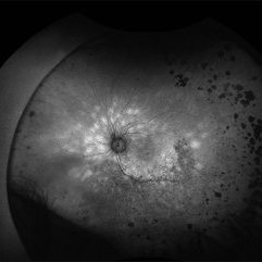
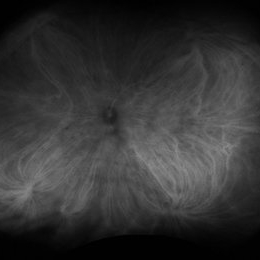
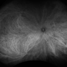
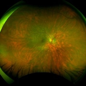
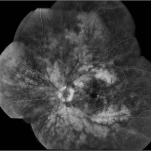
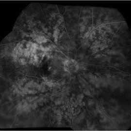
Fundus Autofluorescence of a 86 YO female patient diagnosed with Birdshot Retinochoroidopathy.
Photographer: @eyemissu2
Imaging device: California ICG OPTOS
Condition/keywords: birdshot retinochoroidopathy
-
 Birdshot Retinochoroidopathy
Jun 18 2025 by César Adrián Gómez Valdivia, MD
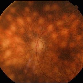
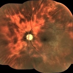
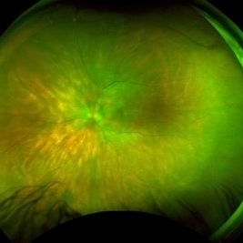
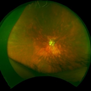
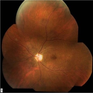
Fundus photograph of a 86 YO female patient diagnosed with Birdshot Retinochoroidopathy. Characteristically multifocal cream-colored or yellow-orange, oval or round lesions that emerge from around the optic nerve can be appreciated.
Photographer: @eyemissu2
Imaging device: TOPCON TRC-50DX
Condition/keywords: Birdshot Retinochoroidopathy
-
 Birdshot Retinochoroidopathy
Jun 18 2025 by César Adrián Gómez Valdivia, MD
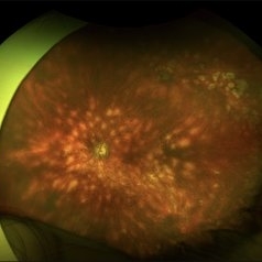
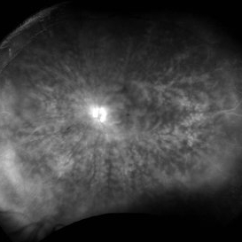
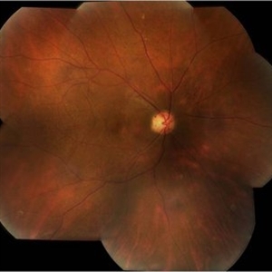
Fundus photograph of a 86 YO female patient diagnosed with Birdshot Retinochoroidopathy. Characteristically multifocal cream-colored or yellow-orange, oval or round lesions that emerge from around the optic nerve can be appreciated.
Photographer: @eyemissu2
Imaging device: California ICG OPTOS
Condition/keywords: Birdshot Retinochoroidopathy
-
 Birdshot Retinopathy
May 9 2023 by JEFFERSON R SOUSA, Tecg.º (Biomedical Systems Technology)
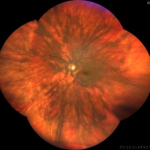
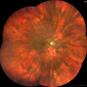
Female patient, 41 years old, with progressive low visual acuity, progressive history of autoimmune disease. In the multimodal retinal fundoscopic evaluation, important characteristics compatible with "Birdshot Retinopathy" were observed. Birdshot retinopathy, also known as birdshot chorioretinopathy or birdshot uveitis, is a rare, chronic inflammatory disorder that affects the retina and the choroid of the eye. It typically develops in adults between the ages of 30 and 60 years, and is more common in women than men. The name "birdshot" refers to the small, round, yellow-white spots that appear on the retina, which resemble the pattern of a shotgun blast. These spots are caused by inflammation in the eye, and can lead to vision loss if left untreated. Symptoms of birdshot retinopathy include blurred vision, floaters, loss of night vision, and difficulty adapting to changes in lighting. The condition can also cause inflammation in other parts of the eye, leading to redness, pain, and sensitivity to light. The exact cause of birdshot retinopathy is unknown, but it is believed to be an autoimmune disorder, in which the body's immune system mistakenly attacks the retina and choroid. Treatment typically involves the use of immunosuppressive medications, such as corticosteroids or biologic agents, to reduce inflammation and preserve vision. Close monitoring by an ophthalmologist is important, as the disease can progress even with.
Photographer: JEFFERSON ROCHA DE SOUSA - Retinal Department at Institute Dr. Suel Abujamra Sao Paulo-Brazil
Imaging device: Clarus 700 - Zeiss, composite of four 135 degree images.
Condition/keywords: bilateral chorioretinal folds, birdshot, birdshot chorioretinopathy, birdshot choroidopathy, birdshot retinochoroidopathy
-
 Birdshot Retinopathy
May 9 2023 by JEFFERSON R SOUSA, Tecg.º (Biomedical Systems Technology)
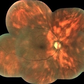
Female patient, 41 years old, with progressive low visual acuity, progressive history of autoimmune disease. In the multimodal retinal fundoscopic evaluation, important characteristics compatible with "Birdshot Retinopathy" were observed. Birdshot retinopathy, also known as birdshot chorioretinopathy or birdshot uveitis, is a rare, chronic inflammatory disorder that affects the retina and the choroid of the eye. It typically develops in adults between the ages of 30 and 60 years, and is more common in women than men. The name "birdshot" refers to the small, round, yellow-white spots that appear on the retina, which resemble the pattern of a shotgun blast. These spots are caused by inflammation in the eye, and can lead to vision loss if left untreated. Symptoms of birdshot retinopathy include blurred vision, floaters, loss of night vision, and difficulty adapting to changes in lighting. The condition can also cause inflammation in other parts of the eye, leading to redness, pain, and sensitivity to light. The exact cause of birdshot retinopathy is unknown, but it is believed to be an autoimmune disorder, in which the body's immune system mistakenly attacks the retina and choroid. Treatment typically involves the use of immunosuppressive medications, such as corticosteroids or biologic agents, to reduce inflammation and preserve vision. Close monitoring by an ophthalmologist is important, as the disease can progress even with.
Photographer: JEFFERSON ROCHA DE SOUSA - Retinal Department at Institute Dr. Suel Abujamra Sao Paulo-Brazil
Imaging device: Clarus 700 - Zeiss, composition of five 135 degree images.
Condition/keywords: bilateral chorioretinal folds, birdshot, birdshot chorioretinopathy, birdshot choroidopathy, birdshot retinochoroidopathy
-
 Birdshot Retinochoroidopathy
Jan 22 2021 by Renata Garcia Franco, Md
50 -year-old female with history of floaters and hazy vision. Yellow-white lesions in the fundus, retinal vasculitis and 2+ vitreous haze.
Photographer: Fatima Hernandez, Instituto de la Retina del Bajio SC
Imaging device: Zeiss
Condition/keywords: birdshot retinochoroidopathy
-
 Birdshot Retinochoroidopathy
Jan 22 2021 by Renata Garcia Franco, Md
50-year-old female with history of floaters and hazy vision. Yellow-white lesions in the fundus, retinal vasculitis, epiretinal membrane and cystoid macular edema.
Photographer: Fatima Hernandez, Instituto de la Retina del Bajio SC
Imaging device: Zeiss
Condition/keywords: birdshot retinochoroidopathy
-
 Birdshot ICG OS
Feb 24 2016 by Armando L. Oliver, MD
Birdshot chorioretinopathy.
Photographer: Moises Castro
Imaging device: Optos California
Condition/keywords: birdshot, birdshot chorioretinopathy, birdshot retinochoroidopathy
-
 Birdshot ICG OD
Feb 24 2016 by Armando L. Oliver, MD
Birdshot chorioretinitis.
Photographer: Moises Castro
Imaging device: Optos California
Condition/keywords: birdshot, birdshot chorioretinopathy, birdshot retinochoroidopathy
-
 Birdshot IVFA OS
Feb 24 2016 by Armando L. Oliver, MD
Birdshot chorioretinitis.
Photographer: Moises Castro
Imaging device: Optos California
Condition/keywords: birdshot, birdshot chorioretinopathy, birdshot retinochoroidopathy
-
 Birdshot IVFA OD
Feb 24 2016 by Armando L. Oliver, MD
Birdshot chorioretinopathy.
Photographer: Moises Castro
Imaging device: Optos California
Condition/keywords: birdshot, birdshot chorioretinopathy, birdshot retinochoroidopathy
-
 Birdshot OS
Feb 24 2016 by Armando L. Oliver, MD
Birdshot chorioretinitis
Photographer: Moises Castro
Imaging device: Optos California
Condition/keywords: birdshot, birdshot chorioretinopathy, birdshot retinochoroidopathy
-
 Birdshot OD
Feb 24 2016 by Armando L. Oliver, MD
Birdshot chorioretinitis.
Photographer: Moises Castro
Imaging device: Optos California
Condition/keywords: birdshot, birdshot chorioretinopathy, birdshot retinochoroidopathy
-
 Birdshot Retinopathy 6
Jan 15 2016 by Raj K. Maturi, MD
Fundus image of an 64-year-old white male.
Photographer: Tom Steele, CRA Midwest Eye Institute Indianapolis, Indiana
Imaging device: Optos
Condition/keywords: birdshot retinochoroidopathy, color fundus photograph
-
 Birdshot Case #2, HVF 60-4, OS
May 1 2013 by Armando L. Oliver, MD
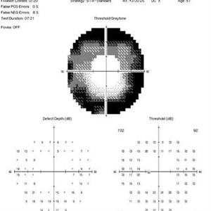
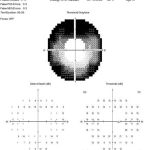
57-year-old woman with known history of Birdshot Chorioretinopathy. Despite 20/20 best corrected visual acuity. The 60-4 Humphrey Visual Field reveals the extensive peripheral visual field loss characteristic of the condition.
Imaging device: Humphrey Visual Field 60-4
Condition/keywords: birdshot, birdshot chorioretinopathy, birdshot retinochoroidopathy
-
 Birdshot Case # 2, Humphrey Visual Vield 60-4, OD
May 1 2013 by Armando L. Oliver, MD
57-year-old woman with known history of Birdshot Chorioretinopathy. Despite 20/20 best corrected visual acuity. The 60-4 Humphrey Visual Field reveals the extensive peripheral visual field loss characteristic of the condition.
Imaging device: Humphrey Visual Field
Condition/keywords: birdshot, birdshot chorioretinopathy, birdshot retinochoroidopathy
-
 Birdshot Case # 2, Color OS
May 1 2013 by Armando L. Oliver, MD
57-year-old Puerto Rican woman presented for evaluation given history of chronic macular edema. Fundus examination revealed oval orange lesions dispersing from the disk. The patient has history of Type 2 DM. Work-up revealed that she was HLA-A29 positive. The patient had a negative CXR, FTA-Abs and RPR.
Photographer: Hector Mendez Caratini, Hyde Park Ophthalmology Group, San Juan, PR
Imaging device: Topcon TRC 50DX
Condition/keywords: birdshot, birdshot chorioretinopathy, birdshot retinochoroidopathy
-
 Birdshot Case # 2, Color OD
May 1 2013 by Armando L. Oliver, MD
57-year-old Puerto Rican woman presented for evaluation given history of chronic macular edema. Fundus examination revealed oval orange lesions dispersing from the disk. The patient has history of Type 2 DM. Work-up revealed that she was HLA-A29 positive. The patient had a negative CXR, FTA-Abs and RPR.
Photographer: Hector Mendez Caratini, Hyde Park Ophthalmology Group, San Juan, PR
Imaging device: Topcon TRC 50DX
Condition/keywords: birdshot, birdshot chorioretinopathy, birdshot retinochoroidopathy
-
 Birdshot Case # 2, IVFA OS
May 1 2013 by Armando L. Oliver, MD
57-year-old Puerto Rican woman presented for evaluation given history of chronic macular edema. Fundus examination revealed oval orange lessions dispersing from the disk. The patient has history of Type 2 DM. Work-up revealed that she was HLA-A29 positive. The patient had a negative CXR, FTA-Abs and RPR.
Photographer: Hector Mendez Caratini
Imaging device: Topcon TRC 50DX
Condition/keywords: birdshot, birdshot chorioretinopathy, birdshot retinochoroidopathy
-
 Birdshot Case # 2, IVFA OD
May 1 2013 by Armando L. Oliver, MD
57-year-old Puerto Rican Woman presented for evaluation given history of chronic macular edema. Fundus examination revealed oval orange lesions dispersing from the disk. The patient has history of Type 2 DM. Work-up revealed that she was HLA-A29 positive. The patient had a negative CXR, FTA-Abs and RPR.
Photographer: Hector Mendez Caratini
Imaging device: Topcon TRC 50-DX
Condition/keywords: birdshot, birdshot chorioretinopathy, birdshot retinochoroidopathy
-
---thumb.jpg/image-square;max$300,300.ImageHandler) Birdshot Case #1 OS FAF
Birdshot Case #1 OS FAF
May 1 2013 by Armando L. Oliver, MD
64-year-old Puerto Rican woman consulted due to the presence of 1+ vitreous cells. The fundus examination revealed orange to yellow lesions dispersing from the disk. Work-up revealed she was HLA-A29 positive and the suspected diagnosis of Birdshot Chorioretinopathy was made. Chest X-Ray, FTA-Abs and RPR were negative.
Photographer: Moises Castro, Instituto de Ojos y Piel, Carolina, PR
Imaging device: Zeiss Visucam NM/FA
Condition/keywords: birdshot, birdshot chorioretinopathy, birdshot retinochoroidopathy
-
---thumb.jpg/image-square;max$300,300.ImageHandler) Birdshot Case #1 OD FAF
Birdshot Case #1 OD FAF
May 1 2013 by Armando L. Oliver, MD
64-year-old Puerto Rican woman consulted due to the presence of 1+ vitreous cells. The fundus examination revealed orange to yellow lesions dispersing from the disk. Work-up revealed she was HLA-A29 positive and the suspected diagnosis of Birdshot Chorioretinopathy was made. Chest X-Ray, FTA-Abs and RPR were negative.
Photographer: Moises Castro, Instituto de Ojos y Piel, Carolina, PR
Imaging device: Zeiss, Visucam NM/FA
Condition/keywords: birdshot, birdshot chorioretinopathy, birdshot retinochoroidopathy
-
---thumb.jpg/image-square;max$300,300.ImageHandler) Birdshot Case #1 OS Color
Birdshot Case #1 OS Color
May 1 2013 by Armando L. Oliver, MD
64-year-old Puerto Rican woman consulted due to the presence of 1+ vitreous cells. The fundus examination revealed orange to yellow lesions dispersing from the disk. Work-up revealed she was HLA-A29 positive and the suspected diagnosis of Birdshot Chorioretinopathy was made. Chest X-Ray, FTA-Abs and RPR were negative.
Photographer: Moises Castro, Instituto de Ojos y Piel, Carolina, PR
Imaging device: Zeiss, Visucam NM/FA
Condition/keywords: birdshot, birdshot chorioretinopathy, birdshot retinochoroidopathy
-
---thumb.jpg/image-square;max$300,300.ImageHandler) Birdshot Case #1 OD Color
Birdshot Case #1 OD Color
May 1 2013 by Armando L. Oliver, MD
64-year-old Puerto Rican woman consulted due to the presence of 1+ vitreous cells. The fundus examination revealed orange to yellow lesions dispersing from the disk. Work-up revealed she was HLA-A29 positive and the suspected diagnosis of Birdshot Chorioretinopathy was made. Chest X-Ray, FTA-Abs and RPR were negative.
Photographer: Moises Castro, Instituto de Ojos y Piel, Carolina, PR
Imaging device: Zeiss, Visucam NM/FA
Condition/keywords: birdshot, birdshot chorioretinopathy, birdshot retinochoroidopathy
-
---thumb.jpg/image-square;max$300,300.ImageHandler) Birdshot Case #1 OS IVFA
Birdshot Case #1 OS IVFA
May 1 2013 by Armando L. Oliver, MD
64-year-old Puerto Rican woman consulted due to the presence of 1+ vitreous cells. The fundus examination revealed orange to yellow lesions dispersing from the disk. Work-up revealed she was HLA-A29 positive and the suspected diagnosis of Birdshot Chorioretinopathy was made. Chest X-Ray, FTA-Abs and RPR were negative.
Photographer: Moises Castro, Instituto de Ojos y Piel, Carolina, PR
Imaging device: Zeiss, Visucam NM/FA
Condition/keywords: birdshot, birdshot retinochoroidopathy
 Loading…