Search results (275 results)
-
 Retinal Arteriolar Variation
Oct 31 2024 by AVIK DEY SARKAR, MS, FVRS, FAICO(VR)
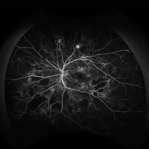
A 43-year-old hypertensive patient, diagnosed with Non-Ischemic Central retinal vein Occlusion in OS, presented with a striking anatomical variation in retinal vasculature. The inferior first-order retinal arteriole after initiating from the optic disc bifurcates, before reaching the fovea, and the superior branch after crossing the midline forms the superior arcade afterwards and produces dichotomous branching as usual. This defies basic anatomical considerations for retinal vasculature as they never cross the midline, also known as the watershed line for retinal vessels.1,2 References: 1. May CA, Rutkowski P. The Horizontal Raphe of the Human Retina and its Watershed Zones. Vision. 2019; 3(4):60. 2. May CA, Rutkowski P. Hypothesis: watershed zones in the human eye are a key for understanding glaucomatous retinal damage. Med Hypotheses. 2017;109:1-5.
Photographer: Dr. Avik Dey Sarkar, MBBS, MS, FVRS, FAICO, Consultant, Department of Vitreoretinal Services, Aravind Eye Hospital, Madurai, India
Imaging device: Wide angled Fundus imaging with Clarus 300
Condition/keywords: background diabetic retinopathy (BDR), Diabetic Retinopathy, retina, vascular anomaly
-
 Proliferative Diabetic Retinopathy Angiography
Jan 22 2024 by Selene Rodríguez-Castro, MD
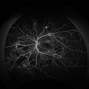
PDR
Photographer: Selene Rodríguez- Castro, APEC
Condition/keywords: background diabetic retinopathy (BDR)
-
 Proliferative Diabetic Retinopathy Angiography
Jan 22 2024 by Selene Rodríguez-Castro, MD
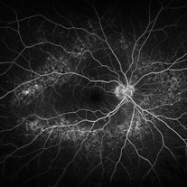
PDR
Photographer: Selene Rodríguez- Castro, APEC
Condition/keywords: background diabetic retinopathy (BDR)
-
 POST CONTROL OF SYSTEMIC DIABETES SHOWING RESOLUTION OF PAPILLOPATHY.
Jun 20 2023 by Deepak Bhojwani, MS
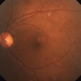
1 MONTH FOLLOW UP FUNDUS IMAGE OF THE SAME PATEINT AFTER GOOD SYSTEMIC CONTROL OF DIABETES. NOTE MARKED REDUCTION OF PAPILLARY RETINOPATHY.
Photographer: DEEPAK BHOJWANI
Condition/keywords: background diabetic retinopathy (BDR)
-
 FELLOW EYE OF DIABETIC PAPILLOPATHY CASE
Jun 20 2023 by Deepak Bhojwani, MS
FUNDUS IMAGE OF FELLOW EYE OF 62 YEAR OLD LADY WITH RIGHT EYE PAPILLOPATHY SHOWING BACKGOUND DIABETIC RETINOPATHY.
Photographer: DEEPAK BHOJWANI
Condition/keywords: background diabetic retinopathy (BDR)
-
 Peripheral Drusen
Jan 19 2022 by Olivia Rainey
Ultra-widefield fluorescein angiogram of an 83-year-old female with Peripheral Drusen affecting both eyes. The patient presented on 1/19/2022 with slightly decreased vision since her last appointment. Her vision was sc20/25-2 in the right eye. The physician did not believe that the peripheral drusen represents AMD and recommended monitoring. The patient also had mild diabetic retinopathy at the time of her visit.
Photographer: Olivia Rainey, OCT-C, COA
Imaging device: Optos California
Condition/keywords: background diabetic retinopathy (BDR), fluorescein angiogram (FA), Optos, Peripheral drusen, staining, ultra-wide field imaging
-
 Diabetic Retinopathy
Mar 14 2021 by Marco Antonio Sauza
DR in a 40-year-old male with DM1.
Photographer: Marco Sauza
Imaging device: Zeiss
Condition/keywords: background diabetic retinopathy (BDR)
-
 Circinate Ring in Diabetic Retinopathy
Mar 27 2019 by Gary R. Cook, MD, FACS
49 year-old white male with moderate NPDR and a circinate lipid ring temporal to the fovea OS; V.A. = 20/25-2.
Imaging device: Topcon VT-50
Condition/keywords: background diabetic retinopathy (BDR), circinate lipid ring, diabetic retinopathy circinate, lipid exudation
-
 Myelinated NFL
Dec 6 2017 by John S. King, MD
Myelinated NFL.
Imaging device: Topcon
Condition/keywords: background diabetic retinopathy (BDR), myelinated nerve fiber layer, myelinated nerve fibers
-
 Diabetic Retinopathy With Laser Photocoagulation.
Sep 10 2017 by JEFFERSON R SOUSA, Tecg.º (Biomedical Systems Technology)
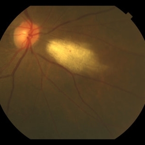
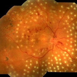
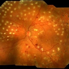
Patient patient 55-year-old, female, attended the clinic with complaint of low visual acuity. It was subjected to laser photocoagulation.
Photographer: JEFFERSON R SOUSA - Study Center and Ophthalmological Research Dr. Andre M V Gomes, Dr. Suel Abujamra Institute São Paulo-Brazil
Imaging device: Topcon TRC-50 DX, Imaginet, 50 degree field. Flash 75, mosaic with eleven images.
Condition/keywords: background diabetic retinopathy (BDR)
-
 Diabetic Retinopathy With Laser Photocoagulation.
Sep 10 2017 by JEFFERSON R SOUSA, Tecg.º (Biomedical Systems Technology)
Patient atient 55-years-old, female, attended the clinic with complaint of low visual acuity. It was subjected to laser photocoagulation.
Photographer: JEFFERSON R SOUSA - Study Center and Ophthalmological Research Dr. Andre M V Gomes, Dr. Suel Abujamra Institute São Paulo-Brazil
Imaging device: Topcon TRC-50 DX, Imaginet, 50 degree field. Flash 75, mosaic with eleven images.
Condition/keywords: background diabetic retinopathy (BDR)
-
 Diabetic Retinopathy Optic Nerve Edema, Fluorescein Angiogram, Stereo
Apr 11 2015 by James B. Soque, CRA, OCT-C, COA, FOPS
Optic Nerve Edema and Leakage on fluorescein angiography in this 48-year-old patient with a 10 year history of diabetes. 50 degree stereo photo fluorescein angiogram.
Photographer: James B. Soque, CRA, COA
Imaging device: Topcon TRC 50 DX, OIS 5 MP Digital Camera, MERGE Software
Condition/keywords: background diabetic retinopathy (BDR), diabetes, disc leakage, fluorescein leakage, optic disc swelling, optic nerve edema, stereo pair
-
 Diabetic Retinopathy, CSME, Exudates, NVD, Color Fundus Photo, Montage
Mar 18 2015 by James B. Soque, CRA, OCT-C, COA, FOPS
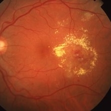
A 58-year-old diabetic male with a longstanding history of diabetic eye disease. Left eye color fundus photo shows extensive CSME, Clinically Significant Macular Edema, with deposits of hard exudates at fixation. There is extensive scattering of hard exudates and sheathing of the vessels.
Photographer: James B Soque, CRA COA
Imaging device: Topcon TRC 50 DX, OIS 5 MP Camera, MERGE software
Condition/keywords: background diabetic retinopathy (BDR), creamy yellow exudates, diabetes, exudates over the posterior pole, neovascularization of the disc (NVD), vessel sheathing
-
 Diabetic Retinopathy, CSME, Color Fundus Photo
Mar 18 2015 by James B. Soque, CRA, OCT-C, COA, FOPS
A 58-year-old diabetic male with a longstanding history of diabetic eye disease. Left eye color fundus photo shows extensive CSME, Clinically Significant Macular Edema, with deposits of hard exudates at fixation. There is extensive scattering of hard exudates and sheathing of the vessels.
Photographer: James B Soque, CRA COA
Imaging device: Topcon TRC 50 DX, OIS 5 MP Camera, MERGE software
Condition/keywords: background diabetic retinopathy (BDR), creamy yellow exudates, diabetes, exudates over the posterior pole, neovascularization of the disc (NVD), vessel sheathing
-
 BDR / Macular Edema / Macroaneurysm
Sep 4 2014 by David Callanan, MD
72-year-old patient, BDR / macular edema / macroaneurysm.
Condition/keywords: background diabetic retinopathy (BDR), macroaneurysm, macular edema
-
 BDR / Macular Edema / Macroaneurysm
Sep 4 2014 by David Callanan, MD
72-year-old patient, BDR / macular edema / macroaneurysm.
Condition/keywords: background diabetic retinopathy (BDR), macroaneurysm, macular edema
-
 BDR / Macular Edema / Macroaneurysm
Sep 4 2014 by David Callanan, MD
72-year-old patient, BDR / macular edema / macroaneurysm.
Condition/keywords: background diabetic retinopathy (BDR), macroaneurysm, macular edema
-
 BDR / Macular Edema / Macroaneurysm
Sep 4 2014 by David Callanan, MD
72-year-old patient, BDR / macular edema / macroaneurysm.
Condition/keywords: background diabetic retinopathy (BDR), macroaneurysm, macular edema
-
 BDR / Macular Edema / Macroaneurysm
Sep 4 2014 by David Callanan, MD
72-year-old patient, BDR / macular edema / macroaneurysm.
Condition/keywords: background diabetic retinopathy (BDR), macroaneurysm, macular edema
-
 BDR / Macular Edema / Macroaneurysm
Sep 4 2014 by David Callanan, MD
72-year-old patient, BDR / macular edema / macroaneurysm.
Condition/keywords: background diabetic retinopathy (BDR), macroaneurysm, macular edema
-
 BDR / Macular Edema / Macroaneurysm
Sep 4 2014 by David Callanan, MD
72-year-old patient, BDR / macular edema / macroaneurysm.
Condition/keywords: background diabetic retinopathy (BDR), macroaneurysm, macular edema
-
 BDR / Macular Edema / Macroaneurysm
Sep 4 2014 by David Callanan, MD
72-year-old patient, BDR / macular edema / macroaneurysm.
Condition/keywords: background diabetic retinopathy (BDR), macroaneurysm, macular edema
-
 BDR / Macular Edema / Macroaneurysm
Sep 4 2014 by David Callanan, MD
72-year-old patient, BDR / macular edema / macroaneurysm.
Condition/keywords: background diabetic retinopathy (BDR), macroaneurysm, macular edema
-
 BDR / Macular Edema / Macroaneurysm
Sep 4 2014 by David Callanan, MD
72-year-old patient, BDR / macular edema / macroaneurysm.
Condition/keywords: background diabetic retinopathy (BDR), macroaneurysm, macular edema
-
 BDR / Macular Edema / Macroaneurysm
Sep 4 2014 by David Callanan, MD
72-year-old patient, BDR / macular edema / macroaneurysm.
Condition/keywords: background diabetic retinopathy (BDR), macroaneurysm, macular edema
 Loading…