Search results (184 results)
-
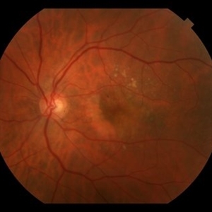
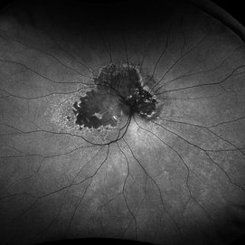
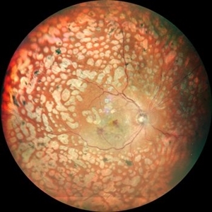
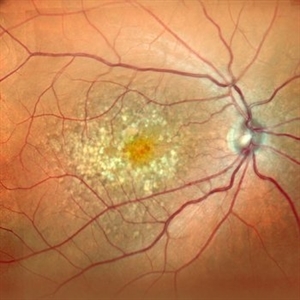
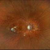 Neovascular AMD with Active CNV
May 22 2025 by Kimberly Wakester
Optomap RGB of an 82-year-old man with Neovascular AMD with Active CNV and Dry AMD in the right eye. There is advanced atrophic changes without subfoveal involvement located temporally to the fovea. Patient is to continue follow up care with dilated exam, repeat OCT, and treatment of intravitreal injection of Vabysmo every 5 weeks at this time.
Photographer: Kimberly Wakester, COA, OCT-C
Imaging device: Optos California
Condition/keywords: advanced geographic atrophy, dry age-related macular degeneration (dry AMD), neovascular age-related macular degeneration (AMD)
-
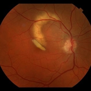
 New Subretinal Hemorrhage in AMD
Jan 8 2025 by Drew Mitchell
HD 1 line 100x OCT scan of a New Subretinal Hemorrhage in a established patient with AMD.
Photographer: Drew Mitchell, OCT-C
Imaging device: Zeiss Cirrus 6000
Condition/keywords: age-related macular degeneration (AMD), OCT, subretinal hemorrhage
-
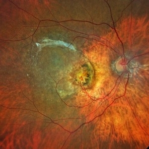
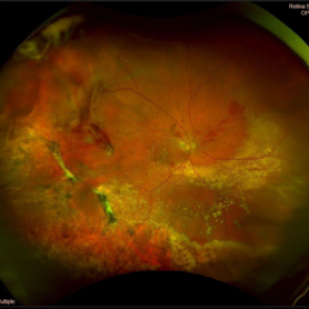 Peripheral Exudative Hemorrhagic Chorioretinopathy
Nov 19 2024 by Toolie Winters
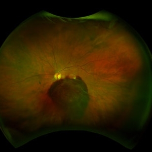
Ultra-wide field fundus photograph of an 85-year-old woman with Peripheral Exudative Hemorrhagic Chorioretinopathy (PECHR) affecting the right eye. Patient presented with a blind spot centrally in the right eye which she first noticed 4 months prior to this image being taken. The patient states that in the month prior to this image, she began noticing bright lights flash across her vision 4-5x/day which last about 15 seconds. The flashes are either black with a blue ring around them or yellow, and their frequency has increased over time. The patient's vision at the time of this appointment was Dcc20/100+1 PHNI. This photo also shows diffuse hemorrhage, lipid, and an eccentric disciform lesion.
Photographer: Toolie Winters
Imaging device: Optos California
Condition/keywords: fundus photography, neovascular age-related macular degeneration (AMD), Optos, OPTOS CALIFORNIA, peripheral exudative hemorrhagic chorioretinopathy (PEHCR), pseudocolor, ultra-wide field imaging, wet age-related macular degeneration (wet AMD)
-
 RPE-Transplantation
Jul 25 2024 by Gabriel Costa Andrade, PhD
Postoperative period of RPE-transplantation in a patient with neovascular AMD after RPE tear.
Photographer: Gabriel Andrade
Condition/keywords: neovascular age-related macular degeneration (AMD), pars plana vitrectomy (PPV), wet age-related macular degeneration (wet AMD)
-
 Drusen
Mar 26 2024 by Akansha Sharma
Autofluorescence photograph of a 52 year old female patient with dry age related macular degeneration.
Photographer: Dr. Akansha Sharma, Bharati Eye Hospital
Condition/keywords: age-related macular degeneration (AMD), drusen, dry age-related macular degeneration (dry AMD)
-
 RPE Rip
Jan 25 2024 by Virginia Gebhart
69 year old female with Neovascular AMD. New RPE rip and increased IRF on OCT 10 weeks s/p Eylea injection. Switched to Vabysmo to extend intervals
Photographer: Virginia Gebhart
Imaging device: Topcon
Condition/keywords: neovascular age-related macular degeneration (AMD)
-
 Dry AMD
Jan 25 2024 by Virginia Gebhart
79 year old female with intermediate dry AMD. Small area of geographic atrophy superior, large drusen and stippled RPE changes. BCVA 20/40
Photographer: Virginia Gebhart
Imaging device: Topcon
Condition/keywords: age-related macular degeneration (AMD), dry age-related macular degeneration (dry AMD), geographic atrophy
-
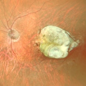
 Hemorrhagic Pigment Epithelial Detachment
Jan 25 2024 by Virginia Gebhart
64 year old male with persistent hemorrhagic PED with oxidized SRH involving the central macula. Continued improvement with 12 week intervals of Eylea. BCVA 20/80
Photographer: Virginia Gebhart
Imaging device: Topcon
Condition/keywords: neovascular age-related macular degeneration (AMD), pigment epithelial detachment (PED)
-
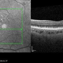 Cuticular Drusen
Jan 17 2024 by John Lee
Heidelberg SD-OCT of a 65-year-old woman with age-related macular degeneration demonstrating classic sawtooth appearance of cuticular drusen.
Photographer: Natasha Vinson
Imaging device: Heidelberg Spectralis
Condition/keywords: age-related macular degeneration (AMD), cuticular drusen
-
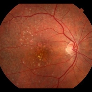
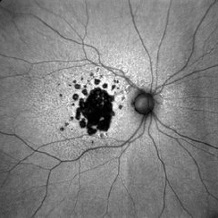
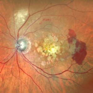
 Geographic Atrophy
Nov 16 2023 by Virginia Gebhart
67 year old female with Neovascular AMD with inactive CNV. Extensive geographic atrophy with minimal foveal sparing. Extensive ectopic CNV just superiorly to ON remains inactive. Discussed with pt treating with Syfovre to slow down GA progression
Photographer: Virginia Gebhart
Imaging device: Optos
Condition/keywords: advanced geographic atrophy, age-related macular degeneration (AMD), dry age-related macular degeneration (dry AMD), geographic atrophy
-
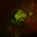 The Phoenix (Mythological)
Aug 23 2023 by Angela Rico
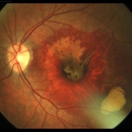
93 year-old female with Neovascular AMD with Active CNV OD
Photographer: Angela Rico M.D.
Imaging device: Optos
Condition/keywords: neovascular age-related macular degeneration (AMD)
-
 AGE RELATED MACULAR DEGENERATION AUTOFLUORESCENCE
Aug 13 2023 by Aditya S Kelkar, MS, FRCS, FASRS,FRCOphth
Autofluorescence fundus photography of an 78-year-old woman diagnosed with age-related macular degeneration.
Photographer: Dr. Harsh Jain, National Institute of Ophthalmology
Imaging device: Clarus 500
Condition/keywords: age-related macular degeneration (AMD)
-
 Retinal angiomatous proliferation (RAP)
Jun 15 2022 by Priyanka Raj, MBBS, MS
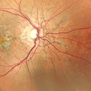
Retinochoroidal anastomosis seen in stage III Retinal angiomatous proliferation (RAP).
Photographer: Sushil Mishra
Imaging device: Zeiss Clarus 500
Condition/keywords: age-related macular degeneration (AMD), retinal angiomatous proliferation (RAP), retinochoroidal anastomosis
-
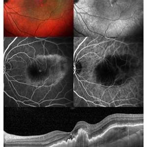
 Macular Degeneration with Extensive Geographic Atrophy
Jan 26 2022 by Olivia Rainey
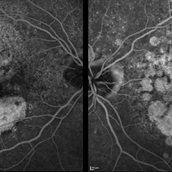
Heidelberg Spectralis fluorescein angiography of a 94-year-old woman with Macular Degeneration affecting both eyes. The FA reveals transmission defects consistent with RPE changes and geographic atrophy of RPE of both eyes, as well as window defects consistent with peripheral scarring in the right eye. The patient's vision was Dcc20/70 in both eyes at the visit the images were taken.
Photographer: Olivia Rainey, OCT-C, COA
Imaging device: Heidelberg Spectralis
Condition/keywords: 30-degrees, choroidal neovascularization (CNV), dry age-related macular degeneration (dry AMD), early phase, fluorescein angiogram (FA), geographic atrophy, heidelberg spectralis, macular degeneration, neovascular age-related macular degeneration (AMD)
-
 Neovascular Age-Related Macular Degeneration (2)
Apr 28 2021 by Ambar Faridi, MD
80-year-old woman with neovascular age-related macular degeneration with interval resolution of large subretinal hemorrhage and vascular exudation followed over time.
Photographer: Jennifer Tu-Bui, VA Portland Health Care System
Condition/keywords: neovascular age-related macular degeneration (AMD), reabsorbing subretinal hemorrhage
-
 Choroidal Neovascular Membrane Evolving With Subretinal Hemorrhage
Apr 23 2021 by Andre Beckenkamp
Wide angle fundus photograph of an 82-year-old woman with dry AMD in her right eye and wet AMD in the left eye, evolving with subretinal hemorrhage and associated serous retinal detachment.
Photographer: Andre Beckenkamp
Imaging device: Optos Daytona
Condition/keywords: age-related macular degeneration (AMD), serous retinal detachment, subretinal hemorrhage, wet age-related macular degeneration (wet AMD)
-
 RPE Tear After Anti-VEGF Injection
Mar 17 2021 by RAFAEL REIS PEREIRA, MD
Retinal pigment epithelium (RPE) tear is a rare devastating complication of age-related macular degeneration (AMD). The believed mechanism lies in an adherence of the neovascularization to the undersurface of the RPE creating a contractile force that increases after VEGF therapy and causes the tear.
Photographer: Rafael Reis, Retina Clinic, São Paulo
Condition/keywords: retinal pigment epithelium (RPE) contracture
-
 Angioid Streaks
Jan 20 2021 by Nivesh Gupta
Fundus photograph of an 51-year-old female patient with angioid streaks with secondary choroidal neovascular membrane.
Photographer: Nivesh Gupta, Retina Fellow, Retina Foundation, Ahmedabad, India
Imaging device: NIDEK SLO MIRANTE
Condition/keywords: age-related macular degeneration (AMD), angioid streaks, choroidal neovascular membrane (CNVM)
-
 CNVM in Pan-retinal Photocoagulated Patient
Dec 30 2020 by ASRS Staff
Wide fundus photograph of 65-year-old, female, diabetic patient.
Imaging device: Nidek Mirante
Condition/keywords: age-related macular degeneration (AMD), diabetes, pan-retinal photocoagulation (PRP)
-
 Neovascular AMD
Jun 27 2020 by Aayesha Khanum
Multicolor image and infra-red image showing fibrovascular pigment epithelial detachment (PED),FFA and ICG confirms the diagnosis., SD-OCT shows notched fibrovascular PED
Photographer: Puttaswamy,Ravikrishna
Imaging device: Heidelberg Spectralis
Condition/keywords: neovascular age-related macular degeneration (AMD)
-
 Dry Age Related Macular Degeneration
Jun 27 2020 by Thirumalesh Mochi Basavaraj, MD
Multiple drusens seen at macula, OCT shows sub RPE deposits of hyper-reflective material.
Photographer: Ravikrishna, Puttaswamy
Imaging device: Heidelberg Spectralis
Condition/keywords: age-related macular degeneration (AMD)
-
 Disciform Scar
Jun 10 2020 by Manish Nagpal, MD, FRCS (UK), FASRS
Disciform scar from wet ARMD.
Photographer: gayathri mohan
Imaging device: nidek slo mirante
Condition/keywords: age-related macular degeneration (AMD), disciform scar
-
 SRNVM
SRNVM
Jun 10 2020 by Manish Nagpal, MD, FRCS (UK), FASRS
Active SRNVM on the temporal edge of drusens.
Photographer: gayathri mohan
Imaging device: nidek slo mirante
Condition/keywords: age-related macular degeneration (AMD), subretinal neovascular membrane
-
 Dry ARMD
Jun 10 2020 by Manish Nagpal, MD, FRCS (UK), FASRS
Drusens in a patient having dry ARMD.
Photographer: gayathri mohan
Imaging device: Nidek Mirante SLO
Condition/keywords: age-related macular degeneration (AMD), drusen
 Loading…