Search results (36 results)
-
 AZOOR
AZOOR
Sep 25 2025 by Hemanth Murthy, MBBS, MD, FASRS
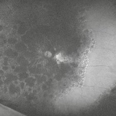
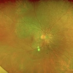
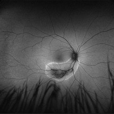
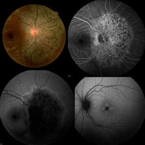
Autofluorescence image of a 72 yr male with history of progressive loss of vision and loos of field of vision more in left eye.It shows an area of normal AF, zone of hypoautofluorescence near the disc and a border of stippled hyper fluorescence.
Photographer: Mr Veda Vyas
Condition/keywords: acute zonal occult outer retinopathy (AZOOR)
-
 AZOOR
AZOOR
Sep 25 2025 by Hemanth Murthy, MBBS, MD, FASRS
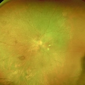
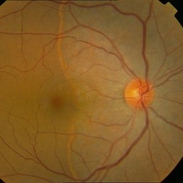
Ultra wide field Fundus image of the left eye of a 72 yr male with history of progressive loss of vision and loos of field of vision more in left eye.
Photographer: Mr Veda Vyas
Condition/keywords: acute zonal occult outer retinopathy (AZOOR)
-
 AZOOR
AZOOR
Sep 25 2025 by Hemanth Murthy, MBBS, MD, FASRS
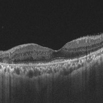
OCT image of left eye of a 72 yr male with history of progressive loss of vision and loos of field of vision more in left eye. The OCT shows a trizonal pattern of outer retinal loss.
Photographer: Mr Veda Vyas
Condition/keywords: acute zonal occult outer retinopathy (AZOOR)
-
 AZOOR
AZOOR
Sep 25 2025 by Hemanth Murthy, MBBS, MD, FASRS
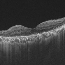
OCT image of right eye of a 72 yr male with history of progressive loss of vision and loos of field of vision more in right eye. It shows a tribunal pattern of outer retina loss
Photographer: Mr Veda Vyas
Condition/keywords: acute zonal occult outer retinopathy (AZOOR)
-
 AZOOR
AZOOR
Sep 25 2025 by Hemanth Murthy, MBBS, MD, FASRS
Ultrawide field Fundus of right eye image of a 72 yr male with history of progressive loss of vision and loos of field of vision more in left eye.
Photographer: Mr Veda Vyas
Condition/keywords: acute zonal occult outer retinopathy (AZOOR)
-
 AZOOR
AZOOR
Sep 25 2025 by Hemanth Murthy, MBBS, MD, FASRS
Autofluorescence image of right eye of a 72 yr male with history of progressive loss of vision and loss of field of vision more in left eye.It shows an area of normal AF, zone of hypoautofluorescence near the disc and a border of stippled hyper fluorescence.
Photographer: Mr Veda Vyas
Condition/keywords: acute zonal occult outer retinopathy (AZOOR)
-
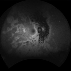
 Acute Zonal Occult Outer Retinopathy (AZOOR) FA, Fluorescein Angiography, Peripheral Vasculitis
Jan 19 2022 by James B. Soque, CRA, OCT-C, COA, FOPS
Acute Zonal Occult Outer Retinopathy (AZOOR). Peripheral Vasculitis OD. Fluorescein angiography showing vasculitis in the far right periphery 8-10 o'clock. 46-year-old white male, VA CC 20/16, 20/12.5, has had recurrent vasculitis for 11 years. No treatment.
Photographer: James Soque, CRA, OCT-C, COA, FOPS, Island Retina, Shirley, NY
Imaging device: Optos California
Condition/keywords: acute zonal occult outer retinopathy (AZOOR), FA early phase, fluorescein angiogram (FA), Peripheral Vasculitis, ultra-wide field imaging
-
 Acute Zonal Occult Outer Retinopathy (AZOOR) FA, Ultra Wide-Field Fluorescein Angiogram Early
Jan 19 2022 by James B. Soque, CRA, OCT-C, COA, FOPS
Acute Zonal Occult Outer Retinopathy, FA, Fluorescein Angiography, OD. 46-year-old white male, VA CC 10/16, 20/12.5, has had recurrent vasculitis for 11 years. No treatment.
Photographer: James Soque, CRA, OCT-C, COA, FOPS, Island Retina, Shirley, NY
Imaging device: Optos California
Condition/keywords: acute zonal occult outer retinopathy (AZOOR), FA EARLY, fluorescein angiogram (FA), ultra-wide field imaging
-
 Acute Zonal Occult Outer Retinopathy, (AZOOR) FAF, Fundus Autofluorescence
Jan 19 2022 by James B. Soque, CRA, OCT-C, COA, FOPS
Acute Zonal Occult Outer Retinopathy, FAF, Fundus Auto Fluorescence, OD. 46-year-old white male, VA CC 10/16, 20/12.5, has had recurrent vasculitis for 11 years. No treatment.
Photographer: James Soque, CRA, OCT-C, COA, FOPS, Island Retina, Shirley, NY
Imaging device: Optos California
Condition/keywords: acute zonal occult outer retinopathy (AZOOR), fundus autofluorescence (FAF), ultra-wide field imaging
-
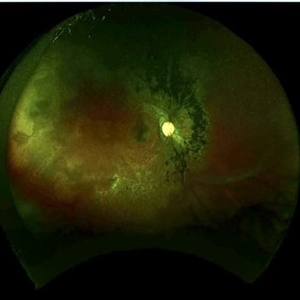
 Acute Zonal Occult Outer Retinopathy (AZOOR)
Jan 19 2022 by James B. Soque, CRA, OCT-C, COA, FOPS
Acute Zonal Occult Outer Retinopathy, OD. Color fundus, Ultra-wide field photograph. 46-year-old white male, VA CC 10/16, 20/12.5, has had recurrent vasculitis for 11 years. No treatment.
Photographer: James Soque, CRA, OCT-C, COA, FOPS, Island Retina, Shirley, NY
Imaging device: Optos California
Condition/keywords: acute zonal occult outer retinopathy (AZOOR), color wide field, optos, ultra-wide field imaging
-
 Acute Zonal Occult Outer Retinopathy
Dec 16 2020 by Robert C Wann, MD
Fundus autofluorescence of a 28-year-old female with AZOOR.
Photographer: Retina Consultants of Alabama
Imaging device: Optos
Condition/keywords: acute zonal occult outer retinopathy (AZOOR)
-
 Acute Zonal Occult Outer Retinopathy
Dec 16 2020 by Robert C Wann, MD
Fundus autofluorescence of a 28-year-old female with AZOOR.
Photographer: Retina Consultants of Alabama
Imaging device: Optos
Condition/keywords: acute zonal occult outer retinopathy (AZOOR)
-
 Acute Zonal Occult Outer Retinopathy
Dec 16 2020 by Robert C Wann, MD
Fundus photo of a 28-year-old female with AZOOR.
Photographer: Retina Consultants of Alabama
Imaging device: Optos
Condition/keywords: acute zonal occult outer retinopathy (AZOOR)
-
 Unilateral AZOOR
May 4 2020 by Iuri Golubev, MD
Color, FA and FAF photos of a 17-year-old female w/h/o unilateral AZOOR OD for 5 years. FAF OS demonstrates an intact fundus.
Condition/keywords: acute zonal occult outer retinopathy (AZOOR)
-
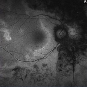
 FA ICG AZOOR
Oct 14 2017 by Navneet Mehrotra, DNB
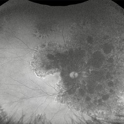
Fundus autofluorescence OS showing peripapillary hypoautofluorescence surrounded by an area of hyperautofluorescence with well demarcated margins suggestive of AZOOR.
Photographer: Ashish jain, Retina Foundation, Ahmedabad
Imaging device: Heidelberg spectralis
Condition/keywords: acute zonal occult outer retinopathy (AZOOR)
-
 FA ICG AZOOR
Oct 14 2017 by Navneet Mehrotra, DNB
fundus autofluorescence OD showing peripapillary hypoautofluorescence surrounded by an area of hyperautofluorescence with well demarcated margins suggestive of AZOOR.
Photographer: Ashish jain, Retina Foundation, Ahmedabad
Imaging device: Heidelberg spectralis
Condition/keywords: acute zonal occult outer retinopathy (AZOOR)
-
 AZOOR
AZOOR
Mar 19 2015 by Niloofar Piri, MD
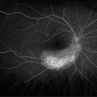
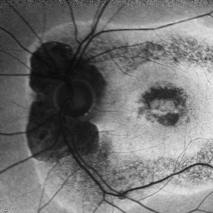
#2 : Fundus autofluorescence OS in the same patient demonstrates more severe changes ; peripapillary hypoAF and concentric rings of hyper and hypo AF in posterior pole
Imaging device: Heidelberg Spectralis
Condition/keywords: acute zonal occult outer retinopathy (AZOOR)
-
 AZOOR
AZOOR
Mar 19 2015 by Niloofar Piri, MD
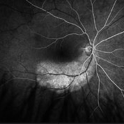
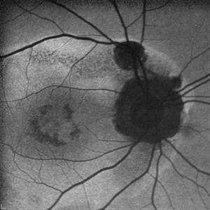
#1: Fundus autofluorescence OD in a patient with AZOOR demonstrates characteristic peripapillary hypoAF as well as concentric rings of hypo and hyper AF in posterior pole .
Imaging device: Heidelberg Spectralis
Condition/keywords: acute zonal occult outer retinopathy (AZOOR)
-
 AZOOR vs. AAOOR
Mar 19 2014 by Ali Tavallali, MD, FASRS
FAF of a 47-year-old female with 20/20 VA of both eyes, note the progression of demarcation line after 4 months
Photographer: Neda Sheibani, Dr. Khodadoust Eye Hospital, Shiraz, Iran
Condition/keywords: acute zonal occult outer retinopathy (AZOOR)
-
 AZOOR vs. AAOOR
Mar 19 2014 by Ali Tavallali, MD, FASRS
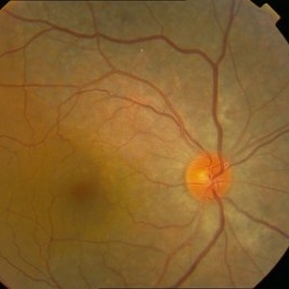
Color fundus photograph of a 47-year-old female with 20/20 VA of both eyes, note the progression of demarcation line after 4 months
Photographer: Neda Sheibani, Dr. Khodadoust Eye Hospital, Shiraz, Iran
Condition/keywords: acute zonal occult outer retinopathy (AZOOR)
-
 AZOOR vs. AAOOR
Mar 19 2014 by Ali Tavallali, MD, FASRS
Color fundus photograph of a 47-year-old female with 20/20 VA of both eyes, note the demarcation line
Photographer: Neda Sheibani, Dr. Khodadoust Eye Hospital, Shiraz, Iran
Condition/keywords: acute zonal occult outer retinopathy (AZOOR)
-
 Autofluorescence 10-14-13 AZOOR
Dec 14 2013 by Robert T. Wendel, MD
AF image
Imaging device: Heidelberg
Condition/keywords: acute zonal occult outer retinopathy (AZOOR)
-
 Autofluorescence 12-5-13 AZOOR
Dec 14 2013 by Robert T. Wendel, MD
AF 12-5-16
Condition/keywords: acute zonal occult outer retinopathy (AZOOR), autofluorescence imaging
-
 Autofluorescence 10-14-13 AZOOR
Dec 14 2013 by Robert T. Wendel, MD
Autofluorescence 10-14-13 AZOOR
Condition/keywords: acute zonal occult outer retinopathy (AZOOR), autofluorescence imaging
-
 Autofluorescence 12-5-13 AZOOR
Dec 14 2013 by Robert T. Wendel, MD
Autofluorescence 12-5-13 AZOOR
Condition/keywords: acute zonal occult outer retinopathy (AZOOR), autofluorescence imaging
 Loading…