Search results (87 results)
-
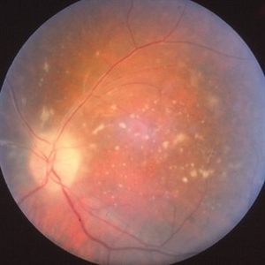
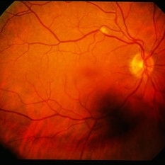
 Acute Retinal Necrosis (resolved)
Aug 21 2025 by Virginia Gebhart
31 year old female with resolved Acute Retinal Necrosis. Pt was positive for HSV2 in 2019, received intravitreal Ganciclovir x3. Both eyes remain stable with no active inflammation, retinitis remains inactive with pigmented edges. Pt continues taking Valtrex qday for maintenance. BCVA 20/40
Photographer: Virginia Gebhart, Retina Consultants of Carolina
Imaging device: Optos California
Condition/keywords: acute retinal necrosis, retinitis
-
 Acute Retinal Necrosis (ARN)
Jul 3 2025 by Heitor Nogueira
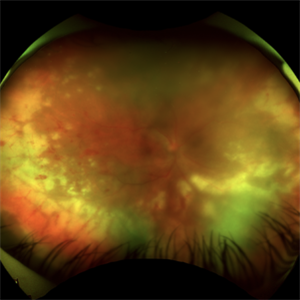
Fundus photograph of an 63-year-old woman who reported unilateral visual acuity loss for 10 days associated with ocular pain. He presented conjunctival hyperemia with temporal and nasal nodular scleritis, anterior chamber reaction 2+/4+, Koeppe nodules, granulomatous PKs, vitreitis 2+/4+, multiple areas of vasculitis in the arcades and periphery, associated with hemorrhages and necrotizing retinitis in the temporal, inferior and nasal periphery. Positive serology for Herpes Virus
Photographer: Heitor Nogueira, Penido Burnier Institute, Campinas, São Paulo, Brazil
Imaging device: Optos Daytona
Condition/keywords: ARN complications, Herpes, progressive outer retinal necrosis (PORN), Uveitis
-
 Acute Retinal Necrosis
Jul 3 2025 by Heitor Nogueira
Fundus photograph of an 53-year-old woman with patient who reported unilateral visual acuity loss for 10 days associated with ocular pain. She presented conjunctival hyperemia with temporal and nasal nodular scleritis, anterior chamber reaction 2+/4+, Koeppe nodules, granulomatous PKs, vitritis 2+/4+, multiple areas of vasculitis in arcades and periphery, associated with hemorrhages and necrotizing retinitis in temporal, inferior and nasal periphery. patient who reported unilateral visual acuity loss for 10 days associated with ocular pain. He presented conjunctival hyperemia with temporal and nasal nodular scleritis, anterior chamber reaction 2+/4+, Koeppe nodules, granulomatous PKs, vitreitis 2+/4+, multiple areas of vasculitis in the arcades and periphery, associated with hemorrhages and necrotizing retinitis in the temporal, inferior and nasal periphery. Positive serology for Herpes Virus.
Photographer: Heitor Nogueira, Penido Burnier Institute and CHOV, Campinas, São Paulo, Brazil
Imaging device: Optos Daytona
Condition/keywords: ARN complications, Herpes, progressive outer retinal necrosis (PORN)
-
 Pigmented KPs
Dec 8 2023 by Nassim Alejandro Abreu Arbaje, MD
Anterior segment photograph of a 27 year old female diagnosed with an Acute Retinal Necrosis. In the picture we can see mutton fat keratic precipitates already pigmented.
Photographer: Nassim Abreu
Imaging device: Alcon NGenuity Systems
Condition/keywords: Acute Retinal Necrosis, mutton-fat keratic precipitates (KP), Uveitis
-
 Acute Retinal Necrosis with Retinal Detachment
Jan 13 2022 by Tuan Tran, MBBS, MMed (OphthSc), FRANZCO, DRCPSC
Acute Retinal Necrosis with Retinal Detachment.
Photographer: Tuan Tran
Condition/keywords: Acute Retinal Necrosis with Retinal Detachment, retinal necrosis
-
 Left Acute Retinal Necrosis
Jan 11 2022 by Tuan Tran, MBBS, MMed (OphthSc), FRANZCO, DRCPSC
84 year-old gentleman presenting with left acute retinal necrosis.
Photographer: Tuan Tran
Imaging device: Optos widefield
Condition/keywords: ARN complications
-
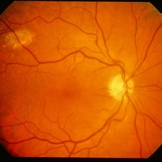
 Acute Retinal Necrosis
May 31 2021 by Aditya S Kelkar, MS, FRCS, FASRS,FRCOphth
Fundus photograph of 43-year-old female with left eye acute retinal necrosis.
Imaging device: Clarus 500
Condition/keywords: acute retinal necrosis
-
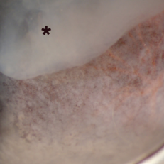 Toxoplasmic Acute Retinal Necrosis
May 18 2020 by McGill University Health Centre
Toxoplasmosis can have several manifestations in the eye, of which toxoplasmic acute retinal necrosis has the worst prognosis. This enucleation specimen shows extensive retinal necrosis with multiple coalescent foci. The vitreous is hazy (*).
Condition/keywords: acute retinal necrosis, toxoplasmosis
-
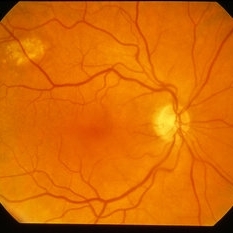
 Primary Intraocular Lymphoma
Nov 20 2019 by McGill University Health Centre
73-year-old man with retinal vasculitis and acute retinal lesions of the left eye. Optic nerve and retinal infiltrates consistent with acute retinal necrosis.
Condition/keywords: acute retinal necrosis, primary intraocular lymphoma
-
 ARN (#3) This is comparison between the latest visit (left) and one week prior (which is the right photo, and same one as photo #2)
May 27 2019 by John S. King, MD
60-year-old African American female who had been treated for iridocyclitis for at least a week sent in for vitritis and a nasal fundus lesion. Complaints included redness, floaters, photophobia, and decreased vision. Husband had recent shingles. Acuity was 20/60-2 with IOP of 12, and small KP in Art's triangel, 1-2+ a/c cell, 2-3+ ant vit cell, diffuse arteriolar sheathing, multiple areas of retinal whitening in periphery and mid-periphery (see Photo #1). PCR of a/c was performed, and intravitreal GCV administered, and VACV 2g qid and ASA started.... PCR positive for HZV, pred taper was started two days after presentation as the infection had begun to stablize..... Five days from presentation the vision was 20/60, inflammation and areas of retinal whitening had improved (see Photo #2).... One week later acuity was 20/30, the a/c was quiet and KP resolved; ant vitreous cell decreased; and there was further improvement in retinal appearance without any signs of retinal holes or detachment; she is now on low dose maint VACV (see photo#3)
Photographer: Maysee Yang
Imaging device: Optos CA
Condition/keywords: acute retinal necrosis, Herpes zoster
-
 ARN (#2) Five Days Since Initial Visit
May 27 2019 by John S. King, MD
60-year-old African American female who had been treated for iridocyclitis for at least a week sent in for vitritis and a nasal fundus lesion. Complaints included redness, floaters, photophobia, and decreased vision. Husband had recent shingles. Acuity was 20/60-2 with IOP of 12, and small KP in Art's triangel, 1-2+ a/c cell, 2-3+ ant vit cell, diffuse arteriolar sheathing, multiple areas of retinal whitening in periphery and mid-periphery (see Photo #1). PCR of a/c was performed, and intravitreal GCV administered, and VACV 2g qid and ASA started.... PCR positive for HZV, pred taper was started two days after presentation as the infection had begun to stablize..... Five days from presentation the vision was 20/60, inflammation and areas of retinal whitening had improved (see Photo #2).... One week later acuity was 20/30, the a/c was quiet and KP resolved; ant vitreous cell decreased; and there was further improvement in retinal appearance without any signs of retinal holes or detachment; she is now on low dose maint VACV (see photo#3)
Photographer: Maysee Yang
Imaging device: Optos CA
Condition/keywords: acute retinal necrosis, Herpes zoster
-
 ARN (#1) Initial Photo
May 27 2019 by John S. King, MD
60-year-old African American female who had been treated for iridocyclitis for at least a week sent in for vitritis and a nasal fundus lesion. Complaints included redness, floaters, photophobia, and decreased vision. Husband had recent shingles. Acuity was 20/60-2 with IOP of 12, and small KP in Art's triangel, 1-2+ a/c cell, 2-3+ ant vit cell, diffuse arteriolar sheathing, multiple areas of retinal whitening in periphery and mid-periphery (see Photo #1). PCR of a/c was performed, and intravitreal GCV administered, and VACV 2g qid and ASA started.... PCR positive for HZV, pred taper was started two days after presentation as the infection had begun to stablize..... Five days from presentation the vision was 20/60, inflammation and areas of retinal whitening had improved (see Photo #2).... One week later acuity was 20/30, the a/c was quiet and KP resolved; ant vitreous cell decreased; and there was further improvement in retinal appearance without any signs of retinal holes or detachment; she is now on low dose maint VACV (see photo#3)
Photographer: Maysee Yang
Imaging device: Optos CA
Condition/keywords: acute retinal necrosis, Herpes zoster
-
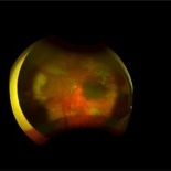
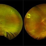
 Acute Retinal Necrosis with Proliferative Vitreoretinopathy and Total Retinal Detachment
Mar 26 2019 by Gary R. Cook, MD, FACS
Same WF patient 9 weeks after initial presentation with Acute Retinal Necrosis now with proliferative vitreoretinopathy and a total combined traction & rhegmatogenous retinal detachment
Imaging device: Topcon VT-50
Condition/keywords: acute retinal necrosis, proliferative vitreoretinopathy (PVR), tractional retinal detachment
-
 Acute Retinal Necrosis
Mar 26 2019 by Gary R. Cook, MD, FACS
Left eye of same patient with acute retinal necrosis who developed rhegmatogenous RD seven weeks after presentation.
Imaging device: Topcon VT-50
Condition/keywords: acute retinal necrosis
-
 Acute Retinal Necrosis
Mar 26 2019 by Gary R. Cook, MD, FACS
Middle-aged white female with peripheral retinal lesions of acute retinal necrosis OS at presentation.
Imaging device: Topcon VT-50
Condition/keywords: acute retinal necrosis
-
 Acute Retinal Necrosis
Mar 26 2019 by Gary R. Cook, MD, FACS
Middle-aged white female with ARN OS showing additional involvement within 2 weeks. Patient was seen prior to the availability of anti-viral therapies.
Imaging device: Topcon VT-50
Condition/keywords: acute retinal necrosis
-
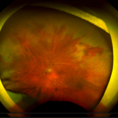
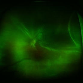
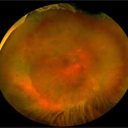 Acute Retinal Necrosis secondary to Herpes Zoster Ophthalmicus
Jan 9 2018 by Olivia Rainey
Ultra-wide field Optos pseudocolor montage of an 40-year-old female presenting with acute retinal necrosis secondary to herpes zoster ophthalmicus affecting her right eye.
Photographer: Olivia Rainey
Imaging device: Optos California
Condition/keywords: acute retinal necrosis, color fundus photograph, Herpes zoster, montage, Optos, ultra-wide field imaging
-
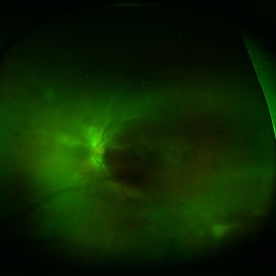
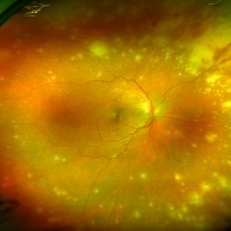 Acute Retinal Necrosis (ARN)
Dec 13 2017 by Gabriel Costa Andrade, PhD
Healthy 47-year-old patient presenting with subacute decline in vision, vitritis, periphlebits, and necrotizing retinitis.
Photographer: Gabriel Andrade, RETINA CLINIC, SP
Imaging device: Optos Wide Field Camera
Condition/keywords: acute retinal necrosis, uveitis, vasculitis
-
 Acute Retinal Necrosis (ARN)-Detached
Dec 30 2015 by Nader Moinfar, MD, MPH, FACS, FASRS
Same patient with PCR-diagniosed VZV ARN presenting with RRD six weeks after initiation of therapy (systemic avcylovir, intravitreal foscarnet, prednisone, aspirin and 360 laser barricade).
Imaging device: Optos Wide Field Fundus Camera
Condition/keywords: acute retinal necrosis
-
 ARN-Regressing
Dec 30 2015 by Nader Moinfar, MD, MPH, FACS, FASRS
Same patient, one month after initiation of therapy of PCR-proven VZV ARN; treatment included iv acyclovir, intravitreal foscarnet, systemic steroids, aspirin and prophylactic 360 laser barricade.
Imaging device: Optos Wide Field Camera
Condition/keywords: acute retinal necrosis
-
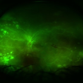 Acute Retina Necrosis-Active
Dec 30 2015 by Nader Moinfar, MD, MPH, FACS, FASRS
Healthy 55-year-old patient presenting with subacute decline in vision, vitritis, periphlebits, and necrotizing retinitis.
Imaging device: Optos Wide Field Camera
Condition/keywords: acute retinal necrosis
-
 CMV Retinitis vs. Possible ARN
Apr 7 2014 by David Callanan, MD
62-year-old Hispanic male.
Condition/keywords: acute retinal necrosis, CMV retinitis
-
 CMV Retinitis vs. Possible ARN
Apr 7 2014 by David Callanan, MD
62-year-old Hispanic male.
Condition/keywords: acute retinal necrosis, CMV retinitis
-
 CMV Retinitis vs. Possible ARN
Apr 7 2014 by David Callanan, MD
62-year-old Hispanic male.
Condition/keywords: acute retinal necrosis, CMV retinitis
-
 CMV Retinitis vs. Possible ARN
Apr 7 2014 by David Callanan, MD
62-year-old Hispanic male.
Condition/keywords: acute retinal necrosis, CMV retinitis
 Loading…